Brain restoration with plasmalogens

In this 2023 presentation for a professional audience, Dr. Dayan Goodenowe showed an example of what could be done (in the form of what he personally did at ages 53-54) to restore and augment brain structure and function over a 17-month period by taking plasmalogens and supporting supplements:

https://drgoodenowe.com/recording-of-dr-goodenowes-presentation-from-the-peptide-world-congress-2023-is-now-available/

Follow the video along with its interactive transcript. Restorative / augmentative supplements included:

1. Nutritional Supplementation Strategy

Forms of MRI used to document brain structure and function changes were:

2. Advanced MRI Technologies

Brain volume decreases are the rule for humans beginning at age 40. Dr. Goodenowe documented brain volume increases, which aren’t supposed to happen, but did per the below slide of overall results:

3. Reversing Brain Shrinkage

“From a global cortical volume and thickness perspective, 17 months of high-dose plasmalogens reversed ~15 years of predicted brain deterioration.”


Specific increased adaptations in brain measurements over 17 months included:

  1. Cortical thickness .07/2.51 = +3%.
  2. White matter microstructure fractional anisotropy +8%.
  3. Nucleus accumbens volume +30%.
  4. Dopaminergic striatal terminal fields’ volume +18%.
  5. Cholinergic cortical terminal fields’ volume +10%.
  6. Occipital cortex volume +10%.
  7. Optic chiasm volume +225%.
  8. Nucleus basalis connectivity.
  9. Neurovascular coupling signal controlled by noradrenaline integrity.
  10. Amygdala volume +4% and its connectivity to the insula, indicating ongoing anxiety and emotional stress response.
  11. Parahippocampus volume +7%.
  12. Hippocampus fractional anisotropy +5%.

No changes:

  1. Amygdala connectivity to the ventral lateral prefrontal cortex, the same part of the brain that relates to placebo effect.
  2. Hippocampus connectivity.

Decreased adaptations in brain measurements included:

  1. White matter microstructure radial diffusivity -10%.
  2. Amygdala connectivity to the anterior cingulate cortex to suppress / ignore / deny anxiety response.
  3. Amygdala connectivity to the dorsal lateral prefrontal cortex.
  4. Entorhinal cortex volume -14%.
  5. Hippocampus volume -6%.
  6. Hippocampus mean diffusivity (white matter improved, with more and tighter myelin) -4%.

The other half of this video was a lively and wide-ranging Q&A session.


The referenced 2023 study of 653 adults followed over ten years showed what brain deterioration could be expected with no interventions. Consider these annual volume decrease rates to be a sample of a control group:

etable 3

https://jamanetwork.com/journals/jamanetworkopen/fullarticle/2806488 “Characterization of Brain Volume Changes in Aging Individuals With Normal Cognition Using Serial Magnetic Resonance Imaging”

Also see a different population’s brain shrinkage data in Prevent your brain from shrinking.


The daily plasmalogen precursor doses Dr. Goodenowe took were equivalent to 100 mg softgel/kg, double the maximum dose of 50 mg softgel/kg provided during the 2022 clinical trial of cognitively impaired old people referenced in Plasmalogens Parts 1, 2, and 3.

He mentions taking 5 ml in the morning and 5 ml at night because he used the Prodrome oil products. 1 ml of a Prodrome oil plasmalogen precursor product equals 900 mg of their softgel product.


“My brain is trying to minimize long-term effects of pain/stress by suppressing my memory of it. But this can only go on for so long before it becomes an entrenched state.

I have solved the sustenance side of the equation. I need to work harder to solve the environmental side.”

While I agree that we each have a responsibility to ourselves to create an environment that’s conducive to our health, the above phenomenon isn’t necessarily resolvable by changing an individual’s current environment. My understanding is that long-term effects of pain, stress, and related human experiences are usually symptoms of causes that started much earlier in our lives.

Adjusting one’s present environment may have immediate results, but probably won’t have much therapeutic impact on long-term issues. Early life memories and experiences are where we have to gradually go in order to stop being driven by what happened back then.

See Dr. Arthur Janov’s Primal Therapy for its principles and explanations. I started Primal Therapy at a similar age, 53, and continued for three years.
Ergothioneine dosing

Four 2023 papers that outlined or used different ergothioneine doses, starting with a human/rodent study:

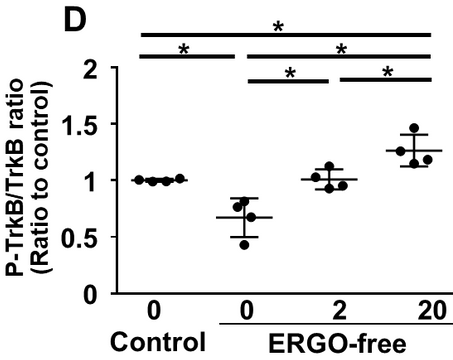
“We found that cognitive function and hippocampal neurogenesis were lower in mice fed an ERGO-free diet than in those fed the control diet. Mice fed an ERGO-free diet were orally administered ERGO (0, 2, and 20 mg/kg) for two weeks which reversed these effects.

trkb ratio

Phosphorylated brain-derived neurotrophic factor receptor TrkB, the activated form of TrkB, was also detected in extracellular vesicles (EVs) derived from serum samples of 52 volunteers who had been orally administered ERGO-containing tablets (5 mg/day for 12 weeks). The ratio of serum EV-derived phosphorylated TrkB was significantly higher in the ERGO-treated group than in the placebo-treated group and was positively correlated with both serum ERGO concentrations and several cognitive domain scores from Cognitrax.

cognitrax

The ratio of p-TrkB to TrkB in serum EVs was proposed as a quantitative diagnostic marker of long-term ERGO-induced cognitive improvement.”

https://www.researchsquare.com/article/rs-2626422/v1 “TrkB phosphorylation in serum extracellular vesicles correlates with cognitive function enhanced by ergothioneine in humans”

Human equivalents of all rodent ergothioneine doses were higher than the 5 mg/day for 12 weeks 2020 human study, cited as Reference 21. I couldn’t access that paper, so here’s its Abstract:

Effect of ergothioneine on the cognitive function improvement in healthy volunteers and mild cognitive impairment subjects – a randomized, double-blind, parallel-group comparison study

“These results indicate that continuous intake of ergothioneine improves cognitive function in healthy subjects.”


A rodent study compared effects of a fermented product with 0.1 and 1.0 mg/g (human equivalent 6 mg (1 mg x .081) x  70 kg) ergothioneine doses:

“Our present study demonstrated for the first time the preventive effect of Rice-koji fermented extracts made by Aspergillus oryzae on anxiety, impaired recognition, and nociception using a psychophysically stressed model. Our results also demonstrated preventive effects of ergothioneine (EGT) on stress-induced anxiety- and pain-like behaviors.

Daily administration of High dose Rice-koji or 0.1 mg/kg EGT decreased anxiety- and pain-like behaviors. These findings suggest that inhibitory effects of Rice-koji on psychological stress might be mediated through the actions of EGT.”

https://www.mdpi.com/2072-6643/15/18/3989 “Preventive Roles of Rice-koji Extracts and Ergothioneine on Anxiety- and Pain-like Responses under Psychophysical Stress Conditions in Male Mice”


Here’s one of several reviews that cited a 2017 clinical trial (duplicately Reference 39 and 61 for some reason) of 5 and 25 mg ergothioneine doses:

“In this pharmacokinetic study, forty-five healthy humans received placebo, 5, or 25 mg encapsulated ergothioneine/d for 7 d and were followed up for an additional 4 weeks. Ergothioneine was rapidly absorbed and largely retained by the body, with large increases in plasma ergothioneine levels and only minimal increases (<4 %) in urinary excretion observed. While plasma levels of ergothioneine decreased when supplementation was withdrawn, levels in whole blood continued to increase in a dose–response fashion, reaching maximal levels 3 weeks after withdrawal of supplement, which were sustained at 4 weeks follow-up.

A large difference in basal concentrations of ergothioneine in whole blood was observed. Participants with the highest basal levels of ergothioneine also appeared to take up more of supplemented ergothioneine.”

https://www.cambridge.org/core/journals/british-journal-of-nutrition/article/ergothioneine-an-underrecognised-dietary-micronutrient-required-for-healthy-ageing/92CED7FF201A9FB23BEAFF0D3EAD7316 “Ergothioneine: an underrecognised dietary micronutrient required for healthy ageing?”


Wrapping up with a deep dive into seven mushroom varieties’ compounds:

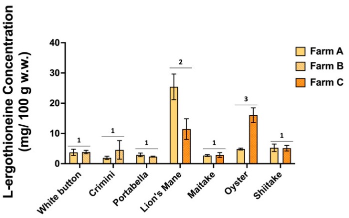
“Mushrooms contain multiple essential nutrients and health-promoting bioactive compounds, including amino acid L-ergothioneine. We compared metabolomes of fresh raw white button, crimini, portabella, lion’s mane, maitake, oyster, and shiitake mushrooms using untargeted liquid chromatography mass spectrometry (LC/MS)-based metabolomics.

Results indicate significantly higher concentrations of L-ergothioneine in lion’s mane and oyster mushrooms compared to the remaining five mushroom varieties, which had concentrations ranging from 1.94 ± 0.55 to 5.26 ± 1.23 mg/100 g wet weight (mean ± SD). There was also variability in concentration of L-ergothioneine between mushroom varieties of the same farm. Different numbers denote significance (p < 0.05).

foods-12-02985-g008

Mushrooms and their bioactive extracts are considered functional foods. Mushrooms have several bioactive compounds, including polysaccharides, lectins, terpenoids, sterols, and alkaloids, among others, which may positively impact health.

Cell walls of mushrooms contain polysaccharides, including β-glucans and chitin, which positively affect health, through modulating the immune system and protecting the cardiovascular system through improvements in glucose and lipid metabolism. Effects on the cardiovascular system are also attributable to lovastatin and polyphenols, known for their lipid-lowering and antioxidant properties, respectively.

While the 1344 compounds in common among the seven mushroom varieties support some level of similarity, detection of hundreds of unique-to-mushroom-variety compounds and differences in amino acid profiles indicate that not all mushrooms are chemically comparable. Given detection of >400 unique-to-mushroom-variety compounds in lion’s mane, maitake, oyster, and shiitake mushrooms, we suggest further targeted investigations on compounds detected and potential health benefits.”

https://www.mdpi.com/2304-8158/12/16/2985 “Metabolomics Profiling of White Button, Crimini, Portabella, Lion’s Mane, Maitake, Oyster, and Shiitake Mushrooms Using Untargeted Metabolomics and Targeted Amino Acid Analysis”

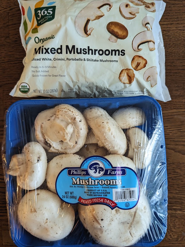
I eat around 200 grams of mushrooms daily, having temporarily overridden the boredom of eating AGE-less chicken vegetable soup every day. I prep all the top package’s frozen umami bomb (283 grams) and half of the bottom’s fresh mushrooms (340 grams) into the soup:

PXL_20230921_193708552

It makes servings for three days, including one for prep day dinner. I’d guess from “concentrations ranging from 1.94 ± 0.55 to 5.26 ± 1.23 mg/100 g (mean ± SD)” that my daily mushroom ergothioneine dose is around 7 mg ((1.94 mg + 5.26 mg) / 2) = 3.6 mg per 100 grams x 2 (for 200 grams).

Continued in Part 2.

Nrf2 Week #2: Neurons

To follow the Nrf2 Week #1 suggestion that Nrf2 target neurological disorders, this 2023 cell study investigated Nrf2 expression in neurons:

“Oxidative metabolism is inextricably linked to production of reactive oxygen species (ROS), which have the potential to damage all classes of macromolecules. Yet ROS are not invariably detrimental. Several properties make ROS useful signaling molecules, including their potential for rapid modification of proteins and close ties to cellular metabolism.

We used multiple single cell genomic datasets to explore Nrf2 expression and regulation in hundreds of neuronal and non-neuronal cell types in mouse and human. With few exceptions, Nrf2 is expressed at far lower levels in neurons than in non-neuronal support cells in both species.

This pattern is maintained in multiple disease states, and the chromatin accessibility landscape at the Nrf2 locus parallels these expression differences. These results imply that Nrf2 activity is limited in almost all neurons of the mouse and human central nervous system (CNS).

nrf2 expression

We separated cell types into neuron or non-neuronal ‘support’ cell categories. The general ‘support’ term is not meant to minimize the functional relevance of non-neuronal cells in the CNS, but is an umbrella term meant to cover everything from glial cell types (astrocytes, microglia, oligodendrocytes) to endothelial cells.

It is not clear why an important, near ubiquitous cytoprotective transcription factor like Nrf2 remains off in mature neurons, especially considering oxidative stress is a driver of many diseases. The simplest explanation is that Nrf2 activity also disrupts normal function of mature neurons.

ROS play a key role in controlling synaptic plasticity in mature neurons. These activity-dependent changes in synaptic transmission, which are important for learning and memory, are disrupted by antioxidants.

A subset of important Nrf2-targeted antioxidant genes (e.g., Slc3a2, Slc7a11, Nqo1, Prdx1) are also low in neurons. So it is likely that these and/or other Nrf2 targets must remain low or non-ROS-responsive in mature neurons. Future work exploring why this expression pattern persists in mature neurons will inform our models on roles of antioxidant genes in normal neuronal physiology and in neurological disorders.

https://www.biorxiv.org/content/10.1101/2023.05.09.540014v1.full “Limited Expression of Nrf2 in Neurons Across the Central Nervous System”


PXL_20230520_182827767

Taurine week #7: Brain

Finishing a week’s worth of 2022 taurine research with two reviews of taurine’s brain effects:

“We provide a overview of brain taurine homeostasis, and review mechanisms by which taurine can afford neuroprotection in individuals with obesity and diabetes. Alterations to taurine homeostasis can impact a number of biological processes such as osmolarity control, calcium homeostasis, and inhibitory neurotransmission, and have been reported in both metabolic and neurodegenerative disorders.

Models of neurodegenerative disorders show reduced brain taurine concentrations. On the other hand, models of insulin-dependent diabetes, insulin resistance, and diet-induced obesity display taurine accumulation in the hippocampus. Given cytoprotective actions of taurine, such accumulation of taurine might constitute a compensatory mechanism that attempts to prevent neurodegeneration.

nutrients-14-01292-g003

Taurine release is mainly mediated by volume-regulated anion channels (VRAC) that are activated by hypo-osmotic conditions and electrical activity. They can be stimulated via glutamate metabotropic (mGluR) and ionotropic receptors (mainly NMDA and AMPA), adenosine A1 receptors (A1R), and metabotropic ATP receptors (P2Y).

Taurine mediates its neuromodulatory effects by binding to GABAA, GABAB, and glycine receptors. While taurine binding to GABAA and GABAB is weaker than to GABA, taurine is a rather potent ligand of the glycine receptor. Reuptake of taurine occurs via taurine transporter TauT.

Cytoprotective actions of taurine contribute to brain health improvements in subjects with obesity and diabetes through various mechanisms that improve neuronal function, such as:

  • Modulating inhibitory neurotransmission, which promotes an excitatory–inhibitory balance;
  • Stimulating antioxidant systems; and
  • Stabilizing mitochondria energy production and Ca2+ homeostasis.”

https://www.mdpi.com/2072-6643/14/6/1292/htm “Taurine Supplementation as a Neuroprotective Strategy upon Brain Dysfunction in Metabolic Syndrome and Diabetes”


A second review focused on taurine’s secondary bile acids produced by gut microbiota:

“Most neurodegenerative disorders are diseases of protein homeostasis, with misfolded aggregates accumulating. The neurodegenerative process is mediated by numerous metabolic pathways, most of which lead to apoptosis. Hydrophilic bile acids, particularly tauroursodeoxycholic acid (TUDCA), have shown important anti-apoptotic and neuroprotective activities, with numerous experimental and clinical evidence suggesting their possible therapeutic use as disease-modifiers in neurodegenerative diseases.

Biliary acids may influence each of the following three mechanisms through which interactions within the brain-gut-microbiota axis take place: neurological, immunological, and neuroendocrine. These microbial metabolites can act as direct neurotransmitters or neuromodulators, serving as key modulators of the brain-gut interactions.

The gut microbial community, through their capacity to produce bile acid metabolites distinct from the liver, can be thought of as an endocrine organ with potential to alter host physiology, perhaps to their own favour. Hydrophilic bile acids, currently regarded as important hormones, exert modulatory effects on gut microbiota composition to produce secondary bile acids which seem to bind a number of receptors with a higher affinity than primary biliary acids, expressed on many different cells.

40035_2022_307_Fig1_HTML

TUDCA regulates expression of genes involved in cell cycle regulation and apoptotic pathways, promoting neuronal survival. TUDCA:

  • Improves protein folding capacity through its chaperoning activity, in turn reducing protein aggregation and deposition;
  • Reduces reactive oxygen species production, leading to protection against mitochondrial dysfunction;
  • Ameliorates endoplasmic reticulum stress; and
  • Inhibits expression of pro-inflammatory cytokines, exerting an anti-neuroinflammatory effect.

Although Alzheimer’s disease, Huntington’s disease, Parkinson’s disease, amyotrophic lateral sclerosis (ALS), and cerebral ischemia have different disease progressions, they share similar pathways which can be targeted by TUDCA. This makes this bile acid a potentially strong therapeutic option to be tested in human diseases. Clinical evidence collected so far has reported comprehensive data on ALS only.”

https://www.ncbi.nlm.nih.gov/pmc/articles/PMC9166453/ “Tauroursodeoxycholic acid: a potential therapeutic tool in neurodegenerative diseases”

Coffee improves information’s signal-to-noise ratio

This 2022 rodent study investigated caffeine’s effects:

“A majority of molecular and neurophysiological studies explored the impact of acute rather than repeated exposure to caffeine. We show that, in bulk tissue analysis, chronic caffeine treatment reduced metabolic processes related to lipids, mitochondria, and translation in mouse hippocampus. In sharp contrast to what was observed in bulk tissue, we found that caffeine induced a neuronal autonomous epigenomic response related to synaptic plasticity activation.

149371-JCI-RG-RV-3_ga_591026

Regular caffeine intake exerts a long-term effect on neuronal activity/plasticity in the adult brain, lowering metabolic-related processes, and simultaneously finely tuning activity-dependent regulations. In non-neuronal cells, caffeine decreases activities under basal conditions, and improves signal-to-noise ratio during information encoding in brain circuits, contributing to bolster salience of information.

Overall, our data prompt the novel concept that regular caffeine intake promotes a more efficient ability of the brain to encode experience-related events. By coordinating epigenomic changes in neuronal and non-neuronal cells, regular caffeine intake promotes a fine-tuning of metabolism in resting conditions.”

https://www.jci.org/articles/view/149371 “Caffeine intake exerts dual genome-wide effects on hippocampal metabolism and learning-dependent transcription”


PXL_20220514_181401668

Brain changes

This 2022 human study investigated healthy young adult brain changes using MRI and epigenetic clock technologies:

“We aimed to characterize the association of epigenetic age (i.e. estimated DNA methylation age) and its acceleration with surface area, cortical thickness, and volume in healthy young adults. It is largely unknown how accelerated epigenetic age affects multiple cortical features among young adults from 19 to 49 years. Prior findings imply not only that these dynamic changes reveal different aspects of cortical aging, but also that chronological age itself is not a reliable factor to understand the process of cortical aging.

accelerated epigenetic age vs brain features

Seventy-nine young healthy individuals participated in this study. Findings of our study should be interpreted within the context of relatively small sample size, without older adults, and with epigenetic age assessed from saliva.

Additional and unique regional changes due to advanced and accelerated epigenetic age, compared to chronological age-related changes, suggest that epigenetic age could be a viable biomarker of cortical aging. Longitudinal and cross-sectional studies with a larger sample and wider age range are necessary to characterize ongoing effects of epigenetic cortical aging, not only for healthy but also for pathological aging.”

https://doi.org/10.1093/cercor/bhac043 “The effects of epigenetic age and its acceleration on surface area, cortical thickness, and volume in young adults” (not freely available) Thanks to Dr. Yong Jeon Cheong for providing a copy.

State-dependent memory

This 2021 review by two coauthors of What can cause memories that are accessible only when returning to the original brain state? provided evidence for alternative interpretations of memory experiments:

“Memory consolidation hypotheses postulate a long series of various and time consuming elaborate processes that come to protect memory from disruption after various periods of time. For more than fifty years, consolidation hypotheses led to the idea that:

  1. Memories are fragile and can easily be disrupted; and
  2. Memories require several hours to be encoded (Cellular Consolidation), and extensive periods of time (days to weeks and even months and years), to be definitely stabilized (Systems Consolidation).

Although these views rely on well substantiated findings, their interpretation can be called into question.

An alternative position is that amnesia reflects retrieval difficulties due to contextual changes. This simple explanation is able to account for most, if not all, results obtained in consolidation studies.

memory state dependency

Systems Consolidation can be explained in terms of a form of state-dependency.

Recent memory remains detailed, context-specific (in animals), and vivid (in humans) and very susceptible to contextual changes. With the passage of time, memories become less precise, and retention performance less and less affected by contextual changes.”

https://www.sciencedirect.com/science/article/abs/pii/S0149763421005510 “Revisiting systems consolidation and the concept of consolidation” (not freely available)


I came across this review while trying to understand why a 2022 rodent study felt wrong. That study followed the standard memory paradigm, and I appreciate its lead author providing a copy since it wasn’t otherwise available.

But those researchers boxed themselves in with consolidation explanations for findings. They used drugs to change subjects’ memories’ contexts between training and testing. They didn’t see that tested memories were dependent on subjects’ initial brain states.

This review cited a paper abstracted in Resiliency in stress responses, namely Neurobiological mechanisms of state-dependent learning.


Crab for lunch

PXL_20220419_190655701

Eat broccoli sprouts for depression, Part 2

Here are three papers that cited last year’s Part 1. First is a 2021 rodent study investigating a microRNA’s pro-depressive effects:

“Depressive rat models were established via chronic unpredicted mild stress (CUMS) treatment. Cognitive function of rats was assessed by a series of behavioral tests.

Nrf2 CUMS

Nrf2 was weakly expressed in CUMS-treated rats, whereas Nrf2 upregulation alleviated cognitive dysfunction and brain inflammatory injury.

Nrf2 inhibited miR-17-5p expression via binding to the miR-17-5p promoter. miR-17-5p was also found to limit wolfram syndrome 1 (Wfs1) transcription.

We found that Nrf2 inhibited miR-17-5p expression and promoted Wfs1 transcription, thereby alleviating cognitive dysfunction and inflammatory injury in rats with depression-like behaviors. We didn’t investigate the role of Nrf2 in other depression models (chronic social stress model and chronic restraint stress model) and important brain regions other than hippocampus, such as prefrontal cortex and nucleus accumbens. Accordingly, other depression models and brain regions need to be designed and explored to further validate the role of Nrf2 in depression in future studies.”

https://link.springer.com/article/10.1007/s10753-021-01554-4 “Nrf2 Alleviates Cognitive Dysfunction and Brain Inflammatory Injury via Mediating Wfs1 in Rats with Depression‑Like Behaviors” (not freely available)

This study demonstrated that activating the Nrf2 pathway inhibited brain inflammation, cognitive dysfunction, and depression. Would modulating one microRNA and one gene in vivo without Nrf2 activation achieve similar results?


A 2021 review focused on the immune system’s role in depression:

“Major depressive disorder is one of the most common psychiatric illnesses. The mean age of patients with this disorder is 30.4 years, and the prevalence is twice higher in women than in men.

Activation of inflammatory pathways in the brain is considered to be an important producer of excitotoxicity and oxidative stress inducer that contributes to neuronal damage seen in the disorder. This activation is mainly due to pro-inflammatory cytokines activating the tryptophan-kynurenine (KP) pathway in microglial cells and astrocytes.

Elevated levels of cortisol exert an inhibitory feedback mechanism on its receptors in the hippocampus and hypothalamus, stopping stimulation of these structures to restore balance. When this balance is disrupted, hypercortisolemia directly stimulates extrahepatic enzyme 2,3-indolimine dioxygenase (IDO) located in various tissues (intestine, placenta, liver, and brain) and immune system macrophages and dendritic cells.

Elevation of IDO activities causes metabolism of 99% of available tryptophan in the KP pathway, substantially reducing serotonin synthesis, and producing reactive oxygen species and nitrogen radicals. The excitotoxicity generated produces tissue lesions, and activates the inflammatory response.”

https://academic.oup.com/ijnp/article/25/1/46/6415265 “Inflammatory Process and Immune System in Major Depressive Disorder”

This review highlighted that stress via cortisol and IDO may affect the brain and other parts of the body.


A 2022 review elaborated on Part 1’s findings of MeCP2 as a BDNF inhibitor:

“Methyl-CpG-binding protein 2 (MeCP2) is a transcriptional regulator that is highly abundant in the brain. It binds to methylated genomic DNA to regulate a range of physiological functions implicated in neuronal development and adult synaptic plasticity.

Ability to cope with stressors relies upon activation of the hypothalamic–pituitary–adrenal (HPA) axis. MeCP2 has been shown to contribute to early life stress-dependent epigenetic programming of genes that enhance HPA-axis activity.

We describe known functions of MeCP2 as an epigenetic regulator, and provide evidence for its role in modulating synaptic plasticity via transcriptional regulation of BDNF or other proteins involved in synaptogenesis and synaptic strength like reelin. We conclude that MeCP2 is a promising target for development of novel, more efficacious therapeutics for treatment of stress-related disorders such as depression.”

https://www.mdpi.com/2073-4409/11/4/748/htm “The Role of MeCP2 in Regulating Synaptic Plasticity in the Context of Stress and Depression”


Osprey lunch

PXL_20220221_192924474

Treating psychopathological symptoms will somehow resolve causes?

This 2020 Swiss review subject was potential glutathione therapies for stress:

“We examine available data supporting a role for GSH levels and antioxidant function in the brain in relation to anxiety and stress-related psychopathologies. Several promising compounds could raise brain GSH levels by either increasing availability of its precursors, or expression of GSH-regulating enzymes through activation of Nrf2.

GSH is the main cellular antioxidant found in all mammalian tissues. In the brain, GSH homeostasis has an additional level of complexity in that expression of GSH and GSH-related enzymes are not evenly distributed across all cell types, requiring coordination between neurons and astrocytes to neutralize oxidative insults.

Increased energy demand in situations of chronic stress leads to mitochondrial ROS overproduction, oxidative damage, and exhaustion of brain GSH pools.

Several compounds can function as precursors of GSH by acting as cysteine (Cys) donors such as taurine or glutamate (Glu) donors such as glutamine (Gln). Other compounds stimulate synthesis and recycling of GSH through activation of the Nrf2 pathway including sulforaphane and melatonin. Compounds such as acetyl-L-carnitine can increase GSH levels.”

https://www.sciencedirect.com/science/article/abs/pii/S0149763419311133 “Therapeutic potential of glutathione-enhancers in stress-related psychopathologies” (not freely available)


Many animal studies of “stress-related psychopathologies” were cited without noting applicability to humans. These reviewers instead had curious none-of-this-means-anything disclaimers like:

“Comparisons between studies investigating brain disorders of such different nature such as psychiatric disorders or neurodegenerative diseases, or even between brain or non-brain related disorders, should be made with caution.”

Regardless, this paper had informative sections for my 27th week of eating broccoli sprouts every day.

1. I forgot to mention in Broccoli sprout synergies that I’ve taken 500 mg of trimethyl glycine (aka betaine) twice a day for over 15 years. Section 3.1.2 highlighted amino acid glycine:

“Endogenous synthesis is insufficient to meet metabolic demands for most mammals (including humans) and additional glycine must be obtained from diet. While most research has focused on increasing brain cysteine levels in order to drive GSH synthesis, glycine supplementation alone or in combination with cysteine-enhancing compounds are gaining attention for their ability to enhance GSH.”

2. Taurine dropped off my supplement regimen last year after taking 500 mg twice a day for years. It’s back on now after reading Section 3.1.3:

“Most studies that reported enhanced brain GSH following taurine treatment were performed under a chronic regimen, and used in age-related disease models.

Such positive effects of taurine on GSH levels may be explained by the fact that cysteine is the essential precursor to both metabolites, whereby taurine supplementation may drive metabolism of cysteine towards GSH synthesis.”

3. A study in Upgrade your brain’s switchboard with broccoli sprouts was cited for its potential:

“Thalamic GSH values significantly correlated with blood GSH levels, suggesting that peripheral GSH levels may be a marker of brain GSH content. Studies point to the capacity of sulforaphane to function both as a prophylactic against stress-induced behavioral changes, and as a positive modulator in healthy animals.”


Sunrise minus 5 minutes

Are sulforaphane supplements better than microwaved broccoli sprouts?

Armando asked a good question in Upgrade your brain’s switchboard with broccoli sprouts:

“Is there any way to consume sulphorafane in a supplement form? Rather than have to jump so many hops to consume it from broccoli.”

That blog post referenced a 2017 study, whose sulforaphane amount was:

“100 µmol [17.3 mg] sulforaphane as standardized broccoli sprout extract in the form of 2 gel capsules.”

One answer in A pair of broccoli sprout studies was No:

  • “Plasma and urinary levels of total SFN [sulforaphane] metabolites were ~3–5 times higher in sprout consumers compared to BSE [broccoli sprout extract] consumers.
  • In sprout consumers, plasma concentrations were 2.4-fold higher after consuming the second dose than after the first dose.
  • Calculated SFN bioavailability from broccoli sprouts exceeded 100%.”

That study was from 2015, though. Are better products than broccoli sprout extracts available now?


Image from the US Library of Congress

During Week 5 of Changing an inflammatory phenotype with broccoli sprouts, back in May when I still believed impossible things like we would:

I contacted a distributor of a dried broccoli sprout powder for evidence of their claim:

“Independent assays confirm that EnduraCELL yields more Sulforaphane per gram and per dose than any other broccoli sprout ingredient available! These assays showed that EnduraCell yields around 3.5 times more SULFORAPHANE than the next highest broccoli sprout product.”

I’ve asked three times for the lab assays. They declined each time to provide the data. In correspondence the company founder said:

“Each 700 mg capsules yields around 15mg sulforaphane.”

The company founder has written several reviews, one of which is entitled Sulforaphane and Other Nutrigenomic Nrf2 Activators: Can the Clinician’s Expectation Be Matched by the Reality? In Section 6.5 Sulforaphane it stated:

“By calculation, MYR [myrosinase]-active whole broccoli sprout supplement yielding 1% SFN could deliver 10 mg SFN per gram of powder, corresponding to ~12 grams of fresh broccoli sprouts (dried powder retains ~8% moisture).

The 2017 study’s dosage of “100 µmol [17.3 mg] sulforaphane as standardized broccoli sprout extract” weighed a gram or less, for a 1.73% sulforaphane yield. A broccoli sprout powder may have a 15 mg / 700 mg = 2.14% sulforaphane yield.

Using calculations from Estimating daily consumption of broccoli sprout compounds and Our model clinical trial for Changing to a youthful phenotype with broccoli sprouts, I eat 131 grams of 3-day-old broccoli sprouts daily. That would be 131 g / 12 = 10.9 grams of a broccoli sprout powder.

The equivalent sulforaphane dosage would be 10.9 g x 21.4 mg per gram = 233.3 mg! That’s obviously too high. What isn’t right?

Subsequent investigation of a distributor’s site found this table:

autism sprout powder

The study referenced for equivalence was Sulforaphane treatment of autism spectrum disorder (ASD). Calculations:

  • The 100 µmol sulforaphane amount for 90 kg participants weighed 17.73 mg per https://pubchem.ncbi.nlm.nih.gov/compound/sulforaphane.
  • The equivalent broccoli sprout powder sulforaphane yield is 0.01773 / 3.6 g = 0.4925%. That’s 5 mg of sulforaphane per gram of broccoli sprout powder.
  • 0.4925% / 2.14 % = 0.23. Decrementing the above sulforaphane weight gives 233.3 mg x .23 = 54 mg.

The answer to my question What isn’t right? I relied on private correspondence rather than what a vendor publicly disclosed.


I’m not particularly concerned about analytical uncertainties for myself. Whatever the numbers are, microwaving techniques for fresh broccoli sprouts increase them.

I immerse 3-day-old broccoli sprouts in 100 ml distilled water, then microwave them on 1000W full power for 35 seconds to ≤ 60°C (140°F) per Microwave broccoli to increase sulforaphane levels. Worst-case estimates are 52 mg sulforaphane with microwaving.


My answer to Armando’s question would be No for sulforaphane supplements. I’d consider a whole broccoli sprout powder after lab assays were personally verified.

Day 70 results from Changing to a youthful phenotype with broccoli sprouts

Here are my Day 70 measurements* to follow up Our model clinical trial for Changing to a youthful phenotype with broccoli sprouts, which had these findings:


Keep in mind that I’m not in the population represented by the clinical trial sample:

  1. My chronological age is above their inclusion range;
  2. My BMI is below their inclusion range; and
  3. I take supplements and meet other exclusion criteria.

I also didn’t take Day 0 measurements.

June 2019 BMI: 24.8

June 2020 BMI: 22.4

2020 IL-6: 1.0 pg / ml. See Part 2 of Rejuvenation therapy and sulforaphane for comparisons.

2020 C-reactive protein: < 1 mg / l.

IL-6 2020

2019 and 2020 No biological age measurements. Why aren’t epigenetic clocks standard and affordable?


I’ve made four lifestyle “interventions” since last summer:

  1. In July 2019 I started to reduce my consumption of advanced glycation end products after reading Dr. Vlassara’s AGE-Less Diet: How a Chemical in the Foods We Eat Promotes Disease, Obesity, and Aging and the Steps We Can Take to Stop It.
  2. In September I started non-prescription daily treatments of Vitamin D, zinc, and DHEA per clinical trial Reversal of aging and immunosenescent trends.
  3. Also in September, I started non-prescription intermittent quercetin treatments of Preliminary findings from a senolytics clinical trial.
  4. I started eating broccoli sprouts every day eleven weeks ago.

1. Broccoli sprouts oppose effects of advanced glycation end products (AGEs) provided examples of Items 1 and 4 interactions.

2. Two examples of Item 2 treatment interactions with Item 4 are in Reversal of aging and immunosenescent trends with sulforaphane:

  • “The effects of the combined treatment with BSE [broccoli sprout extract] and zinc were always greater than those of single treatments.” [Zinc and broccoli sprouts – a winning combination]
  • “Vitamin D administration decreased tumor incidence and size, and the co-administration with SFN [sulforaphane] magnified the effects. The addition of SFN decreased the activity of histone deacetylase and increased autophagy.”

3. How broccoli sprout compounds may complement three supplements I take was in a 2020 review Central and Peripheral Metabolic Defects Contribute to the Pathogenesis of Alzheimer’s Disease: Targeting Mitochondria for Diagnosis and Prevention:

“The nutrients benefit mitochondria in four ways, by:

  • Ameliorating oxidative stress, for example, lipoic acid;
  • Activating phase II enzymes that improve antioxidant defenses, for example, sulforaphane;
  • Enhancing mitochondrial remodeling, for example, acetyl-l-carnitine; and
  • Protecting mitochondrial enzymes and/or stimulating mitochondrial enzyme activities, for example, enzyme cofactors, such as B vitamins and coenzyme Q10 .

In addition to using mitochondrial nutrients individually, the combined use of mitochondrial nutrients may provide a better strategy for mitochondrial protection.”

The review provided a boatload of mitochondrial multifactorial analyses for Alzheimer’s. But these analyses didn’t include effective mitochondrial treatments of ultimate aging causes. I didn’t see evidence of why, after fifteen years of treating mitochondrial effects with supplements, treating one more effect could account for my Week 9 vastly different experiences.


I nod to An environmental signaling paradigm of aging explanations. Its Section 10 reviewed IL-6, C-reactive protein, senescence, and NF-κB in terms of feedback loops, beginning with:

“It is clear that the increasing number of senescent cells depends on the post-adult developmental stage rather than chronological age. The coincidence that these processes result in particular forms of impairment in old age does not seem to be random as it is present in all mammals, and may be causative of many aspects of aging.”

A derived hypothesis: After sufficient strength and duration, broccoli sprout compounds changed my signaling environment, with appreciable effects beginning in Week 9.

I offered weak supporting evidence in Upgrade your brain’s switchboard with broccoli sprouts where a study’s insufficient one week duration of an insufficient daily 17.3 mg sulforaphane dosage still managed to change a blood antioxidant that may have changed four thalamus-brain-area metabolites. For duration and weight comparisons, I doubled my daily amount of broccoli seeds from one to two tablespoons just before Week 6 (Day 35), and from that point onward consumed a estimated 52 mg sulforaphane with microwaving 3-day-old broccoli sprouts every day.

Maybe a promised “In a submitted study, we will report that peripheral GSH levels may be correlated with cognitive functions” will provide stronger evidence? I’m not holding my breath for relevant studies because:

  • There wouldn’t be potential payoffs for companies to study any broccoli sprout compound connections with research areas such as aging, migraines, etc. Daily clinically-relevant broccoli sprout dosages can be grown for < $500 a year.
  • Sponsors would have to change paradigms, a very-low-probability event. They’d have to explain why enormous resources dedicated to current frameworks haven’t produced effective long-term treatments.

What long-term benefits could be expected if I continue eating broccoli sprouts every day?

The longest relevant clinical trial I’ve seen – referenced in Part 2 of Reversal of aging and immunosenescent trends with sulforaphane – was twelve weeks. Part 2 also provided epigenetic clock examples of changes measured after 9 months, which accelerated from there to the 12-month end-of-trial point.

Reviewing clinical trials of broccoli sprouts and their compounds pointed out:

“Biomarkers of effect need more time than biomarkers of exposure to be influenced by dietary treatment.”


A contrary argument: Perhaps people don’t require long durations to effectively change their signaling environments?

I apparently didn’t start eating an effective-for-me daily broccoli sprouts dosage until Day 35, when I changed from one to two tablespoons of broccoli seeds a day. If so, Weeks 6 through 8 may account for my substantial responses during Week 9.

  • Could eating broccoli sprouts every day for four weeks dramatically change a person’s signaling environment?
  • Do you have four weeks and $38 to find out? Two tablespoons of broccoli seeds = 21.4 g x 30 days = .642 kg or 1.42 lbs.

This is what twice-a-day one-tablespoon starting amounts of broccoli seeds look like through three days:


Maintaining the sprouting process hasn’t been a big effort compared with the benefits.

In the absence of determinative evidence, I’ll continue eating broccoli sprouts every day. Several areas of my annual physical have room for improvements. Extending my four lifestyle “interventions” a few more months may also provide hints toward inadequately researched connections.

* Results may not be extrapolatable to other people, to any specific condition, etc.

Upgrade your brain’s switchboard with broccoli sprouts

Further investigating A claim of improved cognitive function, Part 3 of Rejuvenation therapy and sulforaphane offered:

“Improving brain function does not depend on neurogenesis as much as it does on synapse formation and factors such as NMDA receptors which decline in density with age.”

A PubMed “sulforaphane NMDA receptors” search turned up a 2019 cell study The glutathione cycle shapes synaptic glutamate activity:

Sulforaphane is a potent inducer of the Nrf2 transcription factor, has blood–brain barrier penetration, and might expand the size of the glutathione reservoir by our observation that it increases expression of GCL [glutamate cysteine ligase], the rate-limiting step in glutathione biogenesis. Our recent study in human subjects revealed that sulforaphane elevates peripheral glutathione levels and those of other brain metabolites.”

The referenced study was a 2017 Sulforaphane Augments Glutathione and Influences Brain Metabolites in Human Subjects: A Clinical Pilot Study:

“We found that the naturally occurring isothiocyanate sulforaphane increased blood GSH levels in healthy human subjects following 7 days of daily oral administration. In parallel, we explored the potential influence of sulforaphane on brain GSH levels in the anterior cingulate cortex, hippocampus, and thalamus via 7-T magnetic resonance spectroscopy.

A significant positive correlation between blood and thalamic GSH post- and pre-sulforaphane treatment ratios was observed, in addition to a consistent increase in brain GSH levels in response to treatment. The sulforaphane response in brain GSH levels is not influenced by age, sex, or race.

The participants were given 100 µmol sulforaphane as standardized broccoli sprout extract in the form of 2 gel capsules, and instructed to ingest the extract each morning for 1 week.

Following sulforaphane administration, the increase in blood GSH was positively correlated with GABA, Gln [glutamine], Glu [glutamate], and GSH in the THAL [thalamus]. Although these correlations were not significant following multiple comparison, they remain suggestive. Power analysis calculations suggest that a sample size of n = 50 would yield a significant result, and this will be the focus of a future study.

As has been reported for cardiovascular and cerebrovascular diseases, longer treatment duration and/or higher dosages may be warranted. In a submitted study, we will report that peripheral GSH levels may be correlated with cognitive functions.”


One week of consuming sulforaphane wasn’t long enough to achieve much. Not enough subjects and “higher dosages may be warranted” were also thrown in to explain the lack of significant results.

Sulforaphane: Its “Coming of Age” as a Clinically Relevant Nutraceutical in the Prevention and Treatment of Chronic Disease estimated the “100 µmol sulforaphane” dosage to be 17.3 mg. Worst-case estimates made in Estimating daily consumption of broccoli sprout compounds are that since doubling the starting amount of broccoli seeds from one to two tablespoons in Week 6, I’ve consumed 52 mg sulforaphane with microwaving 3-day-old broccoli sprouts every day.

Something happened where the promised “In a submitted study, we will report that peripheral GSH levels may be correlated with cognitive functions” either wasn’t performed or wasn’t published. The follow-on 2019 study became a cell study instead of a 50+ person study.


The study’s thalamus findings provided plausible explanations for why eating a clinically relevant amount of broccoli sprouts every day since at least Week 6, Week 9 was so much different from the others. Sulforaphane changed a blood antioxidant which may have changed four thalamus metabolites.

The thalamus part of our brain is analogous to a switchboard. Signals pass through it to and from other brain areas.

Signals can be routed better when we clean up and upgrade wiring, and lower circuit resistance. Connections within our brains become less inhibited, and external connections concordantly become more apparent.

Clearing out the 2019 queue of interesting papers

I’m clearing out the below queue of 27 studies and reviews I’ve partially read this year but haven’t taken the time to curate. I have a pesky full-time job that demands my presence elsewhere during the day. :-\

Should I add any of these back in? Let’s be ready for the next decade!


Early life

https://link.springer.com/article/10.1007/s12035-018-1328-x “Early Behavioral Alterations and Increased Expression of Endogenous Retroviruses Are Inherited Across Generations in Mice Prenatally Exposed to Valproic Acid” (not freely available)

https://www.sciencedirect.com/science/article/pii/S0166432818309392 “Consolidation of an aversive taste memory requires two rounds of transcriptional and epigenetic regulation in the insular cortex” (not freely available)

https://www.nature.com/articles/s41380-018-0265-4 “Intergenerational transmission of depression: clinical observations and molecular mechanisms” (not freely available)

mother

https://www.ncbi.nlm.nih.gov/pmc/articles/PMC6454089/ “Epigenomics and Transcriptomics in the Prediction and Diagnosis of Childhood Asthma: Are We There Yet?”

https://www.ncbi.nlm.nih.gov/pmc/articles/PMC6628997/Placental epigenetic clocks: estimating gestational age using placental DNA methylation levels”

https://www.ncbi.nlm.nih.gov/pmc/articles/PMC6770436/ “Mismatched Prenatal and Postnatal Maternal Depressive Symptoms and Child Behaviours: A Sex-Dependent Role for NR3C1 DNA Methylation in the Wirral Child Health and Development Study”

https://www.sciencedirect.com/science/article/pii/S0889159119306440 “Environmental influences on placental programming and offspring outcomes following maternal immune activation”

https://academic.oup.com/mutage/article-abstract/34/4/315/5581970 “5-Hydroxymethylcytosine in cord blood and associations of DNA methylation with sex in newborns” (not freely available)

https://physoc.onlinelibrary.wiley.com/doi/full/10.1113/JP278270 “Paternal diet impairs F1 and F2 offspring vascular function through sperm and seminal plasma specific mechanisms in mice”

https://onlinelibrary.wiley.com/doi/full/10.1111/nmo.13751 “Sex differences in the epigenetic regulation of chronic visceral pain following unpredictable early life stress” (not freely available)

https://www.ncbi.nlm.nih.gov/pmc/articles/PMC6811979/ “Genome-wide DNA methylation data from adult brain following prenatal immune activation and dietary intervention”

https://link.springer.com/article/10.1007/s00702-019-02048-2miRNAs in depression vulnerability and resilience: novel targets for preventive strategies”


Later life

https://www.ncbi.nlm.nih.gov/pmc/articles/PMC6543991/ “Effect of Flywheel Resistance Training on Balance Performance in Older Adults. A Randomized Controlled Trial”

https://www.mdpi.com/2411-5142/4/3/61/htm “Eccentric Overload Flywheel Training in Older Adults”

https://www.nature.com/articles/s41577-019-0151-6 “Epigenetic regulation of the innate immune response to infection” (not freely available)

https://link.springer.com/chapter/10.1007/978-981-13-6123-4_1 “Hair Cell Regeneration” (not freely available)

https://www.ncbi.nlm.nih.gov/pmc/articles/PMC6422915/Histone Modifications as an Intersection Between Diet and Longevity”

https://www.sciencedirect.com/science/article/abs/pii/S0306453019300733 “Serotonin transporter gene methylation predicts long-term cortisol concentrations in hair” (not freely available)

https://www.sciencedirect.com/science/article/abs/pii/S0047637419300338 “Frailty biomarkers in humans and rodents: Current approaches and future advances” (not freely available)

https://onlinelibrary.wiley.com/doi/full/10.1111/pcn.12901 “Neural mechanisms underlying adaptive and maladaptive consequences of stress: Roles of dopaminergic and inflammatory responses

https://www.ncbi.nlm.nih.gov/pmc/articles/PMC6627480/ “In Search of Panacea—Review of Recent Studies Concerning Nature-Derived Anticancer Agents”

https://www.sciencedirect.com/science/article/abs/pii/S0028390819303363 “Reversal of oxycodone conditioned place preference by oxytocin: Promoting global DNA methylation in the hippocampus” (not freely available)

https://www.futuremedicine.com/doi/10.2217/epi-2019-0102 “Different epigenetic clocks reflect distinct pathophysiological features of multiple sclerosis”

https://www.ncbi.nlm.nih.gov/pmc/articles/PMC6834159/ “The Beige Adipocyte as a Therapy for Metabolic Diseases”

https://www.sciencedirect.com/science/article/abs/pii/S8756328219304077 “Bone adaptation: safety factors and load predictability in shaping skeletal form” (not freely available)

https://www.nature.com/articles/s41380-019-0549-3 “Successful treatment of post-traumatic stress disorder reverses DNA methylation marks” (not freely available)

https://www.sciencedirect.com/science/article/abs/pii/S0166223619301821 “Editing the Epigenome to Tackle Brain Disorders” (not freely available)

A review of fetal adverse events

This 2019 Australian review subject was fetal adversities:

“Adversity during the perinatal period is a significant risk factor for the development of neurodevelopmental disorders long after the causative event. Despite stemming from a variety of causes, perinatal compromise appears to have similar effects on the developing brain, thereby resulting in behavioural disorders of a similar nature.

These behavioural disorders occur in a sex‐dependent manner, with males affected more by externalizing behaviours such as attention deficit hyperactivity disorder (ADHD) and females by internalizing behaviours such as anxiety. The term ‘perinatal compromise’ serves as an umbrella term for intrauterine growth restriction, maternal immune activation, prenatal stress, early life stress, premature birth, placental dysfunction, and perinatal hypoxia.

The above conditions are associated with imbalanced excitatory-inhibitory pathways resulting from reduced GABAergic signalling. Methylation of the GAD1/GAD67 gene, which encodes the key glutamate‐to‐GABA synthesizing enzyme Glutamate Decarboxylase 1, resulting in increased levels of glutamate is one epigenetic mechanism that may account for a tendency towards excitation in disorders such as ADHD.

The posterior cerebellum’s role in higher executive functioning is becoming well established due to its connections with the prefrontal cortex, association cortices, and limbic system. It is now suggested that disruptions to cerebellar development, which can occur due to late gestation compromises such as preterm birth, can have a major impact on the region of the brain to which it projects.

Activation of the maternal hypothalamic-pituitary adrenal (HPA) axis and placental protection. Psychological stress is perceived by the maternal HPA axis, which stimulates cortisol release from the maternal adrenal gland.

High levels of maternal cortisol are normally prevented from reaching the fetus by the 11β-hydroxysteroid dehydrogenase 2 (HSD11B2) enzyme, which converts cortisol to the much less active cortisone. Under conditions of high maternal stress, this protective mechanism can be overwhelmed, with the gene encoding the enzyme becoming methylated, which reduces its expression allowing cortisol to cross the placenta and reach the fetus.”


The reviewers extrapolated many animal study findings to humans, although most of their own work was with guinea pigs. The “suggest” and “may” qualifiers were used often – 22 and 37 times, respectively. More frequent use of the “appears,” “hypothesize,” “propose,” and “possible” terms was justified.

As a result, many reviewed items such as the above graphic and caption should be viewed as hypothetical for humans rather than reflecting solid evidence from quality human studies.

The reviewers focused on the prenatal (before birth) period more than the perinatal (last trimester of pregnancy to one month after birth) period. There were fewer mentions of birth and early infancy adversities.

https://onlinelibrary.wiley.com/doi/abs/10.1111/jne.12814 “Perinatal compromise contributes to programming of GABAergic and Glutamatergic systems leading to long-term effects on offspring behaviour” (not freely available)

A mid-year selection of epigenetic topics

Here are the most popular of the 65 posts I’ve made so far in 2018, starting from the earliest:

The pain societies instill into children

DNA methylation and childhood adversity

Epigenetic mechanisms of muscle memory

Sex-specific impacts of childhood trauma

Sleep and adult brain neurogenesis

This dietary supplement is better for depression symptoms than placebo

The epigenetic clock theory of aging

A flying human tethered to a monkey

Immune memory in the brain

The lack of oxygen’s epigenetic effects on a fetus