Nrf2 Week #8: Epithelium

A 2023 review of Nrf2 regulating repair of epithelial cells in the skin, eye, lung, liver, and kidney:

“Major functions of epithelial cells include secretion/excretion of material, absorption of nutrients, as well as filtration. Some epithelial cells also act as a barrier to, and sensor of, the external environment, and are actively involved in inflammatory processes.

The epithelium is equipped with efficient protective capabilities to handle diverse environmental challenges while maintaining its function, or in the case of injury, mounting an effective repair response. It coordinates a combination of proliferation, migration, cell spreading, and differentiation to restore the lost tissue and its functionality. Defects in any of these cellular processes can result in chronic tissue damage as seen, for example, in chronic skin ulcers.

m_bst-2022-0228c.02

We summarize evidence for a direct involvement of NRF2 in repair processes after injury has occurred and relevant NRF2 target genes whose function extend beyond cytoprotection. We report on tissues and organs for which such data are available, including skin, eye, lung, liver, and kidney. Roles of NRF2 in repair of additional epithelial tissues are likely, but remain to be determined.

A beneficial effect of NRF2 activation on epithelial repair was confirmed in multiple studies. However, prolonged activation negatively impacted repair of the lung, liver, and kidney under certain conditions.

Compounds or treatment regimens that allow a precise timing of the extent and duration of NRF2 activation are required for promoting tissue repair. Identification of further NRF2 target genes and their function could help predict for what tissues or injury situations NRF2 activation may offer the greatest benefit.”

https://portlandpress.com/biochemsoctrans/article/51/1/101/232562/Targeting-NRF2-to-promote-epithelial-repair “Targeting NRF2 to promote epithelial repair”

Nrf2 Week #7: Immunity

Two reviews of Nrf2 relationships with our two immune systems, starting with adaptive immunity:

“We highlight recent findings about the influence of Keap1 and Nrf2 in development and effector functions of adaptive immune cells, T lymphocytes and B lymphocytes. We summarize Nrf2 research potential and targetability for treating immune pathologies.

Immune cells have mechanisms in place to strike a perfect redox balance, and to modulate levels of ROS differentially during their naive, activated, and effector stages for tailored immune responses. Cells of the lymphoid lineage (T, B, and NK cells) and myeloid lineage (macrophages, granulocytes, dendritic cells, and myeloid-derived suppressor cells) are generated from self-renewing progenitors, hematopoietic stem cell (HSCs) in the bone marrow.

Nrf2 activation in HSCs skews hematopoietic differentiation toward the myeloid lineage at the cost of the lymphoid lineage cells. Nrf2 does not participate in late T cell development leading to generation of single-positive CD4 and CD8 T cells.

ih2200061f2

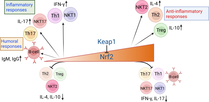
  • Nrf2 activation supports differentiation of the Th2 subset, regulatory T cells (Tregs), and the NKT2 subset while inhibiting differentiation of Th1, Th17, NKT1, and NKT17 subsets.
  • The absence of or low Nrf2 results in enhanced proinflammatory responses, characterized by differentiation of Th1, Th17, NKT1, and NKT17 subsets, and subdued generation of Th2, Treg, and NKT2 subsets.

Nrf2 activation levels also influence generation of humoral responses.

  • Low Nrf2 levels favor T cell–dependent production of IgG and IgM Abs by activated B cells.
  • High Nrf2 suppresses B cell responses such as differentiation of germinal center B cells and plasma cells.

Nrf2 negatively regulates T–cell mediated inflammatory responses and T-dependent B cell responses.

https://journals.aai.org/immunohorizons/article/7/4/288/263657/Beyond-Antioxidation-Keap1-Nrf2-in-the-Development “Beyond Antioxidation: Keap1–Nrf2 in the Development and Effector Functions of Adaptive Immune Cells”


And our innate immune system:

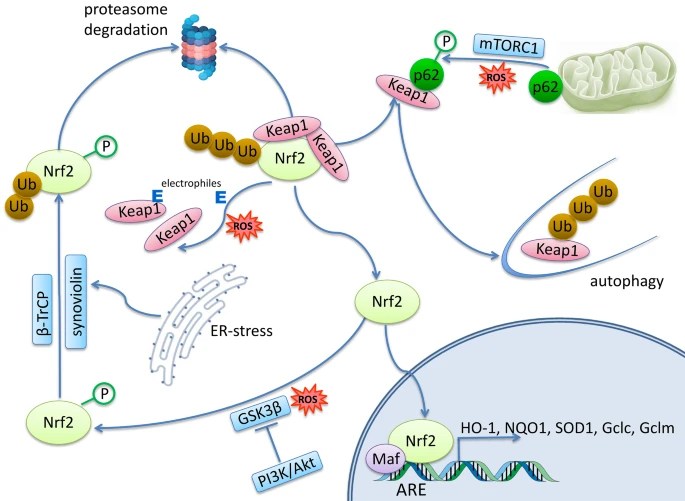
“Nrf2 regulates the immune response by interacting directly or indirectly with one or more of the major innate immune signaling components that maintain cellular homeostasis. Toll-like receptors (TLR) signaling can induce Nrf2 activation, and this is primarily found to be through autophagy-mediated degradation of Keap1.

TLR agonists may be considered as stimuli that induce Nrf2 to reduce stress and inflammation, linking the immune and antioxidant pathways. Conversely, Nrf2 activation may restrain TLR-mediated inflammatory response through induction of antioxidant proteins and inhibition of pro-inflammatory cytokines.

Following LPS stimulation, the NF-κB pathway is engaged to initiate a host of pro-inflammatory responses such as IL-6 and interleukin 1 beta (IL-1β) gene expression. Nrf2 induction inhibits LPS-mediated activation of pro-inflammatory cytokines in macrophages.

Inflammasome activation is an essential component of the innate immune response, and is critical for clearance of pathogens or damaged cells through pro-inflammatory cytokine secretion and/or cell-death induction. While Nrf2 activation is in general associated with an anti-inflammatory state, Nrf2 has also been reported to be required for optimal NLRP3 inflammasome activity.

The type-I interferon (IFN) system constitutes an essential part of innate immunity. Type-I IFNs are produced upon recognition of foreign or self-DNA or RNA, and are best-known for inducing an antiviral state through the induction of interferon-stimulated genes. While Nrf2 interferes with IRF3 activation, STING expression, and type-I IFN signaling, none of these crucial players in innate immunity have been demonstrated to be direct targets of Nrf2.

The antiviral effect of Nrf2 activation by 4-OI may use various pathways to limit viral replication that have not been identified yet. It is important to consider that Nrf2-activating metabolites may also act as immunomodulators in a Nrf2-independent manner.

Anti-inflammatory properties of Nrf2 are independent of redox control. Further mechanistic studies are needed to decipher the exact indirect and/or direct interactions between Nrf2 and innate immune players.”

https://www.sciencedirect.com/science/article/pii/S0952791522000942 “Regulation of innate immunity by Nrf2”

Nrf2 Week #6: Phytochemicals

This 2023 review explored Nrf2 relationships with plant chemicals:

“This review focuses on possible mechanisms of Nrf2 activation by natural phytochemicals in preventing or treating chronic diseases, and regulating oxidative stress. Excess oxidative stress is closely related to many kinds of chronic diseases, such as cardiovascular diseases, cancer, neurodegenerative diseases, diabetes, obesity, and other inflammatory diseases.

Mitochondrial dysfunction and hyperglycemia lead to the massive production of ROS, which triggers molecular damage, inflammation, ferroptosis, insulin resistance, and β-cell dysfunction.

antioxidants-12-00236-g001

Crosstalk between Keap1-Nrf2-ARE pathway and other signaling pathways endows it with high complexity and significance in the multi-function of phytochemicals. Limited human data makes an urgent need to open the new field of phytochemical-original supplement application in human chronic disease prevention.”

https://www.mdpi.com/2076-3921/12/2/236 “The Regulatory Effect of Phytochemicals on Chronic Diseases by Targeting Nrf2-ARE Signaling Pathway”


Top ten mentions, not including references:

  • 21 Sulforaphane
  • 16 Broccoli
  • 9 Curcumin
  • 5 Resveratrol
  • 5 Green tea catechins
  • 4 Luteolin
  • 3 Garlic
  • 3 Soy isoflavones
  • 3 Lycopene
  • 3 Quercetin

PXL_20230518_191008431

Nrf2 Week #5: Elements

Two 2023 papers, starting with a cell study of Nrf2 regulating sulfur:

“We demonstrated that NRF2 increased intracellular persulfides by upregulating cystine transporter xCT encoded by Slc7a11, a well-known NRF2 target gene. Persulfides have been shown to play an important role in mitochondrial function.

Supplementation with glutathione trisulfide (GSSSG), which is a form of persulfide, elevated mitochondrial membrane potential, increased oxygen consumption rate (OCR), and promoted ATP production.

glutathione trisulfide

The sulfur oxidation pathway is thought to protect cells from sulfide toxicity and to support electron transport efficiency. This study clarified that facilitating persulfide production and sulfur metabolism in mitochondria by increasing cysteine availability is one of the mechanisms for NRF2-dependent mitochondrial activation.”

https://www.sciencedirect.com/science/article/pii/S2213231723000253 “Contribution of NRF2 to sulfur metabolism and mitochondrial activity”


The second paper reviewed Nrf2 regulating iron:

“The central role of Nrf2 in dictating multiple facets of cellular stress response has defined the Nrf2 pathway as a general mediator of cell survival. Ferroptosis is an iron- and lipid peroxidation-dependent form of cell death. While Nrf2 was initially thought to have anti-ferroptotic function primarily through regulating antioxidant response, accumulating evidence has indicated that Nrf2 also exerts anti-ferroptotic effects via regulating key aspects of iron and lipid metabolism.

molce-46-3-165-f2

Iron exists in two redox states, ferrous (Fe2+) and ferric (Fe3+). While constant loss or gain of electrons to switch between two redox states makes iron useful for metabolic reactions, generation of free radicals due to an excess of the highly reactive Fe2+ form is toxic to cells. To prevent iron toxicity, free labile iron in the form of (Fe2+) is controlled by multiple systems at both systemic and cellular levels to maintain iron homeostasis.

Nrf2 regulates iron homeostasis by controlling both ferritin synthesis and degradation. Overall, Nrf2 regulation of iron homeostasis is a critical determinant of a cell’s sensitivity or resistance to ferroptosis, which is independent of its antioxidant function.”

https://www.molcells.org/journal/view.html?doi=10.14348/molcells.2023.0005 “Anti-Ferroptotic Effects of Nrf2: Beyond the Antioxidant Response”


PXL_20230520_183122738

Nrf2 Week #4: Aging

Two 2023 reviews of Nrf2 and aging, starting with Nrf2-mitochondria interactions:

“We discuss molecular mechanisms of interactions between Nrf2 and mitochondria that influence the rate of aging and lifespan. Nrf2 activity positively affects both mitochondrial dynamics and mitochondrial quality control.

Nrf2 influences mitochondrial function through regulation of nuclear genome-encoded mitochondrial proteins and changes in the balance of ROS or other metabolites. In turn, multiple regulatory proteins functionally associated with mitochondria affect Nrf2 activity and even form mutual regulatory loops with Nrf2. These loops enable fine-tuning of cellular redox balance and, possibly, of the cellular metabolism as a whole.

mtDNA-encoded signal peptides interact with nuclear regulatory systems, first of all, Nrf2, and are possibly involved in regulation of the aging rate. Interactions between regulatory cascades that link programs ensuring maintenance of cellular homeostasis and cellular responses to oxidative stress are a significant part of both aging and anti-aging programs.

Understanding these interactions will be of great help in searching for molecular targets to counteract aging-associated diseases and aging itself. Future research on Nrf2 signaling and ability of various substances that activate the Nrf2 pathway to prevent age-associated chronic diseases will provide further insight into the role of Nrf2 activation as a possible longevity-promoting intervention.”

https://link.springer.com/article/10.1134/S0006297922120057 “Transcription Factor Nrf2 and Mitochondria – Friends or Foes in the Regulation of Aging Rate” (not freely available) Thanks to Dr. Gregory A. Shilovsky for providing a copy.


The second review evaluated whether Nrf2 is a master regulator of aging:

“This paper briefly presents main mechanisms of mammalian aging and roles of inflammation and oxidative stress in this process. Mechanisms of Nrf2 activity regulation, its involvement in aging and development of the senescence-associated secretory phenotype are also discussed.

The age-related decrease in Nrf2 activity is of universal interspecies character:

  • Rodents with high Nrf2 activity have a longer lifespan than rodents with low activity.
  • Genetic knockout of Nrf2 usually leads to the increased senescent phenotype in a variety of animal organs and tissues, and also reduces lifespan of female mice.
  • There are also opposite examples, where Nrf2 knockout in aging mice reduced iron ions deposition in the brain, lowered the level of oxidative damage in the striatum, and also alleviated age-related motor dysfunction.

10541_2022_2401_Fig2

It would be incorrect to consider the effect of Nrf2 transcription factor at the organism level as exclusively antioxidant, anti-inflammatory, and, ultimately, anti-aging. Nrf2 controls many genes, products of which have complex, pleiotropic effects on the body:

  • No experiments that use Nrf2 chemical inducers as anti-aging drugs have been performed so far.
  • Nrf2 is not involved in life extension caused by caloric restriction.
  • Epigenetic clocks do not reveal transcription factors activity of which changes with aging.

Aging is accompanied by changes in gene expression profiles, which are tissue- and species-specific. These changes only to a small extent include genes controlled by Nrf2. At the moment, it cannot be concluded that Nrf2 is the master regulator of the aging process.”

https://link.springer.com/article/10.1134/S0006297922120045 “Does Nrf2 Play a Role of a Master Regulator of Mammalian Aging?”


PXL_20230520_182643299

Nrf2 Week #3: Epigenetics

To follow the Nrf2 Week #2 finding that chromatin accessibility parallels Nrf2 expression, this 2023 cell study explored how Nrf2 influences other epigenetic processes:

“We identified antioxidant response element sequences in promoter regions of genes encoding several epigenetic regulatory factors, such as histone deacetylases (HDACs), DNA methyltransferases (DNMTs), and proteins involved in microRNA biogenesis.

  • We treated cells with dimethyl fumarate (DMF), an activator of the NRF2 pathway through both the KEAP1 and GSK-3 pathways. NRF2 is able to modulate expression of HDAC1, HDAC2, HDAC3, and SIRT1 in different cell types.
  • DMF treatment induced DNMT1 and DNMT3b at both mRNA and protein levels. For DNTM3a, there was a slight induction of mRNA levels but not at the protein level.

antioxidants-12-00641-g007

  • Our data indicate that of all miRNAs analyzed, only miR-27a-3p, miR-27b-3p, miR-128-3p, and miR-155-5p associate with Nfe2l2 mRNA. NRF2 causes degradation of miR-155-5p, which is implicated in neuroinflammation and other pathologies, and is the main miRNA induced by LPS treatment in microglia. miR-155 alters expression of genes that regulate axon growth, supporting the bioinformatic prediction that miR-155 can regulate expression of genes involved in central nervous system development and neurogenesis.

Todate we only understand how epigenetic modifications affect expression and function of the NRF2 pathway. The fact that NRF2 can promote expression of type I HDACs, DNMTs, and proteins involved in miRNA biogenesis opens new perspectives on the spectrum of actions of NRF2 and its epigenetic influences.”

https://www.mdpi.com/2076-3921/12/3/641 “The Transcription Factor NRF2 Has Epigenetic Regulatory Functions Modulating HDACs, DNMTs, and miRNA Biogenesis”


PXL_20230515_185958612

Nrf2 Week #2: Neurons

To follow the Nrf2 Week #1 suggestion that Nrf2 target neurological disorders, this 2023 cell study investigated Nrf2 expression in neurons:

“Oxidative metabolism is inextricably linked to production of reactive oxygen species (ROS), which have the potential to damage all classes of macromolecules. Yet ROS are not invariably detrimental. Several properties make ROS useful signaling molecules, including their potential for rapid modification of proteins and close ties to cellular metabolism.

We used multiple single cell genomic datasets to explore Nrf2 expression and regulation in hundreds of neuronal and non-neuronal cell types in mouse and human. With few exceptions, Nrf2 is expressed at far lower levels in neurons than in non-neuronal support cells in both species.

This pattern is maintained in multiple disease states, and the chromatin accessibility landscape at the Nrf2 locus parallels these expression differences. These results imply that Nrf2 activity is limited in almost all neurons of the mouse and human central nervous system (CNS).

nrf2 expression

We separated cell types into neuron or non-neuronal ‘support’ cell categories. The general ‘support’ term is not meant to minimize the functional relevance of non-neuronal cells in the CNS, but is an umbrella term meant to cover everything from glial cell types (astrocytes, microglia, oligodendrocytes) to endothelial cells.

It is not clear why an important, near ubiquitous cytoprotective transcription factor like Nrf2 remains off in mature neurons, especially considering oxidative stress is a driver of many diseases. The simplest explanation is that Nrf2 activity also disrupts normal function of mature neurons.

ROS play a key role in controlling synaptic plasticity in mature neurons. These activity-dependent changes in synaptic transmission, which are important for learning and memory, are disrupted by antioxidants.

A subset of important Nrf2-targeted antioxidant genes (e.g., Slc3a2, Slc7a11, Nqo1, Prdx1) are also low in neurons. So it is likely that these and/or other Nrf2 targets must remain low or non-ROS-responsive in mature neurons. Future work exploring why this expression pattern persists in mature neurons will inform our models on roles of antioxidant genes in normal neuronal physiology and in neurological disorders.

https://www.biorxiv.org/content/10.1101/2023.05.09.540014v1.full “Limited Expression of Nrf2 in Neurons Across the Central Nervous System”


PXL_20230520_182827767

Nrf2 Week #1: Targeting

It’s been a while since I curated Nrf2 research. Read almost a dozen relevant 2023 papers last week. Let’s begin with an opinion paper by a highly qualified researcher:

“The inducible transcription factor nuclear factor erythroid 2-related factor 2 (NRF2) regulates expression of several hundred genes encoding proteins with antioxidant, anti-inflammatory, drug metabolising, and other homeostatic functions. Through its transcriptional targets, NRF2 activation orchestrates a comprehensive and long-lasting protection that allows adaptation and survival under diverse forms of cellular and organismal stress.

We highlight three NRF2 activators that have progressed furthest in clinical development. Overall outcomes of clinical trials with sulforaphane-rich preparations have strengthened preclinical evidence that sulforaphane has the potential to prevent toxic and neoplastic effects of environmental carcinogens, as well as to ameliorate conditions characterised by chronic oxidative, metabolic, and inflammatory stress.

Anti-inflammatory effects of most electrophilic NRF2 activators are partly NRF2-independent, and include inhibition of other inflammatory mediators. The majority of non-electrophilic PPI inhibitors are less potent in activating NRF2 in cellular systems than the electrophilic sulforaphane.

It remains to be shown that measurement of NRF2 activation in blood samples can reflect modulation of the pathway in target tissues. The field has yet to reach a consensus on the best approach for monitoring NRF2 activation in humans, including selection of the optimal panel of gene/protein targets.

Even after a single dose of an NRF2 activator, increased levels of the actual protectors (i.e., the downstream transcriptional targets of NRF2) persist over long periods of time (days), exceeding the half-life (hours) of the drug.

target disease

In certain contexts, the role of NRF2 is complex and cell-type-specific, for example, in mouse models of atherosclerosis. Considering that NRF2 activation functions to:

  • Restore cellular redox and protein homeostasis;
  • Preserve mitochondrial function; and
  • Inhibit inflammation.

Perhaps the most logical disease areas are neurological conditions where all of these processes contribute to survival of neurons and astrocytes, as well as metabolic disease and cancer prevention.”

https://www.cell.com/trends/pharmacological-sciences/fulltext/S0165-6147(22)00277-2 “Advances and challenges in therapeutic targeting of NRF2”


PXL_20230520_182543000.MP

Don’t eat yourself into disease, Part 2

This blog’s 1000th curation is a 2023 rodent study associating gut microbiota, behavior, memory, and food reward:

“Energy intake and energy expenditure is regulated by the hypothalamus, and is referred to as homeostatic regulation of food intake. The reward system is the non-homeostatic regulation of food intake – pleasure-related consumption of foods enriched in fat and sugar. The pleasure is encoded by dopamine release from dopaminergic neurons projecting from the ventral tegmental area to the striatum, the nucleus accumbens, and the prefrontal cortex.

Food reward can be divided into three components – liking, wanting, and learning:

  • Liking refers to food hedonic value;
  • Wanting to the motivational process to seek out and consume certain foods; and
  • Learning to reinforcing conditioning behavior associated with food intake stimulus.

We confirmed that obese mice have a dysregulation of the learning and the wanting components of  food reward. Our previous data showed that the liking component was transferable through fecal material transplantation.

We demonstrated that gut microbes play a role in the regulation of food reward, and could be responsible for compulsive behavior and excessive motivation to obtain sucrose pellets. Moreover, obese gut microbes affected dopaminergic and opioid markers involved in reward system.

We identified 33HPP (produced exclusively by gut bacteria) as particularly increased in mice recipients of gut microbes from obese mice. We were able to demonstrate its effects as key mediator of the gut-brain axis controlling the reward response to palatable food.”

https://microbiomejournal.biomedcentral.com/articles/10.1186/s40168-023-01526-w “Obese-associated gut microbes and derived phenolic metabolite as mediators of excessive motivation for food reward”


PXL_20230415_195202937

Don’t eat yourself into disease, Part 1

Starting a sample of 2023 papers with a porcine review:

“Epigenetic programing predisposes pigs to insulin insensitivity, but pigs seem to sense this insensitivity and consequently eat less, preventing obesity. Pigs naturally prefer to eat small breakfasts and large dinners.

Deviating from this eating pattern or providing diets with a high glycemic burden can trigger obesity; however, pigs will restrict food intake to prevent serious obesity. In practice, problems with obesity are rarely seen, even when pigs are fed poorly timed diets similar to junk food, likely because swine diets are balanced for every nutrient.

Feeding pigs diets deficient in micronutrients does trigger obesity. For humans, several micronutrient requirements have not been set officially, and diets optimized for all micronutrients are rarely provided.

metabolites-13-00420-g015

Although we could debate whether this is a cause or effect, the above data on hyper-processed diets fed to pigs would indicate that it is causative. Pigs were fed a diet which included ‘human-targeted junk food’ but was adequate in phosphate, and they experienced no issues.

Controlled human studies are generally conducted with very small populations of subjects for very short durations, as emotions come into play. Humans are hard to persuade to follow a boring diet, especially over a longer period of time, and humans are easily tempted to deviate from a protocol if peer pressure or desires are high.

Even worse, in survey type experiments, people are asked what they ate for the past one or several days, and these data may well be subsequently extrapolated to patterns of behavior and then correlated with developments in health. Recalling what and especially how much a person ate yesterday is already a challenge for many, confounded even further by the desire not to include items that may be considered less acceptable.

On the swine side, knowledge on nutrient yield of foods and nutrient requirement appears further advanced, and controlled feeding trials are much easier to perform. Borrowing pig data is arguably much closer to the truth for humans than having no data at all.”

https://onlinelibrary.wiley.com/doi/10.1002/advs.202205346 “Eat like a Pig to Combat Obesity”


One fish in the gullet, another soon on its way

PXL_20230509_094500357

No exit

This 2023 rodent study investigated aging processes and gut microbiota in crowded conditions:

“Our study provides clear evidence that high-density crowding accelerates the aging process of Brandt’s voles. We also found that ‘high-density microbiota’ promote the aging-related phenotype in voles.

Because we minimized effects of direct fighting on mortality of voles, observed changes in lifespan in this study should mostly represent the natural aging processes of voles.

high-density survival

High density increased the level of stress hormone corticosterone, which disrupted gut microbiota composition by:

  • Decreasing abundance of anti-aging or anti-inflammatory bacterial species; and
  • Increasing the proportion of pathogenic bacteria.

This caused an increase in DNA oxidation and inflammation through upregulation of NF-kB and COX-2 pathways.

Although high-density relief and butyric acid administration interventions could reverse aging-related processes of adult voles, it remains unclear whether they could reverse the aging process in terms of lifespan.

Our results suggest that gut microbiota play a significant role in mediating aging-related processes of voles under high-density conditions, and can be used as a potential therapeutic target for treating stress-related diseases in humans.”

https://onlinelibrary.wiley.com/doi/10.1002/advs.202205346 “Gut Microbiota is Associated with Aging-Related Processes of a Small Mammal Species under High-Density Crowding Stress”


I came across this study by it citing Reversing hair greying for effects of stress interventions.

PXL_20230505_185253518.MP