Assessing a mountain climber’s condition without noticing their empty backpack

A metaphor: for a mountain climber, which point has the most influence on their condition during the climb?

  • The path ahead?
  • The current situation?
  • The recent past?
  • The starting point?
  • The preparations?

Hard to say? Once the climb has started and until it’s finished, though, are there any points at which the preparations have no influence?

Let’s imagine that factors beyond the climber’s control ruined their preparations, leaving them with no reserves and a limited capability to adapt to environmental changes.

Let’s imagine further that researchers take initial physical and psychological measurements of the climber’s condition at an arbitrary point of the ascent or descent. Due to the design of their measurement system, however, they don’t discover that this climber has an empty backpack.

When the researchers interpret the results, will they understand how the climber’s measurements were influenced by the ruined preparations?  end metaphor


A 2014 Israeli study primary finding was of:

“Fear of terror-induced annual increases in resting heart rate.”

The researchers took 325 measurements each “of 17,380 apparently healthy volunteers” who had “consistent exposure to terror threats.”

The study was opaque in some areas. For example, what was the content and handling of a 4-item anxiety questionnaire?

The supplementary material showed that the headlined “fear of terror” term involved three disparate factors:

  • feeling unsafe;
  • fear of crowds; and
  • anxiety about future harm.

I’d like to understand the bases of why the researchers and the reviewer felt it was appropriate that:

“The scores on these items were averaged to yield a continuous FOT [fear-of-terror] score.”


The researchers probably had sufficient measurements of the subjects’ current conditions. They didn’t have a frame of reference that incorporated the present data with contextual information from each individual’s history back to the earliest parts of their life.

Lacking the links provided by such a framework, the researchers likely misassessed measurements that were influenced by how the subjects’ backpacks were packed.

http://www.pnas.org/content/112/5/E467.full “Fear and C-reactive protein cosynergize annual pulse increases in healthy adults”
Are hippocampal place cells controlled by theta brain waves from grid cells?

This 2015 Canadian rodent study tried to establish that grid cells in the medial entorhinal cortex generated brain waves in the theta frequency range that controlled place cells in the hippocampus part of the limbic system.

The researchers stated:

“Our results deviate from the prediction.”

but a commentary Do the spatial frequencies of grid cells mold the firing fields of place cells? said the researchers:

“Obtained fascinating results, largely supporting the model.”

What’s fascinating to me is the volume of studies on the hippocampus that ignore its position as the seat of emotional memories. Human experiments involving the hippocampus are usually designed to not contain any emotional content.

Two studies showed functions of hippocampal place cells:

A summary study of 118 other studies What do grid cells contribute to place cell firing? provided additional information on grid cells and hippocampal place cells, head direction cells, boundary cells, and cells that encode object locations.

The summary study related to the current study by stating that the research through early 2014 arguably found:

“Grid and place cell firing patterns are not successive stages of a processing hierarchy, but complementary and interacting representations that work in combination.”

http://www.pnas.org/content/112/13/4116.full “Place field expansion after focal MEC inactivations is consistent with loss of Fourier components and path integrator gain reduction”

A common dietary supplement that has rapid and lasting antidepressant effects

This 2012 Italian rodent study found that a common dietary supplement had rapid and lasting antidepressant effects:

“Remarkably, L-acetylcarnitine displayed a clear-cut antidepressant effect already after 3 and 7 d[ays] of daily dosing. No tolerance was developed to the action of L-acetylcarnitine. The drug was even more effective after 21 d[ays], and the effect persisted for at least 2 w[ee]k[s] after drug withdrawal.”

The researchers studied stressed mice and rats to determine that:

  1. An effect of the stress was to epigenetically change the hippocampus to produce less of an important molecule – type 2 metabotropic glutamate (mGlu2).
  2. A reduction of the mGlu2 molecule decreased the hippocampus’ regulation of the glutamate neurotransmitter.
  3. Under-regulation of glutamate, in turn, caused symptoms of depression.

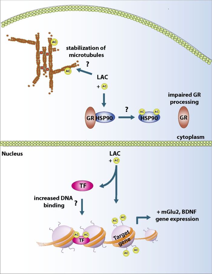
L-acetylcarnitine reversed the immediate causes of stress-induced symptoms by acetylating histone proteins. These control the transcription of the brain-derived neurotrophic factor (BDNF) and mGlu2 receptors in the hippocampus and prefrontal cortex.


LAC putative action

A commentary on this research, Next generation antidepressants, had the above graphic that showed possible mechanisms for the effects of L-acetylcarnitine. Epigenetic histone modifications seem to be more easily reversible than epigenetic DNA methylation.


“Currently, depression is diagnosed only by its symptoms,” Nasca says. “But these results put us on track to discover molecular signatures in humans that may have the potential to serve as markers for certain types of depression.”

It’s tempting to extrapolate this study to humans and test whether depression symptoms could be effectively treated with some multiple of a normal acetyl-L-carnitine dietary supplement dose of 500 mg at $.25 a day. This dietary supplement is better for depression symptoms than placebo analyzed randomized control trials that tested and demonstrated its efficacy.

To cure stress-induced illnesses in humans, though, ultimate causes of stress should be removed or otherwise addressed.

http://www.pnas.org/content/110/12/4804.full “L-acetylcarnitine causes rapid antidepressant effects through the epigenetic induction of mGlu2 receptors”

The hypothalamus’ role in how calorie restriction delays aging

This 2015 Portuguese rodent study showed the underlying mechanism to explain why restricting calories delays aging.

A calorie reduction of 20 to 40% increased production of a normally occurring molecule (neuropeptide Y) in the hypothalamus part of the limbic system. The increased amounts of the molecule stimulated autophagy (the breakdown and recycling of cellular components) in hypothalamic neurons.

“Because both hypothalamic autophagy and neuropeptide Y levels decrease with age..modulation of hypothalamic neuropeptide Y levels may be considered a potential strategy to produce protective effects against hypothalamic impairments associated with age and to delay aging.”

http://www.pnas.org/content/112/13/E1642.full “Neuropeptide Y stimulates autophagy in hypothalamic neurons”

A study of visual perception that didn’t inform us about human conscious awareness

This 2015 Vanderbilt study with a Princeton reviewer stated that they found “compelling evidence” related to:

“How the brain begets conscious awareness.

Identifying the fingerprints of consciousness in humans would be a significant advancement for basic and medical research, let alone its philosophical implications on the underpinnings of the human experience.”

Let’s begin with the “conscious” part of the study’s conscious awareness goal. A summary article of 105 studies entitled Evolution of consciousness: Phylogeny, ontogeny, and emergence from general anesthesia that I curated found:

The core of human consciousness appears to be associated primarily with phylogenetically ancient structures mediating arousal and activated by primitive emotions.”

The current study ignored the evolutionary bases of human consciousness and didn’t include any limbic system and lower brain areas. The researchers’ biases were further indicated by the statement from their press release:

“Focal theories contend there are specific areas of the brain that are critical for generating consciousness, while global theories argue consciousness arises from large-scale brain changes in activity.”

The researchers were in the “global” camp of this unnecessary divide.


Let’s next examine the “awareness” part of the study’s conscious awareness goal. The subjects were 24 students in a visual perception experiment that used fMRI. The visual events that were perceived went into the “aware” bucket and the others into the “unaware” bucket.

The study’s subject selection criteria and experiment seemed a little odd for developing “compelling evidence” related to “how the brain begets conscious awareness.” By equating visual perception with awareness, the researchers excluded the contributions of other senses and methods of awareness.

Would it follow from the study’s methodology that blind people can’t be consciously aware?

The supplementary material showed that 7 of the 24 subjects’ results for one experimental condition, and 12 – half – of the subjects’ results for another condition were excluded because they apparently had problems reporting confidence in their visual perception. I wonder why the reviewer agreed that it was appropriate to discard half of the subjects’ experimental results?

Whatever else it was that the study found, the researchers didn’t reach their goal of developing “compelling evidence” related to “how the brain begets conscious awareness.”

http://www.pnas.org/content/112/12/3799.full “Breakdown of the brain’s functional network modularity with awareness”

Adaptations to stress encourage mutations in a DNA area that causes diseases

This 2015 Baylor human cell study subject was the underlying mechanisms of cellular responses to environmental stressors of cold, heat, hypoxia, and oxidation:

“Because trinucleotide repeats are overrepresented in gene-regulatory proteins, stress-induced trinucleotide repeat mutagenesis may provide a path for the environment to subtly alter gene regulatory networks – with attendant changes in cell behavior – during development, disease, and evolution.”

The study’s overarching framework was that human cells will adapt to best survive in their environment. The study found that the cells’ responses to stress encouraged the creation of mutations in a DNA area that’s:

“The cause of multiple human diseases. This pathway may impact normal cells as they encounter stresses in their environment or during development or abnormal cells as they evolve metastatic potential.”


It’s a logical inference to likewise understand how stressors in a mother’s environment for a developing fetus will cause the fetus to adapt at the cellular level. If, for example, the fetus is stressed by inadequate oxygen – hypoxia – this study shows how cells will adapt in ways that foster mutations and diseases.

When the stressed fetus arrives in a different environment after birth, the newborn’s cells are maladapted to certain aspects of a normal environment – to adequate oxygen in this example. Many of the cells’ adjustments to the old environment persist in the new environment. Pathways epigenetically adapted to best survive during the fetus’ development in the womb may impact the infant’s development in a normal environment.

Researchers could make significant contributions to science by investigating treatments and therapies that potentially reverse causes of epigenetic changes.

http://www.pnas.org/content/112/12/3764.full “Environmental stress induces trinucleotide repeat mutagenesis in human cells”

A study on online cooperation with limited findings

This 2015 Cambridge/Oxford study found:

“Global reputational knowledge is crucial to sustaining a high level of cooperation and welfare.”

Basically, the subjects learned how to “game” a cooperative online game, and the researchers drew up their findings.

To me, the study demonstrated part of the findings of the Reciprocity behaviors differ as to whether we seek cerebral vs. limbic system rewards study, the part where the cerebrum was active in:

“Reputation-based reciprocity, in which they help others with good reputations to gain good reputations themselves.”

The current study ignored how people’s limbic system and lower brain areas may have motivated them to cooperate.

I didn’t see how excluding people’s emotional involvement when cooperating with others improved the potential reach of this study’s findings. Doesn’t a person’s willingness to cooperate in person and in online activities usually also include their emotional motivations?

The findings can’t be applied generally to cooperative motivations and behaviors that the researchers intentionally left out of the study. The study’s findings applied just to the artificial environment of their experiment, and didn’t provide evidence for how:

“Cooperative behavior is fundamental for a society to thrive.”

http://www.pnas.org/content/112/12/3647.full “The effects of reputational and social knowledge on cooperation”


This post has somehow become a target for spammers, and I’ve disabled comments. Readers can comment on other posts and indicate that they want their comment to apply here, and I’ll re-enable comments.

If research doesn’t provide causal evidence for effects, can epigenetics be forced in to explain everything?

This 2015 UK bird study found that older mothers had female children who had fewer offspring than did the rest of the house sparrow population. The finding applied also to older fathers and their male children.

In general, if a study didn’t directly demonstrate cause and effect, it isn’t appropriate to force the use of epigenetics to explain everything. That’s what this study did with epigenetic inheritance.

Did the study:

“Demonstrate that this parental age effect..potentially is epigenetically inherited.”

by analyzing DNA across generations?

No!

The researchers ran some numbers that tested the effect of older foster parents where the model’s only other possible explanation was epigenetic inheritance.

Several other things about this study were off:

  • The researchers used the term “fitness” 28 times as shorthand to mean the number of offspring, but only twice was it explained as “reproductive fitness.” This was potentially misleading in some of the contexts of the term’s other uses. For example, several of the cited references used “fitness” in a different context.
  • The researchers went into a long exposition of telomeres, punctuated by citing 11 references, only to say:

    “However it is unclear how telomere dynamics could affect fitness.”

    The next sentence was:

    “An alternative explanation might be the accumulation of deleterious mutations as individuals age.”

    which was additionally irritating because “alternative” assumed that telomeres presented a factual explanation of the study’s findings in the first place. Was this section an artifact of a struggle with the reviewer?

After forcing epigenetic inheritance as an explanatory factor and potentially misleading readers about reproductive fitness and telomeres, the researchers had little basis to conclude that their research had “important implications.”

http://www.pnas.org/content/112/13/4021.full “Reduced fitness in progeny from old parents in a natural population”

Epigenetic DNA methylation and demethylation with the developing fetus

This extremely dense and informative 2014 UK summary study provided details about genomic imprinting:

“An unusual epigenetic process in that it is heritable and results in autosomal gene expression according to parent of origin.”

Several notes of interest:

  • Figure 3 had a fascinating sketch of how the fetus caused the mother’s hypothalamus to:

    “Determine forward maternal planning by directing/orchestrating maternal physiology and postnatal maternalism to synchronize with development of the fetus.”

  • Figure 4 followed up with a flowchart of how – with a female fetus – coexistence of three matrilineal generations in the pregnant female (her, the fetus, and the grandmother’s influence on the developing fetus’ ovarian oocytes) enabled intergenerational forward planning.
  • The study briefly noted significance of genomic imprinting on male sexual behavior, where, if processes didn’t proceed normally at this early stage of a male fetus’ development, could result in suboptimal adult behavior that didn’t change with experience.

F4.large

I’ll quote a few other unrelated passages that caught my eye.

“Reproductive success of mammals also places a considerable burden on matrilineal time and energy, with some 95% of mammalian female adult life committed to pregnancy, lactation, and maternal care.

Offspring that receive optimal nourishment and improved maternal care will be predisposed to develop a hypothalamus that is both genetically and epigenetically predisposed to this same type of good mothering.

The fetus controls its own destiny in times of acute starvation, especially in the last trimester of pregnancy, by short-term sacrifice of its placenta to preserve resources critical for brain development.”

http://www.pnas.org/content/112/22/6834.full “Genomic imprinting, action, and interaction of maternal and fetal genomes”


This post has somehow become a target for spammers, and I’ve disabled comments. Readers can comment on other posts and indicate that they want their comment to apply here, and I’ll re-enable comments.

Epigenetic DNA methylation of the oxytocin receptor gene affected the perception of anger and fear

This 2015 Virginia human study:

“Reveals how epigenetic variability in the endogenous oxytocin system impacts brain systems supporting social cognition and is an important step to better characterize relationships between genes, brain, and behavior.”

The researchers did a lot of things right:

  • They studied a priori selected brain areas, followed by whole brain analyses;
  • Their subjects were carefully selected

    “Because methylation levels have been shown to differ as a function of race, we restricted our sample to Caucasians of European descent”

    but they didn’t restrict subjects to the same gender;

  • They acknowledged as a limitation:

    “A lack of behavioral evidence to reveal how these epigenetic and neural markers impact the overt social phenotype.”


One thing on which I disagree with the researchers is their assessment of what needs to be done next. Their news release stated:

“When imagining the future possibilities and implications this DNA methylation and oxytocin receptor research may have, the investigators think a blood test could be developed in order to predict how an individual may behave in social situations.”

Nice idea, but the next step should be to complete the research. The next step is to develop evidence for how the oxytocin receptor gene became methylated.

The subjects had a wide range of DNA methylation at the studied gene site – from 33% to 72% methylated!

Why?

At the same gene site:

“There was a significant effect of sex such that females have a higher level of methylation than males.”

Why?

Given these significant effects, why was there no research into likely causes?

Aren’t early periods in people’s lives the most likely times when the “Epigenetic modification of the oxytocin receptor gene” that “influences the perception of anger and fear in the human brain” takes place?

Wouldn’t findings from research on the subjects’ histories potentially help other people?

http://www.pnas.org/content/112/11/3308.full “Epigenetic modification of the oxytocin receptor gene influences the perception of anger and fear in the human brain”

Oxytocin blocks alcohol intoxication symptoms

This joint 2015 Australian/German rodent study found that oxytocin bound to the brain receptors that cause loss of motor control with alcohol intoxication, and prevented rats from displaying these symptoms:

“While oxytocin might reduce your level of intoxication, it won’t actually change your blood alcohol level,” Dr Bowen said. “This is because the oxytocin is preventing the alcohol from accessing the sites in the brain that make you intoxicated, it is not causing the alcohol to leave your system any faster.”

Vasopressin didn’t have the same effect.

The level of alcohol used to produce this finding was roughly equivalent to a human drinking a bottle of wine over a few hours. Oxytocin didn’t prevent loss of motor control when the equivalent of a bottle of vodka was administered because the excess ethanol found its way into other brain receptors and put the rats to sleep.

The study showed oxytocin acting in its original functionalities such as water regulation rather than with its evolved social functions as described in How oxytocin and vasopressin were repurposed through evolution to serve social functions.

http://www.pnas.org/content/112/10/3104.full “Oxytocin prevents ethanol actions at δ subunit-containing GABA-A receptors and attenuates ethanol-induced motor impairment in rats”

Differing characteristics of languages shape people’s brains differently

This 2015 Chinese study found that the differing characteristics of the Chinese and English languages shape people’s brains differently:

“Our results revealed that, although speech processing is largely carried out in the common left hemisphere classical language areas (Broca’s and Wernicke’s areas) and anterior temporal cortex, speech comprehension across different language groups depends on how these brain regions interact with each other.”

For an informed discussion of the study and related issues, visit http://languagelog.ldc.upenn.edu/nll/?p=17949 and comments.

We can infer from the Would you deprive your infant in order to be in a researcher’s control group? study that this shaping process begins during womb life.

http://www.pnas.org/content/112/10/2972.full “Cross-language differences in the brain network subserving intelligible speech”

Would you deprive your infant in order to be in a researcher’s control group?

This 2015 Harvard study found that exposing extremely premature babies to sounds of their mothers enlarged their auditory cortex.

The lead researcher stated:

“Our findings do not prove that the brains of these babies are necessarily better, and we cannot conclude that they will end up with no developmental disabilities.

We don’t know the advantages of having a bigger auditory cortex.”

It’s too bad that studies like this one have to take deprived infants and further deprive them for use as a control group. I suppose it’s possible that the control group members’ development could just be shifted, similar to the Maternal depression and antidepressants epigenetically change infant language development study.

However, given the findings of the Our early experiences are maintained and unconsciously influence us for years, if not indefinitely study, it’s also possible that the last trimester of womb life is a critical period for a child’s auditory cortex. If timely development doesn’t take place within the environment provided by the mother, there may not be another period to fully catch up on growth and learning, even given the effects of neural plasticity.

http://www.pnas.org/content/112/10/3152.full “Mother’s voice and heartbeat sounds elicit auditory plasticity in the human brain before full gestation”

If you had a disease, would you agree to treatment where you became a lab rat?

This 2015 Czech research studied individual neurons in an area of the limbic system of Parkinson’s disease patients. The findings corroborated several findings of previous research, such as:

“We confirm the importance of the subthalamic nucleus as a hub within the limbic circuitry involved in both emotional valence and arousal processing as in two functionally and spatially segregated systems.”

This statement summed up the study for me:

“Several factors could affect our results and reduce the inferences that can be drawn with regards to the physiology of emotional processing and the role of the subthalamic nucleus in the limbic circuits.

One such factor is that the study was conducted with Parkinson’s disease patients, who are known to have a widespread central nervous system pathology and to experience problems in emotional processing.”

The current study referenced The amygdala is where we integrate our perception of human facial emotion study, which similarly used the opportunity of patients with electrodes implanted for deep-brain stimulation to study individual neurons in the amygdala. However, a design difference was that the amygdala study had healthy control subjects in addition to patients, which led to fewer potential limitations on their findings.

Also referenced was a summary study entitled Exploring emotions using invasive methods: review of 60 years of human intracranial electrophysiology. Despite excluding studies of decision making, reward processing, learning, mood disorders, and pain experiences, it didn’t demonstrate that 60 years of experiments using implanted electrodes in the brains of people with epilepsy had substantially advanced science, other than confirming what fMRI and animal research had shown.

I’m not sure how I would feel if I had a disease where the physicians treated my symptoms in such a way that I became a lab rat for research that wasn’t groundbreaking. Do people with epilepsy and Parkinson’s disease have treatments available that factually resolve the underlying causes?

http://www.pnas.org/content/112/10/3116.full “Distinct populations of neurons respond to emotional valence and arousal in the human subthalamic nucleus”

Neural plasticity trumps genetics in the hippocampus part of the limbic system

This 2015 rodent study used a genetic strain of mice that was bred to not express a gene that enabled long-term memory in the hippocampus. The mice were not memory-impaired, however, due to their brains’ neural plasticity.

The researchers found:

“Deletion of genes in organisms does not always give rise to phenotypes because of the existence of compensation.

The current work provides an example of how a complex brain system may adjust to the effects of gene deletion to recover function.”

The Early human brain development can be greatly modified by environmental factors study showed even greater plasticity in another part of the human brain where the people faced much larger obstacles than gene deletion.

I view this finding as a cautionary tale to reference any time a study comes out stating that A and B genes are found to cause X and Y symptoms or behavior. Researchers don’t have enough evidence in 2015 to unequivocally describe what rodent brains are capable of, much less human brains.

The researchers implied how they kept faith in their work with the phrase:

“The compensatory mechanism is imperfect and does not fully restore cGKII-dependent function.”

Is perfection the standard to which their research is also held?

http://www.pnas.org/content/112/10/3122.full “Network compensation of cyclic GMP-dependent protein kinase II knockout in the hippocampus by Ca2+-permeable AMPA receptors”