TMAO and heavy hydrogen

A 2026 review subject was possible involvement of deuterium in TMAO levels, which contrasted with the usual TMAO meme. I’ll curate this paper through an outline of its sections:

“The human gut microbiome plays many essential roles, but an often-overlooked role is to maintain an abundant supply of deuterium depleted (deupleted) nutrients to fuel the host mitochondria. Excess deuterium (heavy hydrogen) damages mitochondrial ATP synthase nanomotors, leading to a decrease in matrix water production with increased reactive oxygen species (ROS) and inefficient ATP production. A microbial metabolite, trimethylamine N-oxide (TMAO) is a powerful signaling molecule whose plasma levels are high in association with many chronic diseases.

In this paper, we present a hypothesis that TMAO is a marker for deuterium overload in the methylation pathway, in addition to its role as an indicator of a disrupted gut microbiome. The original study that brought attention to TMAO involved feeding mice synthetic choline with fully deuterated methyl groups. Fully deuterated TMAO was subsequently detected in the plasma. By contrast, a diet rich in eggs, a natural source of choline (a precursor to TMAO), does not raise TMAO levels. Many of the pathologies that are linked to elevated TMAO can also be viewed as strategies to promote the supply of deupleted water to the mitochondria, systemically.

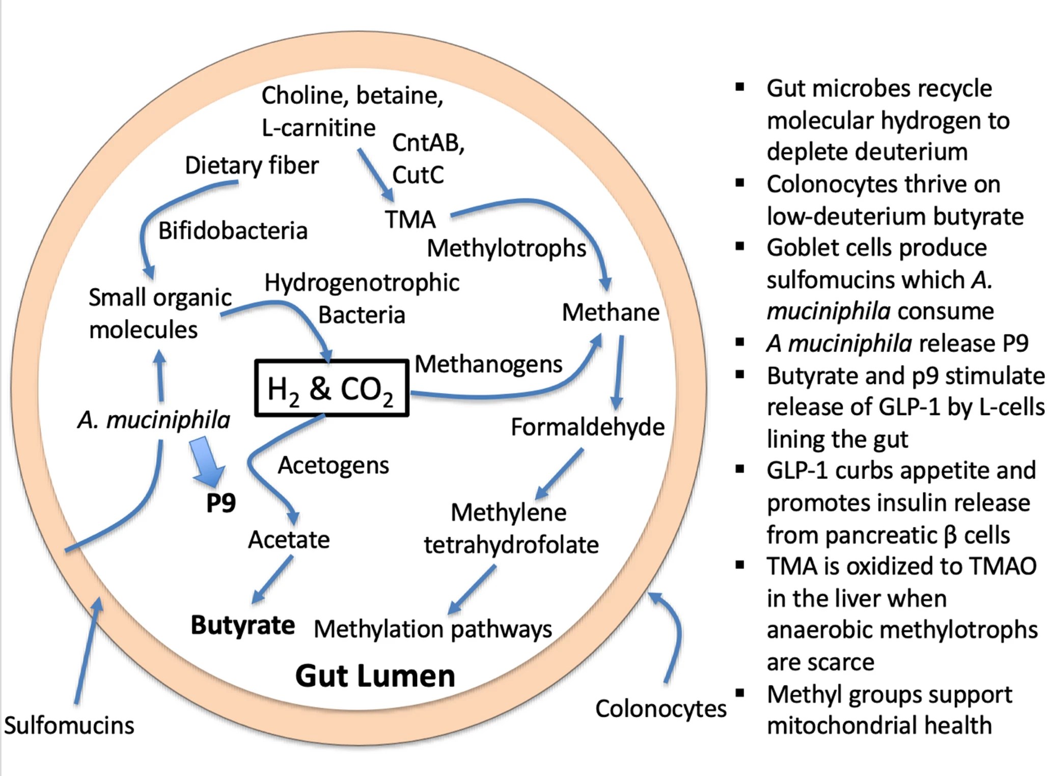
1. Introduction – DNA and histone methylation regulate epigenetic modifications and imprinting. Phosphatidylcholine is a precursor to acetylcholine, an excitatory neurotransmitter in the brain. Trimethylated lysine molecules, recovered during protein metabolism, are precursors to L-carnitine, which facilitates the transport of fatty acids into the mitochondria to be oxidized for fuel production. Dietary phosphatidylcholine and dietary L-carnitine, in addition to endogenous sources, are important nutrients that provide methyl groups to the methylation cycle. Choline and L-carnitine, as well as a close relative, betaine (trimethylglycine), are all precursors to trimethylamine (TMA), a small methylated amine produced through microbial enzymatic action. Obligate anaerobic hydrogen-dependent archaea called methylotrophs in the gut can reduce the three methyl groups in TMA to methane gas.

2. Evidence that deuterium disrupts mitochondrial function – Deuterium (2H) is a heavy isotope of protium (1H; hydrogen), and it is pervasive in nature, found in seawater at a concentration of 155 parts per million (ppm) relative to protium. Deuterium is highly damaging to the F1F0-ATP synthase (ATPase) nanomotors in the mitochondria that produce ATP, the primary fuel source of the cell. Deuterium loading suppresses the activity of many fundamental biologically important hydrolytic enzymes that depend on proton tunneling. It is likely that deuterium increases the frequency of unrepaired nuclear DNA mutations, by suppressing the activity of deuterium-sensitive repair enzymes.

The inherent collective proton tunneling (ICPT) process, which uses membrane-bound ATPase nanomotors in living organisms, is nature’s ultimate tool for discriminating hydrogen isotopes. This is because a deuteron (2H) cannot replace a proton (1H) in its tunnel protein during enzymatic transmembrane transport due to its doubled mass and twice larger atomic nuclear size. The result is large compartmental, inter- and intramolecular deuterium disequilibrium in 2H/1H ratios in all biomolecules, which readily distinguishes respiration from aerobic fermentation with adaptive significance. Deuterons irreversibly clog single proton tunneling ATP synthase nanomotors in mitochondria, resulting in the complete breakdown of ICPT.

This initiates many disease-causing molecular crowding mechanisms, which we review herein from the perspective of prokaryotic proton pumping and H2 gas formation in the organic molecular realm of mitochondrial proton-donating substrates. Understanding at the systems level how humans protect mitochondrial ICPT processes and ATP synthase is a fascinating journey reviewed herein. We hypothesize that the process uses TMAO as the microbial stepping stone, employing deuterium discrimination to become an active player in forming the biological reaction.

2.1 Quantum tunneling and proton-coupled electron transport – ICPT is a theoretical quantum mechanical phenomenon proposing that a single proton spontaneously passes through a potential energy barrier, typically within a hydrogen bond, in a manner that can be functionally irreversible. Unlike classical particles that must surmount energy barriers, protons can ‘tunnel’ through them due to their wave-like nature. Often, this single proton tunneling is part of a larger process where a proton and an electron are transferred simultaneously (or sequentially) as a single kinetic step, often in the presence of strong electric fields that stabilize the transferred state. This process is referred to as ‘proton-coupled electron transport’ (PCET).

Mitochondria exploit PCET to build the proton gradient that powers the nanomotors to produce ATP in the electron transport chain (ETC). Many enzymes, such as dehydrogenases and lipoxygenases, exploit proton tunneling to carry out their reactions. Deuterons are much less capable of tunneling, so this becomes a way to select for substrates containing protons rather than deuterons. The very large kinetic isotope effect (KIE) for soybean lipoxygenase is an example of this phenomenon.

NADH-ubiquinone oxidoreductase (Complex I) couples the transfer of two electrons between NADH and ubiquinone to the translocation of four protons across the membrane. This process provides the driving force for ATP synthase, which harnesses the gradient to produce ATP, but it also assures that few, or no, deuterons arrive on the other (intermembrane) side of the membrane, protecting the ATPase nanomotors.

SAMe, the universal methyl donor, plays a crucial role in regulating oxidative phosphorylation (OXPHOS). It is primarily synthesized in the cytoplasm and imported into the mitochondria via the import protein SAMC. SAMC is the only mitoSAM carrier and is required for OXPHOS and oxidative tricarboxylic acid (TCA) metabolism, showing a strong dependency of mitochondrial health on one-carbon (1C) metabolism. We hypothesize that the importance of SAMe to mitochondrial health is directly linked to the plausible theory that SAMe’s methyl groups are normally highly deupleted.

3. Are microbially synthesized methyl groups and butyrate deuterium depleted? – A careful tracing of multiple metabolic processes taking place in a human cell reveals that they are plausibly designed to greatly restrict the number of deuterons that are in the mitochondrial water. This strategy helps to minimize exposure of the ATPase nanomotors to deuterons. In part, this feat is accomplished through enzymes such as flavoproteins that greatly favor protium over deuterium in their reaction, i.e., that have a high deuterium KIE. The physics usually involves configuring the enzyme to support proton tunneling, since deuterons are much less capable of such tunneling.

Another way to support a reduced deuterium supply to the ATPase nanomotors is to select nutrients that are naturally low in deuterium to feed into the tricarboxylic acid (TCA) cycle. This is what makes the metabolites produced by the gut microbes via hydrogen recycling very significant. The enzyme expressed by anaerobic archaea that metabolize TMA, TMADH, is a flavoprotein with a high deuterium KIE (~ 8.6) due to vibrationally assisted hydrogen tunneling.

This means that the hydrogen recycling that takes place during its metabolism further scrubs deuterium from methylation pathways, while TMA that is left behind becomes enriched in deuterium. This unmetabolized TMA is converted to deuterium-enriched trimethylamine N-oxide (TMAO) in the liver and released into the circulation. Elevated TMAO levels in plasma are associated with increased risk to cardiovascular disease and a long list of other inflammatory diseases, as we will detail in the coming sections.

4. Natural and synthetic choline have different effects on TMAO levels: does deuterium play a role? – The original 2011 paper that first identified TMAO as a risk factor for heart disease, involved feeding mice phosphatidylcholine where all the protons in the methyl groups attached to the nitrogen atom were replaced with deuterium, so that the researchers could trace the products of the nutrient in the body. It turns out that they accidentally conducted an experiment testing what happens when phosphatidylcholine is extremely enriched in deuterium. 🙂 They also determined that supplementation of mice with deuterated choline, TMAO, or betaine resulted in upregulation of multiple macrophage scavenger receptors linked to atherosclerosis. TMAO was not produced if the mice were pretreated with antibiotics or in experiments with germ-free mice, confirming that microbial enzymatic action was a necessary precondition.

A paper in 2021 on human subjects compared choline intake from natural dietary sources with supplemental choline bitartrate and found that the latter but not the former raised blood TMAO levels. Notably, these authors wrote in the conclusion of the abstract: ‘Despite high choline content in egg yolks, healthy participants consuming four eggs daily showed no significant increase in TMAO or platelet reactivity.’ However, TMAO levels rose significantly following synthetic choline bitartrate supplementation. This occurred even though the subjects had normal kidney function, showing that elevated TMAO is not just a consequence of kidney disease.

The authors of the original 2011 study had published a follow-on study on human subjects in 2013, in which they supplemented the subjects with D9-PC, essentially repeating the mouse study but with humans as the subjects. They confirmed that D9-TMAO levels were sharply elevated in the plasma and urine following supplementation. Furthermore, an elevated TMAO level predicted an increased risk of major cardiovascular events, after adjustment for traditional risk factors. This study shows that it may not be phosphatidylcholine vs. choline bitartrate that matters, but rather whether the choline is deuterium depleted or deuterium enriched. By contrast, a survey involving over 14,000 participants found that dietary choline protects from both heart disease and stroke. L-carnitine is also a precursor to TMAO, and a mouse study in which the mice were fed deuterated L-carnitine also showed a sharp increase in plasma TMAO following supplementation, further supporting the idea that deuteration is the primary factor promoting TMAO accumulation.

5. TMAO directs metabolism towards deupleting peroxisomal-mitochondrial crosstalk – The synthesis of TMAO from TMA requires hydrogen peroxide. The source of hydrogen peroxide is via peroxisomal fatty acid chain modifications that utilize molecular oxygen dissolved in plasma to produce SCFAs, ketones, NADH and hydrogen peroxide (H2O2). The resulting metabolic water of the reaction is deupleted, as fatty acids are inherently deupleted molecules in biology. Although peroxisomes do not produce ATP directly, they reduce NAD+ for proton delivery to mitochondria via membrane-based intracellular proton transporters. Although the partial contribution of TMAO synthesis to intermediary metabolism is yet to be determined to efficiently deplete deuterium, it is certain that none of the above works well in the deuterium preserving glucogenic metabolic state. There is a strict dependence of peroxisomes on long chain saturated fatty acid substrates with particularly lower deuterium-related chemical mass.

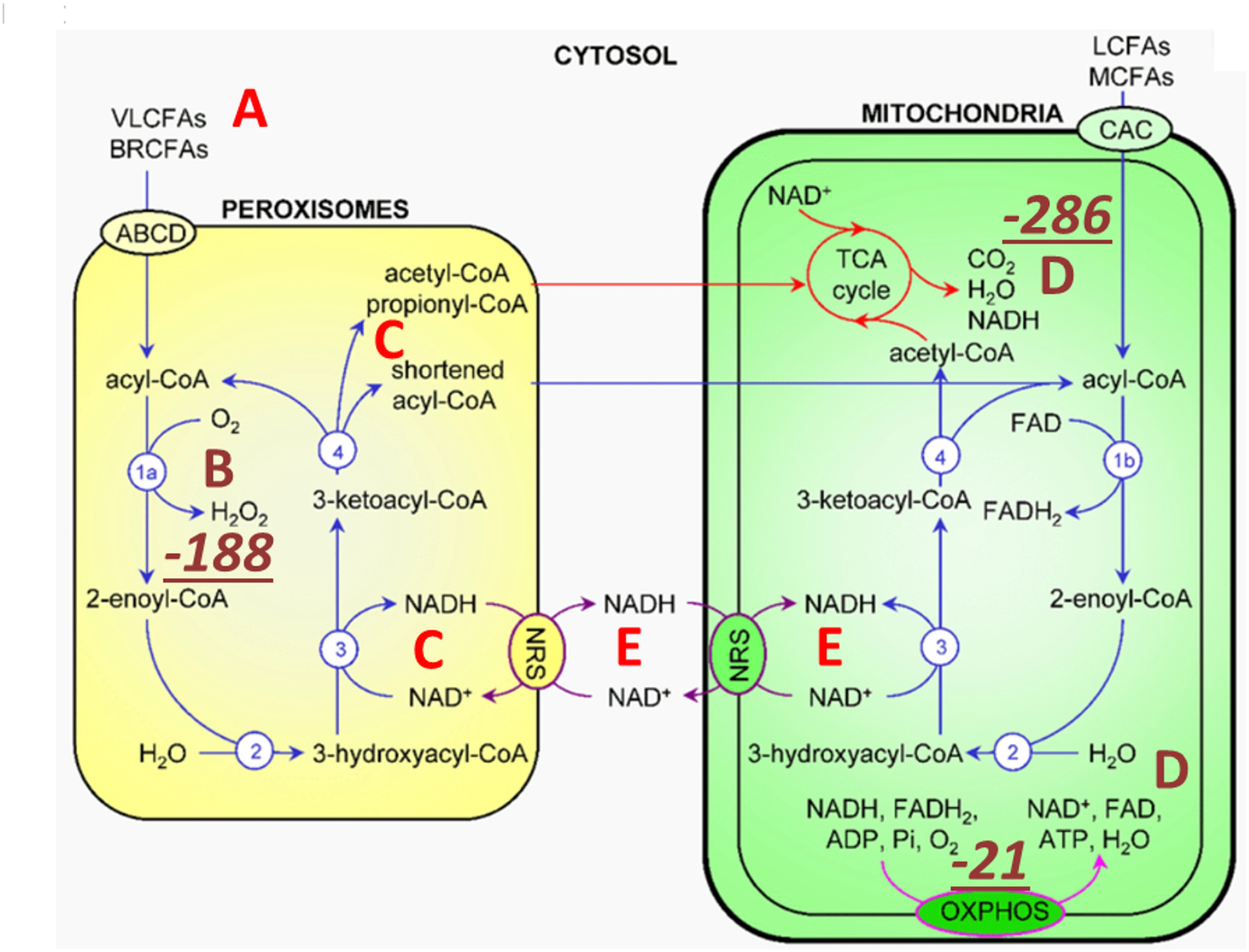
Peroxisomal metabolism triggered by TMAO turnover utilizes very long and branched chain fatty acids (A) as well as dissolved molecular oxygen (B) carried in plasma. Peroxisomes produce SCFAs via β-carbon oxidation, ketones, NADH (C) and hydrogen peroxide (B). H2O2 is rapidly converted to metabolic water by catalase (CAT) that also yields molecular oxygen for the mitochondrial matrix (D) as well as for other cellular compartments. Peroxisomes can also reduce NAD+ for proton delivery to mitochondria via membrane-based intracellular proton transporters. oxidation of very long chain saturated fatty acid β carbons, purportedly of animal source, with the help of molecular oxygen, yields the most deupleted H2O2 by weight. CAT, one of the fastest enzymes in biology with that of isomerases, rapidly and irreversibly produces water from H2O2, while recycling oxygen. Metabolic hydrogen peroxide of fatty acid breakdown with low deuterium consequently provides ATP synthase nanomotor-sparing protons for energy production. High TMAO with hydrogen peroxide turnover can easily depend on CAT-mediated oxygen recycling.

6. Is TMAO an indicator of deuterium overload in the mitochondria?
6.1 TMAO inhibits S-adenosylhomocysteine hydrolase
6.2 TMAO induces reactive oxygen species
6.3 TMAO suppresses autophagy via PI3K/Akt/mTOR activation
7. TMAO and human diseases
8. Does oxidative stress lead to mitochondrial deupletion? – While oxidative stress is a major contributor to cellular damage, the processes involved in resolving ROS are an essential part of the mechanism by which the cell reduces deuterium levels in the mitochondria. Intracellular ROS are derived mainly from NOX, xanthine oxidase, and the mitochondrial electron-transport chain (mETC). Excess mitochondrial deuterium promotes increased ROS generated by the mETC. Superoxide dismutase (SOD) converts ROS to H2O2 , which can release the highly destructive hydroxyl radical in the presence of reduced iron (Fe2+). However, H2O2 is an excellent source of deuterium depleted water (DDW) in the mitochondria, as long as there is sufficient mitochondrial glutathione and both glutathione peroxidase and glutathione reductase are adequately expressed. H2O2 freely crosses the mitochondrial membrane, and, with adequate antioxidant support, it is rapidly converted to two molecules of DDW, catalyzed by glutathione peroxidase.

9. Do lipid-laden foam cells support mitochondrial deupletion?
10. Archaeobiotics
11. A crucial role for A. muciniphila
12. Strategies to lower TMAO levels – It is clear that elevated plasma TMAO is a risk factor for a broad range of chronic diseases, and therefore it is compelling that a strategy that reduces plasma TMAO should show health benefits. However, simply avoiding foods that provide precursors to TMAO is not likely to be productive. Choline, L-carnitine, and betaine are the primary sources that fuel the methylation pathway. Eggs and seafood, rich sources of these nutrients, also contain many valuable micronutrients and healthy fats that are also essential.

Deuterium depleted water (DDW) is commercially available, at dilution levels as low as 5 ppm. It can be mixed with tap water to simulate natural glacier water, typically containing around 100 ppm deuterium. Although the number of studies on the effects of therapeutic deuterium depletion on various health conditions is small, a review paper found that deuterium depletion has shown promise in preventing and treating cancer, improving long-term memory, enhancing sports performance, and reducing symptoms of depression.

It is apparent that the best way to reduce TMAO levels, while simultaneously boosting methylation supplies, is to promote an abundant colonization of anaerobic archaea in the gut, so that they can clear (fully metabolize) the TMA before it has a chance to become TMAO.

13. Discussion – In this paper, we develop the argument that TMAO serves as a marker for excess deuterium in the methylation pathway, and, by extension, in the mitochondria, systemically. While methyl groups have powerful epigenetic effects, the ultimate fate of methyl groups is their metabolism to CO2 and water that is most likely deuterium depleted in the mitochondria. A microbial imbalance leading to reduced colonization by beneficial bacteria and an overgrowth of pathogenic species is the primary cause of overproduction of TMAO.

14. Conclusions – We have shown that TMAO, a causal factor for many diseases, may act as a marker for gut dysbiosis and for excess deuterium load in mitochondria, systemically. We traced through many of the biological pathways involving 1C metabolism and showed the integral role that gut bacteria play in stripping deuterium from methyl groups.”

https://link.springer.com/article/10.1007/s11306-026-02443-3 “The essential role of hydrogen gas recycling by gut microbes in reducing deuterium load in host mitochondria: is trimethylamine oxide a deuterium sensor?”


I take Now brand taurine, acetyl-L-carnitine, flush-free niacin, and betaine. I asked them whether these products are evaluated for their deuterium content. Will update with their response.

Add vitamin C to broccoli sprouts?

A 2026 paper presented results of a clinical trial that selectively added myrosinase enzyme via mustard powder and vitamin C to measure in vivo effects on glucoraphanin conversion to sulforaphane:

“Effects of exogenous myrosinase (Myr) on conversion efficiency of glucoraphanin (GR) to sulforaphane (SF) was compared to gut microbial Myr-like activity. In a randomized, double-blind, crossover study, sixteen subjects (9 F: 7 M) received a single oral dose of GR in 385 mg broccoli seed extract (BSE) with 72.5 mg Myr-containing mustard seed powder, or broccoli seed extract alone, both with 100 mg ascorbic acid.

GR + Myr, on average, doubled the bioavailability of SF (39.8 ± 3.1%) compared to GR alone (18.6 ± 3.1%), and increased the conversion rate in the first 8 h (25.4% ± 2.7%) compared to GR alone (8.0% ± 2.7) based on measurement of urinary metabolites. The majority of subjects given GR as BSE with exogenous Myr and Vit C, converted GR to SF notably faster (e.g., within the first 8 h), than those given GR and Vit C alone.

One of the most likely explanations for the pronounced differences is that when SF was produced as a result of added exogenous Myr, it was produced and absorbed in the small intestine and metabolized primarily to its glutathione (GSH) derivatives and excreted in urine. This is a more rapid process than when GR passes into the large intestine and then is acted upon by the greater bacterial population within that terminal segment of the gastrointestinal system.

No differences were observed in the 8 to 24 h urine collection (Time 24 h) between the two treatments: 11.7 ± 1.3% for GR + Myr vs. 8.9 ± 1.3% for GR alone. Bacterial communities did not differ between low/high GR converters following supplementation.

Many bacteria which persist in the small intestine and upper large intestine, as well as in mucosal-associated fractions, are not well represented in feces. The lack of anatomically specific microbial communities in the human gut limits our knowledge of GR conversion in people.”

https://www.nature.com/articles/s41598-026-39389-4 “Exogenous myrosinase from mustard seed increases bioavailability of sulforaphane from a glucoraphanin-rich broccoli seed extract in a randomized clinical study”


1. Lost in this study’s shuffle was the reason why sulforaphane’s effects are beneficial in the first place. As Switch on your Nrf2 signaling pathway pointed out:

“We use N-acetylcysteine (NAC) in the lab all the time because it stops an Nrf2 activation. So that weak pro-oxidant signal that activates Nrf2, you switch it off by giving a dose of NAC. It’s a potent antioxidant in that right, but it’s blocking signalling. And that’s what I don’t like about its broad use.”

It’s relevant to the Nrf2 activating effects of sulforaphane when antioxidant vitamin C taken to increase myrosinase hydrolyzation of glucoraphanin to sulforaphane if this vitamin C dose may also block Nrf2 activation. An increase of sulforaphane and its metabolites wouldn’t physiologically matter if their beneficial effects were simultaneously blocked.

2. Another indicator that these researchers lost the plot was shown when they asserted: “These data suggest that the present study may not have included sufficient AA to optimize Myr enzyme activity in vivo, another consideration for future studies” based on comparing the in vitro Reference 21 ascorbic acid doses (0, 11, 44, 88, or 154 mg AA/capsule). I don’t have access to Reference 21 to see whether it also didn’t assess Nrf2 activation.

3. For comparison of this study’s 50 mg glucoraphanin dose on two non-consecutive days, the cited Our model clinical trial for Changing to a youthful phenotype with broccoli sprouts provided daily 30 grams of fresh broccoli sprouts that contained an estimated 51 mg of glucoraphanin for ten weeks. Although no broccoli sprout preparation or intake guidelines were enforced, unassisted glucoraphanin conversion to sulforaphane had many beneficial effects in that trial.

4. These researchers stated “No differences were observed in the 8 to 24 h urine collection (Time 24 h) between the two treatments.” Whether the faster small intestine absorption of sulforaphane had benefits over the slower large intestine absorption wasn’t investigated.

5. I’ve previously corresponded with one of this study’s coauthors, and think their research group could do better work without the retired broccoli sprout expert. Maybe they would take more confident ownership of their work if they let him ride off into the sunset, and decide for themselves what predictable findings would be in their research effort’s scope?

Maybe they wouldn’t let fester the same old issues in his papers? It doesn’t say anything good about current research if its main findings just repeat last decade’s findings.

For example, Table S4 has stories that weren’t told. It conforms to the expert’s cited 2015 study findings, but presenting averages doesn’t reveal causes for individual differences. So questions on the individual level continue to be unanswered, such as:

  • Subject 8 had more than a fivefold increase of glucoraphanin-only conversion to glucoraphanin + myrosinase conversion. What blocked the other subjects from achieving similar results?
  • Why was Subject 10 so far behind everyone else’s capabilities? They would have had to increase their glucoraphanin-only conversion by fourfold just to get to the next lowest person’s level, Subject 15, but sixfold to get to Subject 15’s glucoraphanin + myrosinase conversion level.

6. Every researcher wants to have an impact on their field. The time to think over how to newly research possible impactful outcomes is before the study starts.

Since I’m in my seventh year of eating broccoli sprouts every day, I would have paid close attention to more rigorous bioavailability, more exact microbiota collection techniques, or detailed exploration of differences in people’s responses to the same treatments, all of which were predictable issues beforehand. I didn’t really care for listing them in the Discussion section, then dismissing investigations of these findings for some future research to explore.


Self-reinforcing feedback loops of aging

A 2026 rodent study investigated age-associated queuine decline. Queuine is characterized as a “longevity vitamin” in the cited 2018 review Prolonging healthy aging: Longevity vitamins and proteins, along with ergothioneine, astaxanthin, and taurine.

“The contribution of transfer RNA (tRNA)-specific modifications to aging remains largely unexplored. We systematically profile tRNA modifications across multiple organs, species, and senescence models, and identify mannosyl-queuosine (manQ) as the first tRNA-specific modification that consistently declines with age.

Across species, queuine supplementation extends lifespan and enhances healthspan. In naturally aging mice, long-term oral administration beginning at 16-months-old (human equivalent 50 years) extends mean lifespan by 15.3%, reduces DNA methylation age, improves cognitive and motor performance, strengthens antioxidant defenses, remodels the gut microbiota, and alleviates inflammation and metabolic dysfunction without detectable toxicity.

These findings establish tRNA epitranscriptomic remodeling as a previously unrecognized layer of aging regulation, and identify restoration of manQ through queuine supplementation as a multi-system strategy to delay aging.

manQ hypomodification is selective rather than reflecting global tRNA depletion. Aging preferentially reduces the manQ-containing tRNAAsp fragment while leaving the corresponding unmodified tRNAAsp fragment, and other queuosine-modified tRNAs, relatively unchanged.

This pattern supports a regulated defect in modification homeostasis rather than a generalized change in transcript abundance. Such specificity argues that manQ loss is not merely a passive consequence of tissue degeneration, but instead represents a conserved, biologically meaningful aging-associated event with mechanistic impact.

Because proteostasis intersects with multiple canonical hallmarks (e.g. mitochondrial dysfunction, impaired stress resilience, and altered intercellular communication), translation-coupled proteome destabilization offers a unifying explanation for how a single tRNA modification defect can elicit multi-system consequences. In this view, manQ decline is not merely one of many molecular changes observed in aging, but rather a proximate determinant capable of amplifying downstream hallmarks through a common axis of proteome quality control.

Our findings further suggest that manQ depletion may engage self-reinforcing feedback loops that accelerate aging trajectories. This architecture offers a conceptual framework in which aging progressively erodes ‘epitranscriptomic integrity’ at the tRNA level, pushing translation toward an error-prone regime that accelerates proteostatic collapse and functional decline.

A distinctive implication of this work is that queuine introduces a microbiota-host epitranscriptomic axis into aging biology. Queuine is produced by gut microbiota and cannot be synthesized de novo by mammals. These findings expand the conceptual scope of geroscience by placing a microbiota-derived nutrient upstream of translational quality control.

Queuine supplementation offers a distinct therapeutic logic: rather than modulating a single signaling cascade, it restores a tRNA modification state that governs translational fidelity – an upstream determinant of proteome quality that can, in principle, influence multiple downstream hallmarks concurrently. These findings highlight an intervention paradigm centered on restoring molecular fidelity, rather than suppressing a single downstream phenotype, as a strategy to delay systemic aging.”

https://www.biorxiv.org/content/10.64898/2026.03.22.713446v1.full “Evolutionarily Conserved Decline of tRNA Mannosyl-Queuosine Links Translational Regulation to Aging and Is Reversed by Queuine”

Treat your gut microbiota well. Give them what they want, and expect reciprocity.


The hops compound xanthohumol

Two 2026 papers, with the first an in vitro study of over 2000 compounds to select those that best inhibit BACH1:

“BACH1 regulates the cellular oxidative stress responses by suppressing expression of cytoprotective genes. Dysregulated BACH1 activity has been implicated in a range of pathologies, including chronic inflammatory diseases, fibrosis, and cancer, making it a promising therapeutic target.

We identified four structurally distinct compounds that robustly inhibit BACH1 function. Notably, these compounds simultaneously activate transcription factor NRF2, suggesting the potential for a broader modulation of oxidative stress pathways.

However, while NRF2 induces expression of genes that protect against oxidative stress and inflammation and suppress ferroptosis, BACH1 represses them. While NRF2 broadly activates cytoprotective genes, BACH1 inhibition triggers a more restricted response but with a strong upregulation of HMOX1 (significantly stronger than the one obtained upon NRF2 activation).

As such, combined NRF2 activation and BACH1 inactivation is expected to produce a more potent antioxidant and anti-inflammatory effect than targeting either factor alone. Moreover, BACH1 regulates unique targets not shared with NRF2 and can dominantly repress genes even in the presence of active NRF2. This confers BACH1 inhibition distinct therapeutic value, particularly in contexts such as cancer cell invasion, where its suppression yields anti-metastatic effects.

  1. Auranofin is an FDA-approved gold salt used in rheumatoid arthritis whose primary mechanism of action is inhibition of thioredoxin reductases (TrxRs).
  2. Xanthohumol is a natural compound, prenylated chalcone, that belongs to the flavonoid family, with reported antimicrobial, anti-inflammatory, and antioxidant activities, and demonstrated safety in phase I and phase II trials.
  3. Alantolactone is a natural compound, member of the sesquiterpene lactone class with anti-inflammatory and antioxidant effects, and has been tested in several animal models without reported toxicity.
  4. CH55 is a synthetic retinoid with high affinity for RAR-α and RAR-β and antifibrotic activity and has not been yet tested in vivo.

In summary, this work establishes a robust screening platform for identification of functional BACH1 inhibitors, and provides new chemical scaffolds with potential for future therapeutic development.”

https://papers.ssrn.com/sol3/papers.cfm?abstract_id=6439073 “Development of a High-Throughput Screening Platform for Identification of Functional BACH1 Inhibitors Reveals Compounds with Anti-Invasive Potential”

Xanthohumol is ninth on the Nrf2 activator list. BACH1 interactions were also covered in Part 3 of Broccoli sprouts activate the AMPK pathway.


A 2026 paper that was too recent to be referenced in the above study described two xanthohumol clinical trials. The first trial was designed to assess bioavailability in healthy people (6 men and 6 women), and the second was designed to determine bioactivity in 16 healthy women:

“The aim of the present project was to systematically investigate bioavailability of native xanthohumol compared to micellar xanthohumol at two doses (86 mg vs. 172 mg) in a randomized crossover trial. We furthermore examined short-term effects of xanthohumol on resting energy expenditure (REE), blood pressure (BP), and heart rate (HR) in a randomized placebo-controlled crossover study.

Micellar solubilization significantly increased area under the curve (AUC), maximum plasma concentration of xanthohumol (Cmax), timepoint of maximum plasma concentration of xanthohumol (tmax), and apparent bioavailability compared to native xanthohumol. The dose also significantly influenced plasma kinetics, but apparent bioavailability and tmax were dose-independent in contrast to AUC and Cmax. In our subsequent study, xanthohumol did not affect REE, substrate oxidation, BP, or HR.

Two properties of xanthohumol impair its bioavailability. First, xanthohumol is relatively unstable in an acidic environment, and second, xanthohumol is highly lipophilic and hydrophobic, insoluble in the aqueous environment of the intestinal lumen, and poorly absorbed into enterocytes.

In addition to determining typical plasma kinetic parameters (e.g., Cmax, tmax, and AUC), we also calculated the amount of xanthohumol absorbed using maximum plasma xanthohumol concentration. These estimated amounts are minimum quantities of xanthohumol that had to be absorbed to achieve observed plasma xanthohumol concentrations.

Results of these calculations emphasized the relatively poor bioavailability of xanthohumol in humans. Only ∼0.1% and ∼1.2% of native and micellar xanthohumol were absorbed, respectively.

However, a limitation of this plasma estimation is that fractional absorption of xanthohumol was underestimated because distribution of xanthohumol in cells and tissues was not taken into account. It is likely that more xanthohumol was actually absorbed. Whether the bioavailability of xanthohumol is sufficient for physiological efficacy must be investigated further in humans.

In conclusion, oral bioavailability of micellar xanthohumol was higher than that of native xanthohumol. Systemic availability of xanthohumol did not differ between men and women. Our study provides no evidence that xanthohumol acutely affects REE, BP, and HR.”

https://onlinelibrary.wiley.com/doi/10.1002/mnfr.70413 “The Bioavailability of Xanthohumol in Humans and the Influence of Formulation and Dose: Randomized Controlled Trial Data”


Year Six of Changing to a youthful phenotype with sprouts

1. I’ve continued daily practices from Year Five to experience another year without being sick (if I don’t count getting MSG poisoning from Chinese food.) I consequently scheduled a doctor visit next week to get a sumatriptan prescription refilled.

2. Two modifications to what’s mentioned in 2026 diet and supplement changes:

– In that post’s comments, Ole Bisgaard Pedersen asked if I took NAD+ supplements such as NAM, NMN or NR – forms of vitamin B3 that are precursors to NAD+. I didn’t note that last year I started taking Now brand Flush-free niacin 500 mg mid-morning.

Nicotinamide riboside has the most human evidence, including a Tru Niagen clinical trial that showed improvements in peripheral artery disease. It’s too expensive for long-term use, though.

Even if I could afford it, there isn’t a magic bullet for fixing vascular system dysfunction. PAD is just one symptom of a cardiovascular system that needs to be overhauled then maintained at a healthy level. There is no clinical trial that has a logical therapeutic end point to stop treatments where a person could say, “I’ve done enough for my vascular system, my physical and cognitive functions won’t backslide.”

– 2-3 years ago, I changed from microwaving broccoli sprouts in a plastic bag to microwaving them in a small bowl with a small plate covering it to keep them from popcorning out of the bowl. I use a 1000W microwave oven on 80% power for ten seconds.

3. The two vitamin C macaque studies I’ve recently curated both ran for a human equivalent of ten years. I was encouraged that both found Nrf2 activation to be part of their causal beneficial evidence, since vitamin C wasn’t on my radar as a Nrf2 activator.

I expect that in four years I’ll write a Year Ten post on eating microwaved broccoli sprouts. I haven’t seen human evidence for broccoli extracts that bypass small intestine absorption and metabolism per Glucosinolate and isothiocyanate human interventions, or enteric capsules, or nanoformulations as suitable substitutes. Maybe studies on broccoli sprout powder or a Nrf2 activator that tops sulforaphane will be published before then, who knows.

2026 diet and supplement changes

I switch things around pretty often, but I haven’t said much about diet and supplement changes since this time last year. Here’s what I’ve done in terms of changes that I’ve since abandoned or reduced, followed by additions or increases that I’ve kept.

Abandoned and reduced items

1. I stopped using Avena sativa oats to grow 3-day-old oats sprouts. I again ran into the same situation where I got < 10% yield.

The first time this happened in 2023, I related to the Montana farmer that degraded seed vitality was probably caused by the way that Amazon handled their oat products. I’m the customer, though, and I won’t make it my problem if the vendor can’t meet expectations.

I switched to sprouting Avena nuda oats based on Sprouting hulless oats. I’ll note that this Illinois farmer doesn’t let Amazon handle their organic Avena nuda oats, and they add on post office shipping costs. They don’t recommend sprouting, probably because of liability, although I’ve had a 91% germination rate over three days. I might have ordered Avena sativa oats directly from the Montana farmer bypassing Amazon if they were also organic.

2. I stopped taking alpha ketoglutarate. In my view, increasing tricarboxylic acid (TCA) cycle intermediate metabolites such as alpha ketoglutarate and CoQ10 should not be the primary way to improve mitochondrial electron transport chain function.

Instead of biochemical considerations, focus on photon modulation, which precedes biochemical reactions. Which means mitochondrial studies should be controlled for light exposures, and very few of them do that, although it’s the way nature works.

This past winter I increased indoor non-LED light exposure within a circadian rhythm framework. I’ll switch back to walking the beach at sunrise from being out in mid-day sun after it gets a little bit warmer.

3. I’ve taken creatine on and off during the past year. There’s a bit of literature on its use for improving methyltransferase system components like homocysteine.

Stopping creatine fits one of the overall patterns that studies demonstrate – people who are initially deficient in the studied item get a benefit, while people who are initially sufficient don’t benefit from treatment. I’ve always tested mid-range for homocysteine, which is desirable.

4. I had some cocoa powder lying around for a year or two, and I used it this past winter to improve the taste of coffee I bought on sale. Cocoa flavanols are supposed to improve various health measures. But I haven’t been provided access to the most recent human studies, so I won’t repeat their results without reading their details.

5. I saw this at Costco, and picked up a package:

A 2025 review covered pecan research, Pecans and Human Health: Distinctive Benefits of an American Nut. Eating pecans seems to have some health benefits, and they taste alright.

For me, though, the dryness of a chewed pecan bolus creates a swallowing problem that walnuts don’t have. YMMV.

6. I stopped taking 2 g magnesium L-threonate. I’ve always tested high for magnesium without using a specific supplement.

7. I reduced D3 by 25 mcg to a daily 2400 IU. Winter is over.

New and increased items

1. I curated five 2025 ergothioneine studies in Human studies of ergothioneine after stopping mushroom intake via AGE-less chicken soup. I wasn’t thrilled that none of them investigated long-term effects of persistent plasma ergothioneine levels.

This year I decided to start taking the higher 25 mg dose of the first study once a week. That should produce some benefits at a lower ergothioneine blood level than daily doses produce. I’ll check periodically for 2026 research.

2. The only paper I’ve curated on deuterium (heavy hydrogen) is Taurine and mitochondrial health. I started using Icelandic glacier water to make coffee and tea, and for just drinking.

It isn’t advertised as deuterium-depleted water, and it isn’t manufactured as such. But I think any glacier water contains less deuterium than local water. I use local filtered water for sprouting and cooking.

3. Per The return of the free radical theory of aging I started taking extra vitamin C separately from other supplements in the form of Now brand liposomal 1 gram twice daily this past winter. That study found vitamin C to be an anti-aging compound for primates.

Reference 72 of that 2026 paper is a freely available 2025 study https://www.cell.com/cell-stem-cell/fulltext/S1934-5909(25)00339-X “Vitamin C conveys geroprotection on primate ovaries” that used the same vitamin C dose and duration in macaques to find:

“VC slowed aging in various ovarian cell types. Moreover, VC protected human ovarian endothelial and stromal cells (SCs) from aging partially via NRF2 activation. This study establishes a proof-of-concept for delaying primate ovarian aging with a single compound, and provides important insights into preventing and treating degenerative diseases related to ovarian aging.”

4. I restarted taking inulin last year, about 3 grams (a heaping teaspoon) daily after posting Inulin vs. FOS. My 2.5 year-old grandchild takes a level teaspoon daily, as inulin’s beneficial effects aren’t just for old people.

5. I started taking 12 mg astaxanthin twice in the morning. I use Nrf2 activators in the morning because Nrf2 is especially involved in the circadian cycle, as noted in papers such as Broccoli sprouts activate the AMPK pathway, Part 4.

6. I increased daily raw egg consumption from 3 eggs a day to 3 eggs twice daily.

7. This year, Ovega 3 algae oil DHA 420 mg/EPA 140 mg became no longer available AFAIK. I substituted Vegan Omega 3 algae oil DHA 300 mg/EPA 150 mg in the morning and Sports Research Omega 3 fish oil DHA 310 mg/EPA 690 mg in the afternoon.

8. I picked up this Korean seaweed in a 10-pack at Costco. The label doesn’t say what its iodine content is. I eat it as a snack whenever I get a salt craving, maybe once a week.

The return of the free radical theory of aging

A 2026 primate study investigated effects of vitamin C:

“Here, we define a conserved iron-lipid axis driving primate aging, termed ‘ferro-aging.’ Multi-tissue profiling in humans and non-human primates reveals age-progressive iron accumulation, fueling chronic lipid peroxidation orchestrated by acyl-coenzyme A (CoA) synthetase long-chain family member 4 (ACSL 4). Distinct from acute ferroptosis, this ACSL4-mediated process promotes cellular senescence and systemic functional decline.

We identify vitamin C (VC) as a direct inhibitor of ACSL4. Long-term VC administration in aged monkeys for over 40 months potently reduces ferro-aging signatures across tissues, attenuates multi-organ pathology, and improves neurological and metabolic functions. Multi-omic aging clocks indicate the VC-mediated reversal of biological age.

Despite decades of interest in oxidative stress, largely sparked by the free radical theory of aging, efforts to modulate it broadly with antioxidants have yielded inconsistent or neutral outcomes, highlighting the theory’s limitations and underscoring the need to identify more specific, upstream drivers. A critical challenge remains: determining whether the iron-lipid axis constitutes a core upstream driver of aging in primates and, if so, whether it is therapeutically targetable.

In this study, we bridge these gaps. We define an iron-triggered, ACSL4-governed, lipid peroxidation-driven program that escalates with age across diverse cell types and multiple organs in non-human primates.

VC treatment dose-dependently increased Nrf2 phosphorylation and activation. VC orchestrates a dual-defense strategy against ferro-aging: it directly suppresses the pro-aging lipid peroxidation driver ACSL4, while in parallel, it bolsters the cell’s intrinsic antioxidant capacity via Nrf2 pathway activation.

Middle-aged cynomolgus monkeys (12–16 years old, approximating human 40–50 years) received daily oral VC (30 mg/kg group) or a control treatment for 40 months under standardized conditions.

Structural MRI analysis demonstrated that VC intervention counteracted age-related brain atrophy. Using general linear mixed models, we found that VC restored cortical surface area in the frontal lobes of aged monkeys. Regional analysis identified enlargement in four regions of the orbital frontal cortex, an area critical for adaptive behavior.

Diffusion MRI-based connectomics revealed that, compared with young animals, aged monkeys exhibited reduced structural connectivity in 18 brain regions. VC treatment restored connectivity in 9 of these regions, which were predominantly located in the posterior parietal cortex, a hub for spatial awareness and decision-making.

VC exerted robust neuroprotective effects. It attenuated heterochromatin loss (increased H3K9me3) in the prefrontal cortex and hippocampus and reduced abnormal protein aggregates, including cytosolic aggresomes and Aβ. Additionally, VC lowered the abundance of activated microglia and astrocytes and suppressed expression of the innate immune sensor cGAS in the hippocampus.

VC supplementation reduced the estimated biological age across multiple organs. At the epigenetic level, VC lowered DNA methylation age in several tissues, including brain, brown adipose tissue, muscle, skin, aorta, and kidney. In the hippocampus, the most substantial reductions in biological age occurred in microglia, oligodendroglia, and oligodendrocyte precursor cells. In the pancreas, alpha cells, beta cells, and ductal cells showed the greatest rejuvenation.

In summary, chronic VC supplementation inhibits the ferro-aging pathway, reduces multidimensional biological age across primate organs, and ameliorates a spectrum of functional declines in nervous and metabolic systems. Our work establishes ACSL4 inhibition as a promising and translationally relevant therapeutic strategy for mitigating aging-related decline.

A long-term, 40-month intervention study in aged non-human primates is a highly translational model given their shared inability with humans to synthesize VC endogenously. The finding that a single, safe nutrient can reverse multidimensional aging clocks in a primate has profound implications for translational longevity medicine.”

https://www.sciencedirect.com/science/article/abs/pii/S1550413126000537 “Vitamin C inhibits ACSL4 to alleviate ferro-aging in primates” (not freely available) Thanks to Dr. Pradeep Reddy for providing a copy.


Grok’s take on this study:

“For humans (who, like macaques, cannot synthesize vitamin C), the Recommended Dietary Allowance (RDA) is 75–90 mg/day for adults (~1–1.5 mg/kg for a 60–70 kg person) to prevent deficiency. Upper safe intake levels are much higher: up to 2,000 mg/day (Tolerable Upper Intake Level) is considered safe for most adults, with no established adverse effects at that level from food/supplements.

Treated monkeys represent advanced aging stages (likely equivalent to human 50s–70s+ based on ‘aged’ designation and long-term intervention effects), extending the prior 12–16-year monkey range (human ~35–55) to broader anti-aging applications. While human trials are needed, the primate evidence (long-duration, systemic benefits) strengthens the case for high-dose, sustained vitamin C as a strategy against ferro-aging in humans. It elevates vitamin C from a nutrient to a targeted anti-aging compound in primates.”

Coincidentally, I started taking extra vitamin C separately from other supplements in the form of liposomal 1 gram twice daily this past winter. Can’t say that it had any effects on my intended target, avoiding sniffles and sneezing, as allergy season kicked off in early February. With this study’s findings, I’ll continue.

Eat broccoli sprouts for ALS?

A 2026 rodent study investigated sulforaphane’s ability to affect ALS-like symptoms:

“The objective of this study was to evaluate neuroprotective efficacy and safety of sulforaphane (SUFP) in a methylmercury (MMHg⁺)-induced preclinical rat model of amyotrophic lateral sclerosis (ALS). ALS is characterized by progressive motor neuron degeneration and muscle wasting, leading to impairments in gait, swallowing, salivation, and routine motor activities.

64 animals were classified into eight groups: 1st: normal control, 2nd: vehicle control; 3rd: SUFP perse (4 mg/kg, i.p.), 4th: MMHg + (5 mg/kg, p.o.), 5th: MMHg + 5 + SUFP (2 mg/kg, i.p.), 6th: MMHg+ 5 + SUFP (4 mg/kg, i.p.), 7th: MMHg+ 5 + omaveloxolone (OVX) (30 mg/kg, i.p.), and 8th: MMHg + 5 + dimethyl fumarate (DIMT) (50 mg/kg, i.p.). Neurotoxin MMHg + was orally administered at 5 mg/kg for the first 21 days. For the next 22 days, SUFP, OVX, and DIMT were administered intraperitoneally (i.p.).

SUFP modulates neurotransmitter levels such as acetylcholine (A), dopamine (B), GABA (C), glutamate (D), and serotonin (E).

SUFP4 exerted broad neuroprotective effects in ALS pathology by restoring antioxidant proteins (Nrf2, HO-1, SIRT1), suppressing apoptotic (Bax, caspase-3, Bcl-2) and inflammatory markers (TNF-α, IL-1β), and enhancing the anti-inflammatory cytokine IL-10. It also downregulated stress-related signaling pathways (PI3K/Akt, p75NTRECD, MAPKs) associated with neurodegeneration. These molecular effects translated into meaningful functional recovery, as evidenced by improvements in grip strength, locomotor performance, spatial memory, and depressive-like behavior.

Histopathological evaluation demonstrated attenuation of demyelination and preservation of neuronal architecture including the cerebral cortex, hippocampus, striatum, midbrain, and cerebellum. Beyond central neuroprotection, SUFP exerted systemic benefits by normalizing hepatic enzymes, improving skeletal muscle integrity, restoring redox balance, stabilizing neurofilament and myelin-associated proteins, and correcting hematological alterations.

Despite limitations related to study duration and animal sex, this work strongly positions SUFP as a promising, multi-target therapeutic candidate for ALS with both neural and systemic protective efficacy.”

https://link.springer.com/article/10.1007/s12035-026-05683-5 “Sulforaphane-Mediated Multitarget Therapeutic Effects in Methylmercury-Induced ALS-Like Pathology: Comparative Analysis and Multifaceted Approach to Neuroprotection and Systemic Recovery” (not freely available) Thanks to Dr. Sidharth Mehan for providing a copy.


Unlike A Nrf2 treatment for ALS?, this study didn’t present evidence that its treatment compound was effective for preventing ALS. For one thing, currently-known disease factors involving heat shock proteins and associated genes, some of which are Nrf2 targets, weren’t investigated.

Two Nrf2 activators were used in both studies as comparators of Nrf2 activation effects. Neither omaveloxolone nor dimethyl fumarate are ALS causal treatments, though, and have undesirable side effects.

A human equivalent of this study’s higher sulforaphane dose is ((4 mg x .162) x 70 kg) = 45 mg. 45 mg of sulforaphane might be too much to consistently take at one time because of unpalatability. But I documented taking an estimated 52 mg for a year during 2020-2021 by eating microwaved 3-day-old broccoli sprouts twice a day.


Nrf2 and stem cells

A 2026 review subject was mechanisms and therapeutic potential for Nrf2 activators in combination with mesenchymal stem cells:

“Mesenchymal stromal/stem cells (MSCs) are multipotent stem cells that can be isolated from various tissues – such as bone marrow (BM), umbilical cord (UC), adipose tissue (AD), dental pulp (DP), hair follicle (HF), and placenta – and differentiated into multiple lineages under appropriate conditions. Their functional repertoire includes immunomodulation, homing, and differentiation, which collectively help establish a balanced inflammatory and regenerative niche within damaged tissues during severe inflammation. MSCs-derived extracellular vesicles (MSCs-EVs) and conditioned medium (MSCs-CM) play remarkable roles, exhibiting potent anti-inflammatory and antioxidant properties that offer novel therapeutic alternatives for inflammatory diseases.

Therapeutic capacity of MSCs in inflammatory conditions is increasingly attributed to their potent paracrine activity rather than solely to their differentiation potential. A key mechanism underlying this paracrine effect is activation of the Nrf2 antioxidant pathway.

MSCs and their secreted products including exosomes (Exos), EVs, and CM, activate Nrf2 through multi-dimensional/target mechanisms, thereby enhancing cellular antioxidant defenses, modulating immune responses, and promoting tissue repair. It is noteworthy that therapeutic efficacy of MSCs and their derivatives can be enhanced through external modulation, including pretreatment with natural compounds.

Preconditioning refers to brief treatment of MSCs or their derivatives with physical, chemical, or biological factors prior to application, aiming to enhance their ability to counteract oxidative stress and improve their therapeutic efficacy. Flavonoids precondition and prime MSCs via the direct Keap1-Nrf2 pathway or indirect PI3K-Akt pathway, which enhances cellular resilience to adverse conditions by reducing apoptosis and promoting survival. Primed MSCs, in turn, remodel the microenvironment through an altered secretory profile, releasing bioactive factors that create more favorable conditions for their own persistence.

The core logic of these strategies lies in simulating or inducing adaptive stress, such as employing specific chemical molecules or drug stimuli, or utilizing physical / microenvironmental preconditioning to mimic specific physical conditions of the in vivo injury environment. The most straightforward strategy is overexpression of Nrf2 or its key downstream effector molecules.

The majority of existing studies remain at the level of observing correlations with Nrf2 upregulation, and there is still a lack of precise causal validation regarding key upstream signals – such as specific cytokines, miRNAs, or proteins – through which MSCs or derivatives initiate Nrf2 activation. Mechanistic insights are predominantly derived from in vivo or rodent (mouse/rat) model experiments, with a notable absence of clinical validation, insufficient long-term safety and pharmacokinetic data, and a lack of standardization in administration routes and dosages, all of which hinder clinical translation.

The essential role of the Nrf2 pathway has not been rigorously confirmed, as most studies have not employed reverse genetic validation using Nrf2-knockout animals or specific inhibitors. Consequently, it remains unclear whether therapeutic effects are necessarily and exclusively dependent on Nrf2, and potential synergistic contributions from other pathways may have been overlooked.

Most natural flavonoids face challenges such as low oral bioavailability, rapid metabolism, and poor targeting. Numerous challenges remain to be addressed in order to translate these promising preclinical findings into clinical practice. Future research should focus on the following aspects:

  1. Elucidating precise upstream molecular mechanisms by which MSCs activate Nrf2;
  2. Employing more clinically relevant chronic disorder models;
  3. Systematically evaluating long-term safety, optimal delivery strategies (including dosage and route of administration), and immunogenicity of MSCs-based therapies;
  4. Validating selection criteria (optimal source), quality control, batch-to-batch consistency of MSCs, and addressing regulatory and ethical barriers to clinical translation; and
  5. Integrating molecular docking, ADMET (Absorption, Distribution, Metabolism, Excretion, Toxicity) prediction, and in vitro and in vivo validation to further elucidate regulatory effects of flavonoids and enhance understanding of their mechanisms of action.”

https://link.springer.com/article/10.1186/s13287-026-04925-6 “Activation of Nrf2 with natural flavonoids and mesenchymal stromal/stem cells: mechanisms and therapeutic potential for inflammatory diseases” (click pdf)


This paper was overly long at 127 pages, so I focused on the later sections. None of these treatments are currently ready for clinical trials.

I also didn’t mention specific flavonoids as Nrf2 activators. It’s beyond a reviewer’s task to rank Nrf2 activators, and a study’s researchers seldom address why they used a poorly-activating flavonoid instead of a higher-ranked natural plant compound such as sulforaphane.

Polling vs. propaganda

An anonymous doctor’s perspective on what’s permitted to be publicly discussed regarding vaccine injuries:

“Only one polling organization independently investigated it, Rasmussen Reports (a conservative polling organization which has a reputation for getting accurate results due to them having listeners punch answers in response to an automated voice rather than directly talking to someone who may bias them). For American adults, they found:

  1. July 2021: 32% believed public health officials were lying about the safety of COVID-19 vaccines.
  2. December 2022: 56% of 1000 respondents believed the vaccines were effective, 57% were concerned the vaccines had major side effects. Most importantly, 34% of those vaccinated reported minor side effects and 7% reported major side effects (e.g., those seriously impairing their quality of life).
  3. January 2023: 49% believed it is likely that side effects of COVID-19 vaccines have caused a significant number of unexplained deaths and 28% personally knew someone whose death may have been caused by side effects of the COVID-19 vaccines. 57% wanted Congress to investigate how the CDC handled assessing vaccine safety (presumably since many suspected the CDC had covered up the dangers of the COVID vaccination program).
  4. March 2023: 11% of those surveyed reported that they believed a member of their household died from COVID-19, while 10% believed a member of their household died and that their death may have been due to a side effect of the vaccine.
  5. September 2023: 47% of those surveyed stated they did not believe the vaccines were safe and 34% did not believe they were effective. As before, these results also politically stratified as Democrats were less likely to believe the vaccines were unsafe (14% D vs. 51% R) or ineffective (17% D vs. 57% R).
  6. November 2023: 24% personally knew someone they believe died from a COVID vaccine, and of those individuals, 69% would be likely to join a class action lawsuit against the pharmaceutical companies.
  7. January 2024: 53% believe it is likely that side effects of COVID-19 vaccines have caused a significant number of unexplained deaths and 24% personally knew someone whose death may have been caused by side effects of COVID-19 vaccines.
  8. September 2024: 55% surveyed believe it is likely that side effects of COVID-19 vaccines have caused a significant number of unexplained deaths – including 30% who say it’s very likely.
  9. November 2025: 26% reported they had minor side effects from the vaccine and 10% reported major side effects. Additionally, 46% believed it is likely that side effects of COVID-19 vaccines have caused a significant number of unexplained deaths – including 25% who say it’s very likely.

In short, the data shows you aren’t crazy, and while the news is not reporting it, the majority of people are seeing exactly the same thing you are. There is no getting around the fact a lot of people were harmed by these vaccines.”

https://www.midwesterndoctor.com/p/polling-reveals-a-profound-shift “Polling Reveals A Profound Shift on Vaccines: We Can’t Let Pharma Bury It”


I knew two women who died in mid-2022 in their late sixties who probably had vaccine injuries contributing to their shortened lives. The subject wasn’t allowed to be discussed earlier this decade, and it largely still isn’t.

Everything this article says about covid vaccine injuries is applicable to the childhood vaccines. I found out at a house party earlier this week that both subjects are verboten, especially with women who have preschool children, despite their libertarian orientation.

Don’t know what will change public acknowledgement of vaccine harms. Isn’t it obvious that we were and are being lied to? Why still trust untrustworthy professions?

I thought at the beginning of this decade that people would soon see through the propaganda, but that didn’t happen. I also thought that public punishments for crimes against humanity would penetrate people’s awareness that we’d been duped, but the guilty still run free.

Eat broccoli sprouts to prevent or treat obesity

This 2026 rodent study made mice obese with a high-glycemic-index diet, and intervened with different doses of sulforaphane during and after inducing obesity:

“To our knowledge, this is the first study investigating therapeutic effects of sulforaphane (SFN) on obesity resulting from feeding with high-glycemic-index diet (HGID). To evaluate the potential role of SFN on energy metabolism, obesity development, and insulin resistance, effects were tested by administering SFN at different doses [oral 5 mg and 20 mg] in addition to HGID and after animals were made obese with HGID.

Energy, macronutrient, and fiber contents of the HGID used in the experiment and the isocaloric control group feed were kept equal. The only difference between HGID and the control group feed was composition of the starch. While starch in the HGID was a waxy corn starch consisting of 100% amylopectin, it was natural starch (75% amylopectin, 25% amylose) in the control group.

This study is strengthened by its:

  • Experimental design, in which SFN was administered at multiple doses both during exposure to HGID and after development of HGID-induced obesity, allowing for a comprehensive evaluation of its effects on energy metabolism, obesity progression, and insulin resistance.
  • Focus on specific components of HGID. To be able to separate effects of the HGI diet pattern, one of the long-standing criticisms regarding GI, from individual components it contains, especially dietary fiber, we were able to evaluate glycemic index interaction by keeping energy, macronutrient, and fiber contents of feeds equal and the starch composition different.

Several limitations should be acknowledged.

  • Potential adverse effects of the 5 mg/kg/day and 20 mg/kg/day doses of sulforaphane in this study were evaluated in terms of clinical signs. However, systemic adverse effects, particularly those affecting the brain, cardiovascular system, or other organs, were not assessed.
  • The relatively short duration of the SFN intervention (five weeks following development of obesity induced by a HGID) may have limited the ability to fully capture all potential changes in the measured variables. It may be beneficial to observe for a longer period in future studies to provide evidence that SFN reverses HGID-induced obesity.
  • The ideal dose of SFN has not yet been determined. Dose and bioavailability are considered important parameters that need to be clarified for SFN to be considered as an anti-obesity agent.

Results indicate that SFN may provide potential benefits both as a protective agent in the obesity development process and as a therapeutic approach after obesity has developed.

  • While SFN suppresses obesity development by combating increased energy consumption, body weight, deteriorated lipid profile, and decreased insulin sensitivity upon exposure to HGID, it supports obesity treatment with its aspects of reducing food consumption and body weight gain and improving glycemic control.
  • SFN may reverse adverse effects of HGID in a time- and dose-dependent manner by regulating postprandial insulin, restoring IRS1/IRS2 function, inhibiting gluconeogenesis through coordinated activation of signaling between sirtuins and PGC-1α, and shifting liver metabolism from lipid synthesis toward mitochondrial oxidation.”

https://www.mdpi.com/2072-6643/18/4/574 “Sulforaphane Against the Metabolic Consequences of a High-Glycemic-Index Diet: Protective and Therapeutic Mechanisms Associated with Obesity and Insulin Resistance”


A human equivalent to this study’s daily oral low sulforaphane dose is (5 mg x .081) x 70 kg = 28 mg, which is achievable by eating broccoli sprouts every day. People won’t tolerate quadrupling 28 mg to a human equivalent of the study’s 20 mg daily oral sulforaphane dose, so I didn’t curate this study’s high-sulforaphane-dose-specific findings.

Human age equivalents to this study’s 8-week-old, 23-week-old, and 28-week-old mice are respectively 18-25 years, 25-35 years, and 28-38 years.

A 42-month human study of broccoli sprouts’ effects on cognitive function

A 2026 paper provided details of a 2020-2023 human trial of broccoli sprouts:

“In a 42-month randomized, double-blind, placebo-controlled trial, 26 participants aged 63–90  years with memory impairment were randomly assigned to receive either 30 mg/day of glucoraphanin (GLR) or placebo. The primary outcome was the change in Memory Performance Index (MPI) scores from the mild cognitive impairment (MCI) screen. This study evaluated the long-term efficacy of GLR supplementation on cognitive function in older adults at an elevated risk for Alzheimer’s disease (AD), including those with MCI.

Participants were instructed to take three capsules of either the GLR or placebo supplements daily for 42 months. The GLR supplement contained 30 mg of GLR purified from broccoli sprouts, along with 120 mg of mustard powder per three capsules. Mustard powder was included as a source of exogenous active myrosinase to enhance the enzymatic conversion of GLR to sulforaphane. The placebo supplement contained 0 mg of GLR.

No significant group difference was observed in the initial 6 months. A marginal difference in favor of GLR appeared in the later phase (30 and 42 months), including the 42-month endpoint.

The GLR group demonstrated superior performance on immediate recall and delayed free recall tests. MCI participants showed a greater MPI improvement with GLR.

Long-term GLR supplementation may help preserve cognitive function in individuals at elevated risk for AD, particularly those with MCI. Larger trials are warranted to confirm efficacy and clarify underlying mechanisms.”

https://www.frontiersin.org/journals/nutrition/articles/10.3389/fnut.2026.1740494/full “Efficacy of 42-month oral administration of glucoraphanin in preventing cognitive decline in individuals at elevated risk of dementia, including those with mild cognitive impairment: a randomized, double-blind, placebo-controlled pilot study”


Again, 42 is the answer! 🙂

This study was funded by the supplement manufacturer. There was no explanation of what the supplement’s “purified from broccoli sprouts” entails. Also, I didn’t mention results of voluntary group exercise because there was a long gap in the participants’ data due to government response to covid.

  • For comparison of this study’s 30 mg glucoraphanin dose, Our model clinical trial for Changing to a youthful phenotype with broccoli sprouts provided 30 grams of fresh broccoli sprouts that contained an estimated 51 mg of glucoraphanin for ten weeks. That study’s corresponding coauthor said of their 30 gram broccoli sprouts dose in Understanding a clinical trial’s broccoli sprout amount that “When we carried out tests with consumers, previous to the bioavailability studies, higher amounts per day were not easy to consume and to get eaten by participants.” There was no rationale provided for this study’s 30 mg dose other than citing two previous human studies that also used a 30 mg glucoraphanin dose.
  • For comparison of this study’s 120 mg mustard seed powder dose, my daily cruciferous food intake since five and a half years ago includes sprouted yellow mustard seeds started with 3.5 grams of seeds, along with sprouted broccoli and red cabbage started with 3.6 grams of each vegetable’s seeds, all sprouted for three days. I haven’t seen studies that show sprouting has effects on myrosinase enzyme activity.
  • This study cited Does sulforaphane reach the colon? which used 2% mustard seed powder to create sulforaphane from glucoraphanin. This study’s 120 mg mustard seed powder / 30 mg glucoraphanin is a lot more than 2%. My daily sprout intake started from 3.5 g mustard seeds / (3.6 g broccoli seeds +3.6 g red cabbage seeds) is also a lot more than 2%.

I’ve changed some items along the way, switching supplier from True Leaf to Johnny’s for organic broccoli seeds, and from non-organic Red Acre red cabbage seeds to True Leaf organic red cabbage seeds. I recently had to find another supplier of organic yellow mustard seeds when Naturevibe stopped carrying that product. I tried Food to Live, but their yellow mustard seeds when sprouted aren’t mild. I’ll next try Frontier Co-op to see if those are mild as advertised.


No hero will be rescuing your and your children’s neurodegeneration for you

Starting this blog’s twelfth year by curating a poorly-done 2026 review of Nrf2 and its capability to change a person’s development of Parkinson’s disease. I’ll emphasize precedent conditions that if not effectively dealt with in youth, can’t prevent PD from occurring at some later life stage.

“This review explicitly examines how age-associated decline in NRF2 responsiveness intersects with redox imbalance, mitochondrial dysfunction, proteostatic failure, and neuroinflammation, core mechanisms shared between aging and PD. PD unfolds through a complex interplay of cellular stress and immune responses. Oxidative stress, mitochondrial dysfunction, and chronic neuroinflammation converge to damage dopaminergic neurons, with microglia playing a central role in amplifying this injury.

NRF2 emerges as a key regulator of antioxidant defenses, inflammatory balance, and mitochondrial protection, offering a promising target for clinical intervention. NRF2 activity favors the anti-inflammatory microglial over the pro-inflammatory phenotype. Decline in NRF2 inducibility with age impairs microglial clearance, promotes neuroinflammation, and reduces antioxidant defenses, while NRF2 activation restores protective functions and offers a promising therapeutic target.

Strategies aimed at restoring or enhancing NRF2 activity hold significant promise as disease-modifying interventions, not only to slow PD progression but also to promote resilience against the broader spectrum of age-associated neurodegenerative and inflammatory conditions.”

https://www.sciencedirect.com/science/article/pii/S0891584926000316 “NRF2 AT THE CROSSROADS OF PARKINSON’S DISEASE AND AGING: MECHANISTIC INSIGHTS AND TRANSLATIONAL PERSPECTIVES”


This review only gave lip service to PD progression outside of the brain, as if the importance of prodromal factors to a person’s neurodegeneration such as dysfunction in gut, eyes, skin, and olfactory systems can be minimized. But failure to recognize early what will doom a person to be unable to recover health in later decades is disingenuous. Since these reviewers omitted early interventions into PD prodromal factors, the best they came up with was interventions to “slow PD progression.”

Maybe these reviewers felt it would be outside the scope of this review to discuss early non-brain PD factors for more than one sentence? However, while PD is defined by striatal brain neurons, Nrf2 activity is much less in brain and central nervous system neurons than elsewhere in the body per Nrf2 Week #2: Neurons.

I disagree with these reviewers’ self-imposed emphasis on aging. Repeating ‘age-associated’ numerous times seemed as if they wanted to influence the reader into thinking age in and of itself was a cause for PD, rather than an imputed mathematical correlation. Their emphasis led to dumb mentions such as senolytics for no apparent reason than senescence is a ‘hallmark of aging’, and to meaningless ‘diseasome of aging’ characterizations, and to ignoring the existence of early non-age-associated PD diagnoses in 20- and 30-year-olds.

Whatever it takes to get published, I’d guess. Or maybe it’s that the number of omissions and useless points a review paper makes increases with the number of reviewers and their sponsors’ agendas.

For example, why was it permissible to dedicate lip service to ‘exposome’ factors like microplastics, environmental pollution, and viruses, but it’s still not permitted in 2026 to discuss research into the impacts on vascular disease and neurodegeneration of lipid nanoparticles and DNA contamination in what a large number of humans were exposed to by injected pharmaceuticals starting in late 2020? Not to mention two studies published in 2024 of over 2.5 million people whose incidences of neurologic issues, mild cognitive impairment, and Alzheimer’s disease rapidly increased after ‘vaccination’?

I’ve mentioned in this blog many times how it’s every human’s choice whether or not we take responsibility for our own one precious life. I suggest, if it’s not too late, do that for your children’s lives, too.

Celebrate Mozart’s 270th birthday

“Mozart’s motet, Ave Verum Corpus, was composed in June of 1791, for his friend, Anton Stoll, a school teacher and choirmaster of the small parish church of St. Stephan in Baden, a spa-town located near Vienna which was famous for its hot thermal mineral springs. Mozart’s wife, Costanze, was pregnant with their sixth child and was ill, and so she was staying at the Baden spa for treatment (“taking the waters”).
The work was written almost as a payment to Stoll, who had often helped Mozart by making travel arrangements to and from Baden for Constanze. Writing very simply, Mozart was perhaps conscious of the limitations of a small-town choir, the manuscript for this work is dated June 17, 1791, and was presumably intended for the feast of Corpus Christi, which fell that year on June 23.”