Ergothioneine dosing, Part 2

Continuing Part 1 with a 2024 rodent healthspan and lifespan study:

“We investigated the effects of daily oral supplementation of ergothioneine (ERGO) dissolved in drinking water on lifespan, frailty, and cognitive impairment in male mice from 7 weeks of age to the end of their lives. Ingestion of 4 ~ 5 mg/kg/day of ERGO remarkably extended the lifespan of male mice.
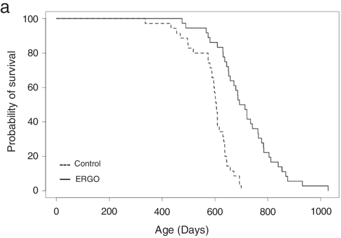
11357_2024_1111_Fig1_HTML

The ERGO group showed significantly lower age-related declines in weight, fat mass, and average and maximum movement velocities at 88 weeks of age. This was compatible with dramatic suppression by ERGO of age-related increments in plasma biomarkers. ERGO also rescued age-related impairments in learning and memory ability.

Ingestion of ERGO may promote longevity and healthy aging in male mice, possibly through multiple biological mechanisms.”

https://link.springer.com/article/10.1007/s11357-024-01111-5 “Ergothioneine promotes longevity and healthy aging in male mice”

Subjects’ plasma ergothioneine levels of an estimated 4 ~ 5 mg/kg daily dose were:
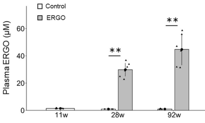
11357_2024_1111_Fig3_HTML

A human equivalent daily dose is an estimated 22 mg to 28 mg (4 or 5 mg x .081 x 70 kg).

The third paper in Part 1 cited a 2017 clinical trial that provided 5 mg and 25 mg ergothioneine doses for 7 days, resulting in these plasma ergothioneine levels:
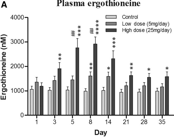
figure 3

The first paper of Part 1 referenced a 2020 human study where the dose was 5 mg/day for 12 weeks, but I don’t have access to it. It’s unclear whether humans could continually raise ergothioneine levels by daily consumption throughout our lives as did this rodent study.


A 2024 paper reviewed the importance of ergothioneine to humans:

“We propose that the diet-derived compound ergothioneine (ET) is an important nutrient in the human body, especially for maintenance of normal brain function, and that low body ET levels predispose humans to significantly increased risks of neurodegenerative and possibly other age-related diseases.

Work by multiple groups has established that low ET levels in humans are associated not only with cognitive impairment/AD but also with other age-related conditions, including frailty, Parkinson’s disease, vascular dementia, chronic renal disease, cardiovascular disease, and macular degeneration. Low ET levels also correlate with increased risk of developing preeclampsia in pregnant women [53].

Plasma ET levels from healthy (age-matched) vs unhealthy individuals in Singapore – Mild cognitive impairment (MCI); Alzheimer’s disease (AD); vascular dementia (VaD); Parkinson’s disease (PD); age-related macular degeneration (AMD):
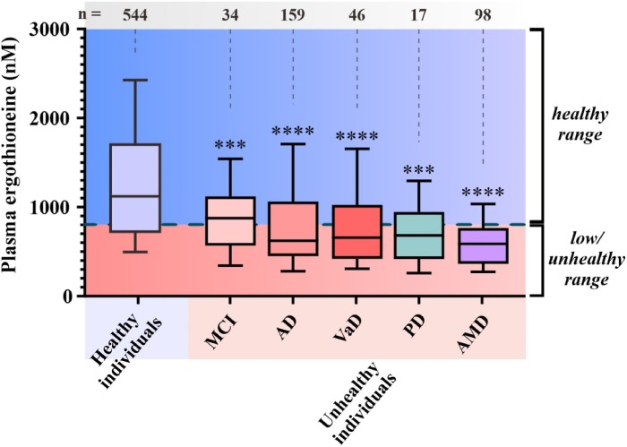
1-s2.0-S0891584924001357-gr2_lrg

  • Does low ET cause or contribute to age-related neurodegeneration, or
  • Does disease cause low ET, or
  • Low ET and increased disease risk are both caused by something else, as yet unidentified?

Prevention of neurodegeneration is especially important, since by the time dementia is usually diagnosed damage to the brain is extensive and likely irreversible.”

https://www.sciencedirect.com/science/article/pii/S0891584924001357 “Are age-related neurodegenerative diseases caused by a lack of the diet-derived compound ergothioneine?”

Whether or not the healthy individuals ate mushrooms daily in the above graphic was lost while conglomerating multiple studies.

Note that scales of the above two human graphics are a thousand times smaller than the above rodent graphic. I thought that maybe the rodent study made a plasma ergothioneine calculation error, but didn’t see one in the provided Supplementary data.


Reference 53 of the second paper is a 2023 human study:

“We analysed early pregnancy samples from a cohort of 432 first time mothers. Of these 432 women, 97 went on to develop pre-term or term pre-eclampsia (PE).

If a threshold was set at the 90th percentile of the reference range in the control population (≥462 ng/ml), only one of these 97 women (1%) developed PE, versus 96/397 (24.2%) whose ergothioneine level was below this threshold. One possible interpretation of these findings, consistent with previous experiments in a reduced uterine perfusion model in rats, is that ergothioneine may indeed prove protective against PE in humans.”

https://portlandpress.com/bioscirep/article/43/7/BSR20230160/233119/Relationship-between-the-concentration-of “Relationship between the concentration of ergothioneine in plasma and the likelihood of developing pre-eclampsia”

Eyeballing the Healthy individuals in the above graphic, none of those 544 people were below this study’s 462 ng threshold.


A 2023 companion article analyzed the third paper’s unusual findings:

“These results suggest that there might be a dichotomized association between ergothioneine concentrations and preeclampsia; and only a high ergothioneine level over 90th percentile of the control population could be protective against preeclampsia.

Univariable results showed that ergothioneine had a significant non-linear association with preeclampsia and it would start to offer protective effect from 300 ng/ml onward. Analysis also confirmed that body mass index was significantly associated with an increased risk of preeclampsia.

A large observational study could strengthen the causal association between ergothioneine and preeclampsia. If confirmed, a randomized controlled trial (RCT) assessing whether ergothioneine supplementation can reduce risk of preeclampsia will be imminently feasible. Ideally, such RCT should compare placebo with a range of different doses of ergothioneine to identify the best or minimal effective dose, given its good safety records, including in pregnancy, with a no-observed-adverse-effect level (NOAEL) of 800 mg/kg body weight per day.”

https://portlandpress.com/bioscirep/article/43/8/BSR20231076/233395/Dose-related-relationship-between-ergothioneine “Dose-related relationship between ergothioneine concentrations and risk of preeclampsia”

My daily mushroom ergothioneine dose is around 7 mg, and I weigh about 70 kg. I don’t think a daily 800 mg/kg ergothioneine dose would be desirable for anybody, regardless of what experts say.

How many times have public health employees been wrong this decade? Would you bet your or your child’s health on their advice?


PXL_20240316_182330822

Take acetyl-L-carnitine if you are healthy

Eight 2023 acetyl-L-carnitine / L-carnitine papers, starting with three healthy human studies:

“Thirty healthy volunteers aged between 19 and 52 years were divided randomly into two equal groups, one of which received 1000 mg of L-carnitine (LC) per day over a 12-week period. Total cholesterol and HDL-C increased significantly after supplementation. LC could be useful in impeding development of heart diseases in subjects with low HDL-C.”

https://journaljammr.com/index.php/JAMMR/article/view/5166 “L-Carnitine Increases High Density Lipoprotein-Cholesterol in Healthy Individuals: A Randomized Trial”

Rationale for dose selection wasn’t provided, and the possibility of limited results due to poor study design wasn’t mentioned.


“This study examined effects of 12 weeks of LC supplementation on bone mineral density (BMD) and selected blood markers involved in bone metabolism of postmenopausal women participating in a resistance training (RT) program. Participants’ diets were supplemented with either 1 g of LC-L-tartrate and 3 g of leucine per day (LC group) or 4 g of leucine per day as a placebo (PLA group), in a double-blind fashion.

Because the study protocol consisted of both exercise and supplementation, some favorable changes in the BMD could be expected. However, it was not possible to detect them in the short study period. No significant modification in BMDs of the spine, hip, and total skeleton and no differences between groups in one-repetition maximum could be due to the relatively short duration of the RT intervention.”

https://nutritionandmetabolism.biomedcentral.com/articles/10.1186/s12986-023-00752-1 “Effect of a 3-month L-carnitine supplementation and resistance training program on circulating markers and bone mineral density in postmenopausal women: a randomized controlled trial”

Same comments as the first study regarding no rationale for dose selection, and no mention that limited results were possibly due to an inadequate dose.


In a letter to the editor, a researcher took issue with a study’s methodology:

“Based on finding that intravenous provision with carnitine alone does not increase muscle carnitine accretion, and on the above-reevaluated data, it appears that the basis for carnitine with caffeine being able to increase muscle carnitine levels, and thereby manipulation of muscle metabolism and exercise performance, is uncertain.

Carnitine bioavailability in any group would have been 9.5%. This assessment would be in line with previously recorded values of 5%–18% carnitine bioavailability. It is firmly believed that low carnitine bioavailability is attributable to the inability of kidneys to reabsorb carnitine when the threshold concentration for tubular reabsorption (about 40–60 μmol/L) has passed this value.

The authors’ proposed long-term use of carnitine supplementation as an aid to improve fat oxidation in type II diabetes also seems to lack provision.”

https://physoc.onlinelibrary.wiley.com/doi/10.14814/phy2.15736 “LTE: Does caffeine truly raise muscle carnitine in humans?”


Two genetic studies:

“Our findings suggest that humans have lost a gene involved in carnitine biosynthesis. Hydroxytrimethyllysine aldolase (the second enzyme of carnitine biosynthesis) activity of serine hydroxymethyl transferase partially compensates for its function.”

https://www.researchsquare.com/article/rs-3295520/v1 “One substrate-many enzymes virtual screening uncovers missing genes of carnitine biosynthesis in human and mouse”


“Reported prevalence of primary carnitine deficiency (PCD) in the Faroe Islands of 1:300 is the highest in the world. The Faroese PCD patient cohort has been closely monitored and we now report results from a 10-year follow-up study of 139 PCD patients.

PCD is an autosomal recessive disorder that affects the function of organic cation transporter 2 (OCTN2) high-affinity carnitine transporters, that localizes to the cell membrane and transport carnitine actively inside the cell. Without proper functioning OCTN2 carnitine transporters, renal reabsorption of carnitine is impaired, and as a consequence, patients suffering from PCD have low plasma levels of carnitine. This can disturb cellular energy production and cause fatigue, but also in extreme cases lead to cellular dysfunction and severe symptoms of coma and sudden death.

PCD patients seem to adhere well to L-carnitine treatment, even though they have to ingest L-carnitine tablets at least three times a day. Overall mean L-carnitine dosage was 66.3 mg/kg/day.”

https://onlinelibrary.wiley.com/doi/10.1002/jmd2.12383 “Patients with primary carnitine deficiency treated with L-carnitine are alive and doing well—A 10-year follow-up in the Faroe Islands”

The average daily dose is (66.3 mg x 70 kg) = 4,641 mg. A third of this dose would be about 1.5 g.

The first study of Acetyl-L-carnitine dosing also suggested dosing L-carnitine three times a day because of 10-20% bioavailability.


A study with unhealthy humans:

“This retrospective study analyzed medical records of adult patients between March 2007 and April 2019, with presenting complaints of fatigue and lethargy. Acetyl-L-carnitine has physiological functions similar to L-carnitine but has higher bioavailability and antioxidant properties. This study confirmed that a triple combination therapy with γ-linolenic acid, V. vinifera extract, and acetyl-L-carnitine can improve arterial stiffness in patients.

Our study had some limitations:

  1. The study population may not be representative of the entire Korean adult population.
  2. The study did not have a medication-free control group. Instead, the comparison group comprised patients with medication compliance <80%.
  3. Drop-out rate of the triple-combination therapy (46.2%, 147/318) was relatively high, indicating the possibility of bias due to loss to follow-up.
  4. The study did not consider lifestyle factors such as smoking, diet, and physical activity level, which may affect arterial stiffness.
  5. The study did not examine interactions among drugs comprising the combination therapy, although all drugs are known to positively impact blood vessels.”

https://onlinelibrary.wiley.com/doi/10.1111/jch.14708 “Efficacy of γ-linolenic acid, Vitis vinifera extract, and acetyl-L-carnitine combination therapy for improving arterial stiffness in Korean adults: Real-world evidence”

This study’s acetyl-L-carnitine dose was 500 mg three times a day.


Wrapping up with two rodent studies:

“Acetyl L-carnitine (ALCAR) has proved useful in treatment of different types of chronic pain with excellent tolerability. The present work aimed at evaluating the anti-hyperalgesic efficacy of ALCAR in a model of persistent visceral pain associated with colitis.

The acetyl group in the ALCAR molecule can enhance cholinergic signalling by promoting synthesis of neurotransmitter acetylcholine, which plays an important role in both the enteric and central nervous systems. Acetylcholine signalling has significant antinociceptive effects in development of visceral pain, so it has been proposed as a therapeutic target.

ijms-24-14841-g001

ALCAR significantly reduced establishment of visceral hyperalgesia in DNBS-treated animals, though the interventive protocol showed a greater efficacy than the preventive one.

  • The interventive protocol partially reduced colon damage in rats, counteracting enteric glia and spinal astrocyte activation resulting from colitis.
  • The preventive protocol effectively protected enteric neurons from inflammatory insult.

These findings suggest the putative usefulness of ALCAR as a food supplement for patients suffering from inflammatory bowel diseases.”

https://www.mdpi.com/1422-0067/24/19/14841 “Anti-Hyperalgesic Efficacy of Acetyl L-Carnitine (ALCAR) Against Visceral Pain Induced by Colitis: Involvement of Glia in the Enteric and Central Nervous System

This study cited multiple animal studies that found acetyl-L-carnitine was effective for different types of pain. I’ve taken it every day for nineteen years, and haven’t noticed that effect.


“Repetitive mild traumatic brain injuries (rmTBI) may contribute to development of neurodegenerative diseases through secondary injury pathways. Acetyl-L-carnitine (ALC) shows neuroprotection through anti-inflammatory effects, and via regulation of neuronal synaptic plasticity by counteracting post-trauma excitotoxicity. This study aimed to investigate mechanisms implicated in etiology of neurodegeneration in rmTBI mice treated with ALC.

ALC is an endogenously produced carnitine metabolite present in tissue and plasma, and readily crosses the blood brain barrier, unlike its unacetylated form. ALC is also a commonly available nutritional supplement, with a known safety profile, and had been well-studied for its role in aiding β-oxidation of long chain fatty acids in the mitochondria.

While some studies have shown promise for improving clinical and psychometric outcomes in individuals with probable Alzheimer’s disease (AD) and mild cognitive impairment, other studies that included participants with moderate AD progression were less conclusive. It may be that this lack of improvement is related to a therapeutic window of opportunity. Once neurodegenerative mechanisms have commenced, a reversal of these processes is not attainable.

There is currently a lack of evidence for safe therapeutics that can be administered long-term to reduce the risk of individuals developing cognitive and neuropsychological deficits after rmTBIs. Prophylactic ALC treatment in a paradigm of neurotrauma may be a way to maximize its therapeutic potential.

While brain structures display differential vulnerability to insult as evidenced by location specific postimpact disruption of key genes, this study shows correlative mRNA neurodegeneration and functional impairment that was ameliorated by ALC treatment in several key genes. ALC may mitigate damage inflicted in various secondary neurodegenerative cascades – confirmed by improvements in behavioral and cognitive function – and contribute to functional protection following rmTBI.”

https://www.frontiersin.org/articles/10.3389/fphar.2023.1254382/full “Repetitive mild traumatic brain injury-induced neurodegeneration and inflammation is attenuated by acetyl-L-carnitine in a preclinical model”

I read many traumatic brain injury papers earlier this year, but only curated two in Brain endothelial cells. I came away thinking that there’s no permanent recovery from TBIs, as just symptoms are effectively treated.

Most TBIs happen to old people who have diminished brain reserves. I didn’t see studies that factored in evidence of what happened earlier in injured people’s lives that created TBI susceptibility but wasn’t remembered.

Unlike other years, I haven’t watched any football this season. It’s unsettling that transient entertainment value continues to take precedence over permanent effects on players’ lives.


PXL_20231223_175628957

Building your plasmalogen savings account

A webinar from earlier this week with Dr. Goodenowe, a clinical trial facilitator, and a physician:

From the Q&A segment:

“Is there a particular age where it’s recommended to test for plasmalogen levels? And what levels would be considered normal?

That’s a good question. That actually raises this whole concept of optimal health and this concept of aging.

The best way to think about it – we talked about this paycheck-to-paycheck situation, where as long as our bills are paid every day, technically we think we’re normal. But we still feel this sense of health anxiety – if you will – like we just don’t know if my car breaks down, or my water heater breaks down, do I have enough money to pay these events in my life?

That’s what health feels like to a lot of people, because they’re just kind of getting by. From a health perspective, they’re considered normal, but they have no reserve capacity, and they have no vitality in terms of health.

Plasmalogens are a type of molecule that you build a savings account of, over years, over decades. Your heart builds them up, your brain builds them up, and you slowly accumulate them. Then when you get an oxidative stress like what’s happening now in today’s world with all the covid and myocarditis and brain fog – a lot of these things are being caused because that reserve of plasmalogens has been depleted.

We want plasmalogens for a longevity perspective. There are other situations that can have low plasmalogens, other things can really knock your plasmalogens down.

So you want to start early, you want to build a savings account, and you want to maintain it. Maintain health and function, and create a sustained surplus for optimal health, for optimal neuromuscular performance.”


PXL_20231207_185012059

A good activity for bad weather days

A free educational series recorded in 2021-2022 available at https://drgoodenowe.com/dr-goodenowes-educational-seminars/ takes the viewer through underlying research and principles of Dr. Goodenowe’s approach to health. It’s advertised as lasting four hours, but took me two days to view.

The series’ discussions and references are background material to better understand later presentations and interviews. Points of interest included:

  • Seminar B100 shows that the metabolomic profile of people who regularly eat broccoli is different than others.
  • B109 clarifies how peroxisomal function is improved through resistance exercise and intermittent fasting.
  • C103 and C104 show how plasmalogens act against neurodegeneration (Parkinson’s disease and multiple sclerosis).

Texts below videos are additional information, not transcripts. C101 text is historically informative.


The B200 ProdromeScan tutorial will take more study. But unlike Labcorp tests, ordering a ProdromeScan requires using a practitioner in Dr. Goodenowe’s network.

I sent the following to Prodrome customer service earlier this month:

Please add me to your approved list for ProdromeScan.

Customer service replied:

“We only add health professionals to an approved list, not individuals.”

I responded:

Good morning. I looked at the websites of doctors who are associated with Dr. Goodenowe who are near me. All of them are too compromised for me to establish a doctor / patient relationship. But I’m glad they left up their blog posts from earlier this decade so I could see who they really were before I reached out to them.

I request an exception to the policy.

Customer service replied:

“There is no exception that can be made to this policy. You need to be a patient of a certified practitioner.”

I’ll escalate my request before my 90-day trial of Prodrome Glia and Neuro products ends so I can get an appropriate metabolomic status. Right now, I won’t involve someone I can’t trust just to know my ProdromeScan information that’s additional to next week’s Labcorp tests.

My treatment-result metabolomic data is probably not mature today on Day 29 of ProdromeGlia and ProdromeNeuro supplementation, resistance exercise, and intermittent fasting. I otherwise wouldn’t have experienced these two events:


I have a quibble with the series’ recommendations for taking N-acetyl cysteine. Relevant views and research:

Switch on your Nrf2 signaling pathway pointed out:

“We use NAC in the lab all the time because it stops an Nrf2 activation. So that weak pro-oxidant signal that activates Nrf2, you switch it off by giving a dose of NAC. It’s a potent antioxidant in that right, but it’s blocking signalling. And that’s what I don’t like about its broad use.”

If someone bombs themself everyday with antioxidants, they’re doing nothing to improve training of their endogenous systems’ defensive functions. What happens when they stop bombing? One example was a 2022 human study that found GlyNAC-induced improvements dissolved back to baseline after supplements stopped.

Also, Precondition your defenses with broccoli sprouts highlighted NAC’s deleterious effects on autophagy and lysosome functions:

“TFEB activity is required for sulforaphane (SFN)-induced protection against both acute oxidant bursts and chronic oxidative stress. SFN-induced TFEB nuclear accumulation was completely blocked by pretreatment of cells by N-acetyl-cysteine (NAC), or by other commonly used antioxidants. NAC also blocked SFN-induced mRNA expression of TFEB target genes, as well as SFN-induced autophagosome formation.”

If a secondary goal of taking NAC per is also necessary for the formation of glutathione, taurine can do that without an antioxidant bomb. Taurine supplementation will free up cysteine to do things other than synthesize taurine, like synthesize glutathione.


PXL_20231123_194849211.MP

Brain restoration with plasmalogens

In this 2023 presentation for a professional audience, Dr. Dayan Goodenowe showed an example of what could be done (in the form of what he personally did at ages 53-54) to restore and augment brain structure and function over a 17-month period by taking plasmalogens and supporting supplements:

https://drgoodenowe.com/recording-of-dr-goodenowes-presentation-from-the-peptide-world-congress-2023-is-now-available/

Follow the video along with its interactive transcript. Restorative / augmentative supplements included:

1. Nutritional Supplementation Strategy

Forms of MRI used to document brain structure and function changes were:

2. Advanced MRI Technologies

Brain volume decreases are the rule for humans beginning at age 40. Dr. Goodenowe documented brain volume increases, which aren’t supposed to happen, but did per the below slide of overall results:

3. Reversing Brain Shrinkage

“From a global cortical volume and thickness perspective, 17 months of high-dose plasmalogens reversed ~15 years of predicted brain deterioration.”


Specific increased adaptations in brain measurements over 17 months included:

  1. Cortical thickness .07/2.51 = +3%.
  2. White matter microstructure fractional anisotropy +8%.
  3. Nucleus accumbens volume +30%.
  4. Dopaminergic striatal terminal fields’ volume +18%.
  5. Cholinergic cortical terminal fields’ volume +10%.
  6. Occipital cortex volume +10%.
  7. Optic chiasm volume +225%.
  8. Nucleus basalis connectivity.
  9. Neurovascular coupling signal controlled by noradrenaline integrity.
  10. Amygdala volume +4% and its connectivity to the insula, indicating ongoing anxiety and emotional stress response.
  11. Parahippocampus volume +7%.
  12. Hippocampus fractional anisotropy +5%.

No changes:

  1. Amygdala connectivity to the ventral lateral prefrontal cortex, the same part of the brain that relates to placebo effect.
  2. Hippocampus connectivity.

Decreased adaptations in brain measurements included:

  1. White matter microstructure radial diffusivity -10%.
  2. Amygdala connectivity to the anterior cingulate cortex to suppress / ignore / deny anxiety response.
  3. Amygdala connectivity to the dorsal lateral prefrontal cortex.
  4. Entorhinal cortex volume -14%.
  5. Hippocampus volume -6%.
  6. Hippocampus mean diffusivity (white matter improved, with more and tighter myelin) -4%.

The other half of this video was a lively and wide-ranging Q&A session.


The referenced 2023 study of 653 adults followed over ten years showed what brain deterioration could be expected with no interventions. Consider these annual volume decrease rates to be a sample of a control group:

etable 3

https://jamanetwork.com/journals/jamanetworkopen/fullarticle/2806488 “Characterization of Brain Volume Changes in Aging Individuals With Normal Cognition Using Serial Magnetic Resonance Imaging”

Also see a different population’s brain shrinkage data in Prevent your brain from shrinking.


The daily plasmalogen precursor doses Dr. Goodenowe took were equivalent to 100 mg softgel/kg, double the maximum dose of 50 mg softgel/kg provided during the 2022 clinical trial of cognitively impaired old people referenced in Plasmalogens Parts 1, 2, and 3.

He mentions taking 5 ml in the morning and 5 ml at night because he used the Prodrome oil products. 1 ml of a Prodrome oil plasmalogen precursor product equals 900 mg of their softgel product.


“My brain is trying to minimize long-term effects of pain/stress by suppressing my memory of it. But this can only go on for so long before it becomes an entrenched state.

I have solved the sustenance side of the equation. I need to work harder to solve the environmental side.”

While I agree that we each have a responsibility to ourselves to create an environment that’s conducive to our health, the above phenomenon isn’t necessarily resolvable by changing an individual’s current environment. My understanding is that long-term effects of pain, stress, and related human experiences are usually symptoms of causes that started much earlier in our lives.

Adjusting one’s present environment may have immediate results, but probably won’t have much therapeutic impact on long-term issues. Early life memories and experiences are where we have to gradually go in order to stop being driven by what happened back then.

See Dr. Arthur Janov’s Primal Therapy for its principles and explanations. I started Primal Therapy at a similar age, 53, and continued for three years.

A smell and taste anecdote

Two 2023 papers, starting with a study of smell and taste disorders:

“This study investigates the impact of etiology on the epidemiologic profile, disease severity, type of treatment, and therapy outcome in smell and taste disorders.

Hyposmia has a prevalence of about 15%, while approximately 5% of the population suffers from anosmia. Multiple innervation of the taste mucosa with fibers from the seventh, ninth, and tenth cranial nerves assures robustness of the gustatory system compared to smell.

Conservative therapy employs corticosteroids, antibiotics, vitamins and and minerals as well as functional rehabilitation by olfactory training. Data regarding outcome of therapy were only available for 71 (26.3%) of patients. Only the sinunasal etiology was significantly more likely to show improvement after therapy (27.4% show improvement vs. 9.6% show no improvement).”

https://link.springer.com/article/10.1007/s00405-023-07967-1 “Characteristics of smell and taste disorders depending on etiology: a retrospective study”

This study was a little light on describing effective treatments for smell and taste problems. For example, olfactory training was said to have good therapeutic response. Looking it up, though, it seems to be whatever each practitioner feels like doing.


A review introduced the subject of olfactory ensheathing cells:

“Olfactory ensheathing cells (OECs) are glial cells of the primary olfactory nervous system, which are composed of the olfactory nerve and outer nerve fiber layer of the olfactory bulb. The primary olfactory nervous system is unique in that it can constantly regenerate.

It is now possible to remove olfactory bulb tissue and olfactory mucosa (outermost layer and lamina propria, which belong to the central nervous system and peripheral nervous system, respectively), which also suggests the potential value of OECs therapy in central nervous system and peripheral nervous system diseases. OECs can survive and renew in the central nervous system, and have been widely used in nerve regeneration and tissue repair.

Schwann cells (SCs) form the myelin sheath of the peripheral nerve, protect and nourish neurons, and play an irreplaceable role in the repair of peripheral nerve injury. There is no transcriptional difference between OECs and SCs. OECs are highly similar to SCs, and express the biomarkers of SCs.

fimmu-14-1280186-g002

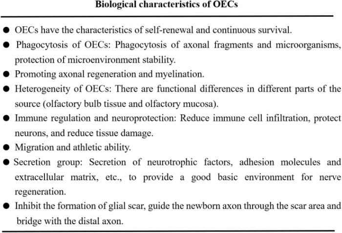
Functional mechanisms of OECs in the treatment of neurological diseases include neuroprotection, immune regulation, axon regeneration, improvement of nerve injury microenvironment and myelin regeneration, which also includes secreted bioactive factors. Results obtained in clinical trials are not very satisfactory, and the effectiveness of these cell-based therapies remains to be proved.”

https://www.frontiersin.org/articles/10.3389/fimmu.2023.1280186/full “Potential therapeutic effect of olfactory ensheathing cells in neurological diseases: neurodegenerative diseases and peripheral nerve injuries”


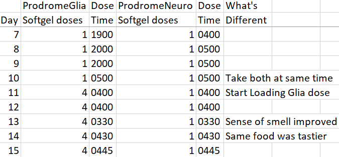
Something interesting may have unexpectedly started with my 90-day trial of Prodrome Glia and Neuro products. Here’s an abbreviated look that omits my intermittent fasting and resistance exercise data:

day 7-15

Both product labels have a loading dose suggestion of 4-8 softgels (2 to 4 times the standard two-softgel dose) for 1-3 months. Two days after I started a Glia loading dose, my sense of smell, then sense of taste, were noticeably better.

I’ll guess that my primary olfactory nervous system glial cells are responding to these changes. At the beginning I thought that my peripheral nervous system Schwann cells might be affected regarding my left ulnar nerve. Since olfactory ensheathing cells are highly similar to Schwann cells, it doesn’t seem to be that much of a stretch to think that they could also be affected by my current regimen.

More testing is warranted, of course. I’ve had diminished smell and taste for decades, though. If the gardenias, roses, magnolias, honeysuckles, and other scents in past summers that had fainter scents than I remembered come across stronger, so much the better.

IMG_20200425_154336

Plasmalogens, Part 3

The 2022 plasmalogen clinical trial mentioned in Parts 1 and 2 bypassed peroxisome metabolism of cognitively impaired people per discussion of the below diagram:

fcell-10-864842-g003

Increasing the body’s fasting state with time-restricted eating, and preventing muscle atrophy with resistance exercise, were offered as the two most important ways to improve peroxisomal function.

I didn’t find any relevant 2023 human studies (where I could access the full study) on different non-drug treatments that I was willing to do. A 2023 review outlined aspects of peroxisomes, to include a few older human studies:

“Peroxisomes are small, single-membrane-bound organelles, which are dynamic and ubiquitous. Peroxisomes directly interact with other organelles, such as endoplasmic reticulum, mitochondria, or lysosomes. Peroxisomes exert different functions in various cells through both catabolic and anabolic pathways.

The main functions of peroxisomes can be categorized as reactive oxygen species (ROS) metabolism, lipid metabolism, and ether-phospholipid biosynthesis. Peroxisomes also play important roles in inflammatory signaling and the innate immune response.”

1-s2.0-S2667325823001425-gr3_lrg

https://www.sciencedirect.com/science/article/pii/S2667325823001425 “Peroxisome and pexophagy in neurological diseases”


1. Since I haven’t recently tried the two main ways to improve peroxisomal function, I’ll give them a go over the next three months:

  • Expect to get my feeding timeframe to within eight hours. Don’t know about making it short like 6 hours, because my first meal of the day is 35 calories of microwaved cruciferous sprouts, then I wait an hour before eating anything else.
  • Resistance exercise progress should be measurable, as I recorded exercises during the first ten weeks of eating broccoli sprouts every day 3.5+ years ago.

2. Don’t know that I’ll recognize any cognitive improvements to the extent I did during Week 9.

  • I don’t have a young brain anymore, and I’m sure some decline could be measured in memory tests. But I’m not going to become a lab rat.
  • There’s an occasional annoyance that’s been going on for some time, especially when I’m distracted. It happens when I think of something to do, and it somehow becomes a short-term memory that I did it, instead of going into a Things To Do queue. It’s largely self-correcting. For example, regardless of what I paid, I’ll drive back to the grocery store self-checkout to retrieve a third bag that didn’t make it home. A pink-haired employee said young people leave their paid-for groceries behind all the time. It’s usually more of a reality disconnect for me than forgetfulness, because I have a memory that I performed the action. Definitely room for improvement.

3. Don’t know that I’d see biochemical changes such as some described in Part 1. Maybe I’ll move up an annual physical to compare it with the last one in May?

  • I already have very little oxidative stress, very little inflammation, low triglycerides, high HDL, and no major improvements are indicated on CBC / CMP / lipid panels.
  • Take supplements to ensure other things like acetylcholine neurotransmitter availability, one-carbon / methylation metabolism, vitamin / mineral adequacy.

4. I started the two Prodrome plasmalogen precursor supplements (ProdromeGlia and ProdromeNeuro) a week ago, and take their standard doses. My thought is that resultant plasmalogens won’t degrade very much if their primary use isn’t to immediately address oxidative stress and inflammation. That could give these extra plasmalogens a chance to make larger homeostatic contributions in myelin and membrane areas.

I don’t expect any particular effects to manifest. But I’m interested to see if these two areas would be affected:

  • My left ulnar nerve has been giving me problems for over five years, and several resistance exercises aggravate it. I’ve had two nerve continuity tests during that time to confirm. Numbness and pain are intermittent, though.
  • I still take acetaminophen several times a day for other pain.

None of the above treatments are specifically indicated. But if time-restricted feeding and/or extra plasmalogens have an effect on left ulnar or other pain, maybe I’ll be able to make better progress on resistance exercise.

Update #1 11/13/2023

Update #2 11/22/2023

Update #3 12/13/2023 comments

Update #4 1/30/2024

Update #5 3/31/2024

Plasmalogens, Part 2

This post compares Dr. Goodenowe’s clinical trial mentioned in Part 1 with other researchers’ human plasmalogen studies this decade. One of its findings was:

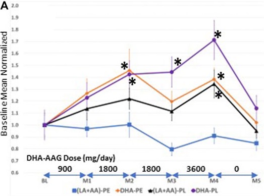
“Figure 1A illustrates that plasmalogen precursor DHA-AAG dose-dependently elevated both direct and indirect target species [DHA-PL, DHA-PE, and (LA + AA)-PL] and had no effect on levels of biochemically unrelated PE species index (LA + AA)-PE.

  • DHA-AAG had a greater elevating effect on its direct target, DHA-PL than its indirect targets.
  • The 1-month washout period resulted in decreased levels of both direct and indirect target species and no effect on unrelated PE species.

Figures 1A,B illustrate that DHA-AAG is converted to its direct and indirect target species in humans as predicted from animal studies on similar AAG plasmalogen precursors (Wood et al., 2011d).”

fcell-10-864842-g001A

Given this century’s background of numerous animal studies, there’s a need to know what translates to humans. Here are the three most recent human plasmalogen studies in descending order where I could access the full study:

2022

“Forty unmarried male students aged 18–22 years (20 in the plasmalogen group and 20 in the placebo group) were randomly allocated to either plasmalogen (2 mg per day) or placebo treatment of 4 weeks’ duration and ingested two capsules of 0.5 mg plasmalogen or placebo twice daily.

  • The primary efficacy outcome was the Total Mood Disturbance (TMD) T-score of POMS 2–Adult Short.
  • Secondary outcomes included the seven individual scales of POMS 2, other psychobehavioral measures (Athens Insomnia Scale and Uchida-Kraepelin test), physical performance test (shuttle run, grip muscle strength, and standing long jump), plasmalogen levels in plasma and erythrocytes, plasma levels of brain-derived neurotrophic factor (BDNF), urinary 8-hydroxy-2′-deoxyguanosine (8-OHdG), body mass index, and percent body fat.

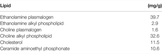
Lipid composition of purified ether phospholipids from scallop is shown below. One capsule contained 0.48 mg of ethanolamine plasmalogen and 0.02 mg of choline plasmalogen. Plasmalogen and placebo capsules were prepared by a manufacturer (B&S Corporation, Tokyo).

fcell-10-894734-t001

There were no between-group differences in physical and laboratory measurements. It is suggested that orally administered plasmalogens alleviate negative mood states and sleep problems, and also enhance mental concentration.”

https://www.frontiersin.org/articles/10.3389/fcell.2022.894734/full “Orally Administered Plasmalogens Alleviate Negative Mood States and Enhance Mental Concentration: A Randomized, Double-Blind, Placebo-Controlled Trial”

There was no dose / response investigation, so there’s no data to corroborate that this 2 mg treatment produced these effects. It isn’t difficult to think of other factors that could influence the primary outcome of a 18-22 year-old unmarried male’s moods.


2020

“Effects of ascidian-derived plasmalogens on cognitive performance improvement were assessed in a randomized, double-blind, placebo-controlled study including Japanese adult volunteers age 45.6 ± 11.1 years with mild forgetfulness. An allocation controller who was not directly involved in the study equally, but randomly, assigned participants to either the intervention group (n=33) or the placebo group (n=33), based on normalized Cognitrax composite memory score (the primary outcome), sex, and age at time of screen. Participants were administered either one active capsule (200 mg medium-chain triglyceride (MCT) oil including ascidian plasmalogen oil) or placebo capsule (200 mg MCT oil) per day with water, any time during the day for 12 weeks.

Ascidian plasmalogen oil was extracted from ascidians (Halocynthia roretzi) and sold by NIHON PHARMACEUTICAL CO., LTD. Based on a previous study, 33% of lipids contained in ascidians are phospholipids, 23% of which are plasmalogens, and fatty acids of the sn-2 position of plasmalogens are mainly EPA, DHA, oleic acid, and arachidonic acid. The active capsule contains 1 mg plasmalogen.

Compared to the placebo group, the intervention group showed a significant increase score in composite memory (eight weeks: 3.0 ± 16.3 points, 12 weeks: 6.7 ± 17.5 points), which was defined as the sum of verbal and visual memory scores. These results indicate consumption of ascidian-derived plasmalogen maintains and enhances memory function.”

https://www.jstage.jst.go.jp/article/jos/69/12/69_ess20167/_article “The Impact of Ascidian (Halocynthia roretzi)-derived Plasmalogen on Cognitive Function in Healthy Humans: A Randomized, Double-blind, Placebo-controlled Trial”

Again no dose / response investigation, so no corroborating data. Standard deviations many times larger than a sample’s mean indicated wild variability (aka noise). Maybe intervention participants experienced memory loss (3.0 mean – 16.3 SD = -13.3; 6.7 mean – 17.5 SD = -10.8)? Yet statistics inferred a signal that allowed interpreting this treatment as producing meaningful positive changes in cognitive function.


“Ten Parkinson’s disease (PD) patients age 67.80 (7.41) years received oral administration of 1 mg/day of purified ether phospholipids derived from scallop for 24 weeks. Clinical symptoms and blood tests were checked at 0, 4, 12, 24, and 28 weeks. Blood levels of plasmalogens in patients with PD were compared with those of 39 age-matched normal controls.

B&S Corporation Co. Ltd. (Tokyo) was involved in provision of capsules containing ether phospholipids derived from scallop. Ethanolamine ether phospholipids (ePE) in plasma from PD and relative composition of ethanolamine plasmalogen (plsPE) of erythrocyte membrane in PD were significantly low as compared to those of age-matched normal controls.

Oral administration of purified ether phospholipids derived from scallop for 24 weeks increased plasma ePE and erythrocyte plsPE to almost normal levels, and concomitantly improved some clinical symptoms of patients with PD. Results indicate the efficacy of oral administration of purified ether phospholipids derived from scallop to some nonmotor symptoms of PD. Physiological mechanisms of the efficacy of purified ether phospholipid derived from scallop remained to be elucidated.”

https://www.hindawi.com/journals/pd/2020/2671070/ “Improvement of Blood Plasmalogens and Clinical Symptoms in Parkinson’s Disease by Oral Administration of Ether Phospholipids: A Preliminary Report

Again no dose / response investigation, so no corroborating data. These researchers asserted their 2017 study to be a plasmalogen gold standard, as did the other two above studies.

Here’s part of what Dr. Goodenowe said about that 2017 study in a 2019 review Plasmalogen deficiency and neuropathology in Alzheimer’s disease: Causation or coincidence?:

“They did not observe a significant elevation of plasma levels of plasmalogens in the treated group relative to the baseline. Lower dose of plasmalogens (1 mg twice daily) and the labile nature of the vinyl-ether bond might have limited absorption of the intact molecule and might have contributed to the lack of response in terms of plasmalogen levels in blood as well as the cognitive function. Reported instability of plasmalogens in acidic environments questions the stability of preformed plasmalogens in gastric juice during digestion which might reduce plasmalogen bioavailability.”

Also see Part 1’s explanation of why using age-matched controls in plasmalogen studies is ridiculous.

Continued in Part 3.

Plasmalogens, Part 1

The person who knows the most about this subject is Dayan Goodenowe, PhD. Some recent publications include:

https://www.frontiersin.org/articles/10.3389/fcell.2022.864842/full “Targeted Plasmalogen Supplementation: Effects on Blood Plasmalogens, Oxidative Stress Biomarkers, Cognition, and Mobility in Cognitively Impaired Persons”

https://www.frontiersin.org/articles/10.3389/fcell.2022.866156/full “Brain ethanolamine phospholipids, neuropathology and cognition: A comparative post-mortem analysis of structurally specific plasmalogen and phosphatidyl species”

plasmalogens and cognition


A sample of links freely available at https://drgoodenowe.com/.

1. Presentations to professional groups. Have your mouse ready to click the pause button.

https://drgoodenowe.com/dr-goodenowe-presents-at-the-iagg2023-in-yokohama-japan/ “A rare children’s disease that may be the key to reversing neurological decline in aging”

Includes videos of a treatment’s effects on a child.

https://neomarkgroup.wistia.com/medias/0qln0wy93t “The most influential biomarkers for aging and disease”

Despite the title, a considerable number of studies were presented on prenatal, infant, and early childhood development. He misspoke a few times, so read the slides.

Phenotype is reality. Genotype is possibility. Communications links between different fields are very poorly connected in science.

Peroxisomes are islands. They don’t have DNA like your mitochondria do. Peroxisomal transport issues are important things to understand.

All aging-related cross-sectional analyses are on the rate of decline. You’re declining from a previous well state. Age-matched controls are the most ridiculous thing to do.”


2. I’ll highlight the longest of several interviews because there was plenty of room to expand on points. Maybe the best detailed explanations came as responses to that interviewer challenging with contrasting AD, traumatic brain injury, and cholesterol paradigms. Its transcript is more accurate than a usual YouTube interpretation, but there are still mistakes such as “fossil lipid” vs. phospholipid.

https://www.betterhealthguy.com/episode186 “Plasmalogens with Dr. Dayan Goodenowe, PhD”

“Science is how do you push things to its failure, until you can’t fail it again. We’ve lost that. It’s become more hypothesis proving.

Plasmalogens levels go up for a different reason than people think. The reason why it peaks in our 40s and 50s is because we’ve been myelinating. The white matter of our brain is still increasing. It’s not because we’re making more plasmalogens. It’s because the lake, the reservoir, gets full. What you’re measuring in blood is overflow from the lake. The lower plasmalogens start trickling down in your blood, the bigger drain that’s occurring on that system.

Low plasmalogens don’t just predict dementia in the elderly population. It predicts the rate of decline of that dementia. It predicts the rate of death.

The biggest drivers of plasmalogen manufacturing and the biggest reasons why they decrease with age, or in other circumstances is two things. One, the failure to maintain a fasting state of the human body. The second one is muscle atrophy.

Amyloid has absolutely nothing to do with Alzheimer’s, or dementia. It’s just a bystander on the road watching an accident happen.

Age-related cognitive decline is clearly where plasmalogens have the greatest impact. You’re always going to have mixed pathologies in the brain.

Nutritional availability of plasmalogens is virtually non-existent. As soon as they hit the hydrochloric acid of your stomach, they’re gone. They don’t make it past the stomach, or the upper intestine.”


I came across Dr. Goodenowe’s work last month from clicking a comment on this blog that linked back to her blog. Always be curious.

Continued in Part 2.

Bridging Nrf2 and autophagy

Three more 2023 papers that cited Precondition your defenses with broccoli sprouts, starting with a review:

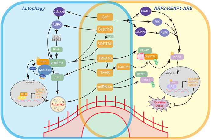
“Examining crosstalk between Nrf2 antioxidant signaling and autophagy provides insights into how they are interconnected and proteins that mediate their communication. These factors are potential therapeutic targets for diseases with both autophagy dysfunction and oxidative stress.

A working model illustrates mechanisms of bridging factors (SQSTM1, TFEB, Sestrin2, TRIM16, Ca2+, and miRNAs) connecting autophagy (left) and the main antioxidant Nrf2-Keap1-ARE pathway (right) and feedback loops between these factors.

fcell-11-1232241-g003

  • A network forms that connects Nrf2, SQSTM1, TFEB, and mTOR.
  • Other non-canonical autophagy regulatory proteins like Sestrin2 and tripartite motif-containing protein 16 (TRIM16) also participate in regulation of Nrf2 and mTOR via direct or indirect interactions.
  • Ca2+ is the most widespread intracellular messenger whose role in autophagy has been studied extensively.
  • At post-transcriptional level, microRNAs have been reported to impact both the regulation of autophagy and Nrf2 antioxidant signaling.

Since these regulatory proteins seem intricately entangled, potential side effects in practical scenarios should also be taken into consideration. Further studies on understanding the complex crosstalk between autophagy and antioxidant pathways are yet to be conducted.”

https://www.frontiersin.org/articles/10.3389/fcell.2023.1232241/full “An update on the bridging factors connecting autophagy and Nrf2 antioxidant pathway”


A second review subject was improving autophagy:

“Lysosomes are crucial degradative organelles that maintain cellular homeostasis. During the pathogenesis of neurodegenerative diseases and aging, functions of lysosomes are impaired, and lysosomal degradative capacity is consequently reduced.

Transcription factor EB-mediated lysosome biogenesis enhances autolysosome-dependent degradation, which subsequently alleviates neurodegenerative diseases. Small-molecule compounds that enhance TFEB activity and lysosome biogenesis are potential therapeutic agents.”

https://journals.lww.com/nrronline/fulltext/2023/11000/enhancement_of_lysosome_biogenesis_as_a_potential.7.aspx “Enhancement of lysosome biogenesis as a potential therapeutic approach for neurodegenerative diseases”


A third review tied mitochondrial participation into these processes:

“Mitochondria play an essential role in neural function, such as supporting normal energy metabolism, regulating reactive oxygen species, buffering physiological calcium loads, and maintaining the balance of morphology, subcellular distribution, and overall health through mitochondrial dynamics. Given recent technological advances in the assessment of mitochondrial structure and functions, mitochondrial dysfunction has been regarded as the early and key pathophysiological mechanism of cognitive disorders.

Mitochondrial dysfunction caused by acute and chronic brain injury is difficult to be distinguished because they may exhibit similar structural and functional impairments. Mitochondrial physiological function and morphology are integral, so when one is damaged, the other is also involved.

We recommend that all of the above methods can be used to explore mitochondrial dysfunction in different pathological pathways of cognitive disorders. Results may be related to special pathological pathways, sensitivity of the method, experiment cost, and degree of proficiency.”

https://journals.lww.com/nrronline/fulltext/2024/04000/latest_assessment_methods_for_mitochondrial.18.aspx “Latest assessment methods for mitochondrial homeostasis in cognitive diseases”


PXL_20231003_110600182

Take Vitamin K2 to protect against aluminum toxicity

This 2023 rodent study investigated relationships of MK-7 menaquinone, aluminum trichloride, and brain health:

“A variety of endogenous and exogenous agents, such as metals and environmental toxins (aluminum, mercury, etc.), can contribute to neurodegeneration, which is of multifactorial clinical occurrence.

The current study showed that Alzheimer’s Disease (AD)-like condition was induced in mice by AlCl3 treatment affecting spatial and recognition memory. Neuropathological alterations included neuroinflammation, oxidative stress, an increase in brain amyloid β levels, and loss of hippocampal neurons.

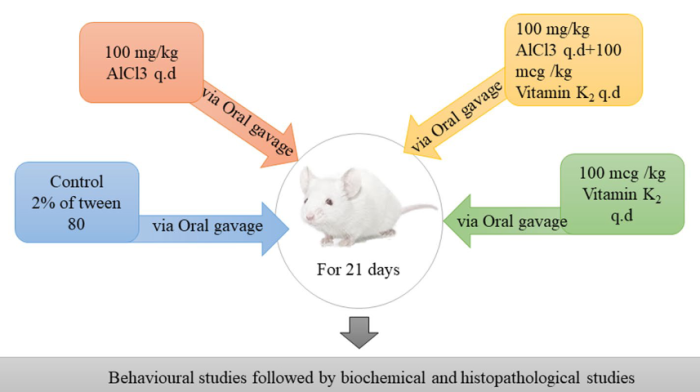
Aluminium chloride (AlCl3; 100 mg/kg for 3 weeks orally) was administered to Swiss albino mice to induce neurodegeneration and Vitamin K2 (100 mcg/kg for 3 weeks orally) was applied as treatment. This was followed by behavioral studies to determine memory changes.

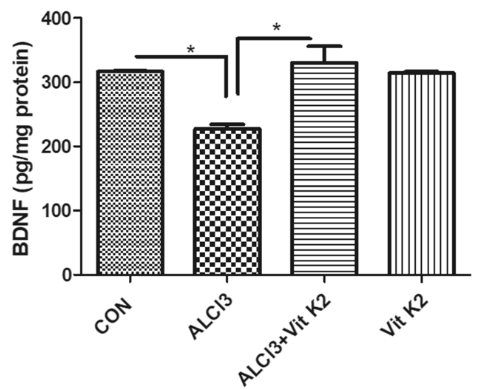
Antioxidants like glutathione and SOD were low compared to the control group, while oxidative stress marker MDA was elevated. BDNF levels increased in the Vitamin K2 treated animals, suggesting its neuroprotective functions.

k2 abstract

vitamin K2 BDNF

Vitamin K2 could partially reverse AlCl3-mediated cognitive decline. It increased hippocampal acetylcholine and BDNF levels while reducing oxidative stress, neuroinflammation, and β-amyloid deposition, protecting hippocampal neurons from AlCl3-mediated damage.

https://link.springer.com/article/10.1007/s10787-023-01290-1 “Vitamin K2 protects against aluminium chloride-mediated neurodegeneration” (not freely available)


This study’s human equivalent Vitamin K2 dose is (100 mcg x .081) x 70 kg = 567 mcg. I’ve taken 600 mcg MK-7 every day for the past two years.

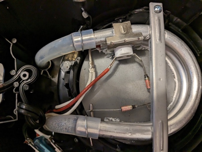
Found out last week that I’ve also been inadvertently dosing myself with aluminum every day. This is the underside of my former 3-year-old drip coffee maker with its cover removed:

PXL_20230813_172709641

I’m certain its aluminum tubing that heats reservoir water started to corrode a long time ago. Currently trying out methods of making aluminum-free coffee.

Neuritogenesis

Three 2023 papers on the initial stage of neuronal differentiation, starting with a rodent study of taurine’s effects:

“We aimed to assess the role of taurine (TAU) in axonal sprouting against cerebral ischemic injury, clarify the function of mitochondria in TAU-induced axonal sprouting, and further determine the underlying potential molecular mechanism.

experiment design

We determined that TAU improved motor function recovery and restored neurogenesis in ischemic stroke. This possibly occurred via improvements in mitochondrial function.

We investigated that the Sonic hedgehog (Shh) pathway exerted an important role in these effects. Our study findings highlighted the novel viewpoint that TAU promoted axonal sprouting by improving Shh-mediated mitochondrial function in cerebral ischemic stroke.”

https://www.scielo.br/j/acb/a/nxKvGXGk9g6gRkHxybMfbYJ/?lang=en “Taurine promotes axonal sprouting via Shh-mediated mitochondrial improvement in stroke”


A rodent study investigated effects of a soy isoflavone gut microbiota metabolite:

“Perinatally-infected adolescents living with HIV-1 (pALHIV) appear uniquely vulnerable to developing substance use disorders (SUD). Medium spiny neurons (MSNs) in the nucleus accumbens core (NAcc), an integrator of cortical and thalamic input, have been implicated as a key structural locus for the pathogenesis of SUD.

Treatment with estrogenic compounds (e.g., 17β-estradiol) induces prominent alterations to neuronal and dendritic spine structure in the NAcc supporting an innovative means to remodel neuronal circuitry. The carcinogenic nature of 17β-estradiol, however, limits its translational utility.

Plant-derived polycyclic phenols, or phytoestrogens, whose chemical structure resembles 17β-estradiol may afford an alternative strategy to target estrogen receptors. The phytoestrogen S-Equol (SE), permeates the blood-brain barrier, exhibits selective affinity for estrogen receptor β (ERβ), and serves as a neuroprotective and/or neurorestorative therapeutic for HIV-1-associated neurocognitive and affective alterations.

Beginning at approximately postnatal day (PD) 28, HIV-1 transgenic (Tg) animals were treated with a daily oral dose of 0.2 mg of SE. The SE dose of 0.2 mg was selected for two primary reasons, including:

  1. A dose-response experimental paradigm established 0.2 mg of SE as the most effective dose for mitigating neurocognitive deficits in sustained attention in the HIV-1 Tg rat; and
  2. The dose, which yielded a daily amount of 0.25–1.0 mg/kg/SE (i.e., approximately 2.5–10 mg in a 60 kg human), is translationally relevant (i.e., well below the daily isoflavone intake of most elderly Japanese.

Daily oral treatment continued through PD 90.

j_nipt-2023-0008_fig_002

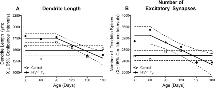
HIV-1 Tg animals exhibited an initial increase in dendrite length (A) and the number of dendritic spines (B) early in development; parameters which subsequently decreased across time. In sharp contrast, dendrite length and the number of dendritic spines were stable across development in control animals.

Targeting these alterations with the selective ERβ agonist SE during the formative period induces long-term modifications to synaptodendritic structure, whereby MSNs in the NAcc in HIV-1 Tg animals treated with SE resemble control animals at PD 180.”

https://www.degruyter.com/document/doi/10.1515/nipt-2023-0008/html “Constitutive expression of HIV-1 viral proteins induces progressive synaptodendritic alterations in medium spiny neurons: implications for substance use disorders”


A rodent brain cell study investigated soy isoflavones’ effects on a different estrogen receptor:

“We evaluated effects of isoflavones using mouse primary cerebellar culture, astrocyte-enriched culture, Neuro-2A clonal cells, and co-culture with neurons and astrocytes. Soybean isoflavone-augmented estradiol mediated dendrite arborization in Purkinje cells.

These results indicate that ERα plays an essential role in isoflavone-induced neuritogenesis. However, G-protein-coupled ER (GPER1) signaling is also necessary for astrocyte proliferation and astrocyte–neuron communication, which may lead to isoflavone-induced neuritogenesis.

We highlight the novel possibility that isoflavones enhance dendritogenesis and neuritogenesis, indicating that they can be a useful supplementary compound during brain development or in the injured brain.”

https://www.mdpi.com/1422-0067/24/10/9011 “Isoflavones Mediate Dendritogenesis Mainly through Estrogen Receptor α”

Brain endothelial cells

Six 2023 papers on the subject, starting with a rodent study:

“One of the primary discoveries of our study is that the endothelial cell (EC) transcriptome is dynamically regulated by both aging and heterochronic parabiosis. We found that ECs, when compared with other brain cell types, exhibited one of the highest fractions of aging-related genes that were rescued after heterochronic parabiosis in the old brain, and similarly, the highest fraction of aging-related genes that were disrupted after heterochronic parabiosis in the young brain. This finding supports our previous research that vasculature is strongly affected by aging and disease, and is capable of regrowth after heterochronic parabiosis or systemic GDF11 treatment.

parabiosis

We observed that a subset of ECs was classified as mitogenic. It is reasonable to speculate that the growth of these cells, which is probably prevented or suspended by the inflammatory environment of the aged brain, may be among the cell populations that respond to these interventions.

Although proteostasis in brain ECs has not been thoroughly investigated, they are apparently long-lived cells and, like neurons, might therefore accumulate protein aggregates with age, potentially compromising their function. ECs become senescent with age, but parabiosis may reverse that phenotype as well.

These findings underline the strong susceptibility and malleability of ECs, which are directly exposed to secreted factors in both brain parenchyma and blood, to adapt to changes in their microenvironment. ECs, despite comprising <5% of the total number of brain cells, are a promising and accessible target for treatment of aging and its associated diseases.”

https://www.nature.com/articles/s43587-023-00373-6 “Heterochronic parabiosis reprograms the mouse brain transcriptome by shifting aging signatures in multiple cell types”


A review elaborated on endothelial cell senescence:

“ECs form highly dynamic and differentiated monolayers arranged in a vascular network. Within brain tissue, the ECs of arteries, capillaries, and veins present different molecular characteristics. The main functions of ECs as a major cellular component of the blood-brain barrier (BBB) are to express cell membrane transport proteins, produce inflammatory mediators, deliver nutrients to brain tissue, and prevent drugs and toxins from entering the central nervous system.

ECs are the first echelons of cells affected at the onset of senescence due to their special structural position in the vascular network. Senescent ECs produce reactive oxygen species (ROS), which directly inhibit smooth muscle potassium channels and cause vasoconstriction.

The vascular endothelium is in a constant process of damage and repair, and once damage occurs, ECs replenish themselves to remove damaged cells. EC senescence makes the endothelium less capable of self-repair. With the decline in endothelial function, excess accumulated senescent cells express senescence-associated secretory phenotypes (SASPs), which result in senescence of adjacent cells, and eventually degeneration of vascular function.”

https://www.aginganddisease.org/EN/10.14336/AD.2023.0226-1 “Endothelial Senescence in Neurological Diseases”


A human study investigated above-mentioned differences in brain endothelial cells:

“We performed single nucleus RNAseq on tissue from 32 Alzheimer’s Disease (AD) and non-AD donors each with five cortical regions: entorhinal cortex, inferior temporal gyrus, prefrontal cortex, visual association cortex, and primary visual cortex. Analysis of 51,586 endothelial cells revealed unique gene expression patterns across the five regions in non-AD donors.

Visual cortex areas, which are affected late in AD progression and experience less neurodegeneration, expressed more genes related to vasculogenesis and angiogenesis. Highly vulnerable areas such as the entorhinal cortex expressed more oxidative stress-related genes in normal aged brain, suggesting endothelial dysfunction in this region even in the absence of severe AD pathology.

The present work shows that senescence-related gene signatures are increased across several brain regions, and confirms these changes in endothelial cells in the absence of other vascular cell types. While endothelial cells are not typically associated with protein aggregation, upregulated protein folding pathways suggest that proteostatic stress is a key pathway in this cell type.”

https://www.biorxiv.org/content/10.1101/2023.02.16.528825v1.full “Endothelial Cells are Heterogeneous in Different Brain Regions and are Dramatically Altered in Alzheimer’s Disease”


A human cell study abstract on above-mentioned blood-brain barrier endothelial cells:

“The BBB is a semi-permeable and protective barrier of the brain, primarily composed of endothelial cells interconnected by tight junction proteins, that regulates movement of ions and molecules between blood and neural matter. In pathological conditions such as traumatic brain injury (TBI), disruption of the BBB contributes to leakage of solutes and fluids into brain parenchyma, resulting in onset of cerebral edema and elevation of intracranial pressure.

The objective of this study was to determine upstream regulators of NLRP3 signaling and BBB hyperpermeability, particularly to determine if extracellular adenosine triphosphate (ATP) via P2X7R, a purinergic receptor, promotes NLRP3 inflammasome activation. Extracellular ATP is a major contributor of secondary injuries following TBI.

Our results suggest that extracellular ATP promotes NLRP3 inflammasome activation. Subsequent caspase-1 and MMP-9-mediated tight junction disorganization are major pathways that lead to BBB dysfunction and hyperpermeability following conditions such as TBI.”

https://journals.physiology.org/doi/abs/10.1152/physiol.2023.38.S1.5732827 “Regulation of Blood-Brain Barrier Endothelial Cell Hyperpermeability by NLRP3 Inflammasome Inhibition”


A human study further investigated effects of traumatic brain injury on brain endothelial cells:

“We previously demonstrated that extracellular vesicles (EVs) released from injured brains led to endothelial barrier disruption and vascular leakage. Here, we enriched plasma EVs from TBI patients (TEVs), detected high mobility group box 1 (HMGB1) exposure to 50.33 ± 10.17% of TEVs, and found the number of HMGB1+TEVs correlated with injury severity. We then investigated for the first time the impact of TEVs on endothelial function using adoptive transfer models.

HMGB1 is secreted by activated cells or passively released by necrotic or injured cells. After post-translational modifications, it interacts with receptors such as toll-like receptors (TLRs; e.g., TLRs 2, 4, and 9) and the receptor for advanced glycation end products (RAGE) to trigger multiple signaling pathways and mediate inflammatory and immune responses. Extracellular HMGB1 promotes endothelial dysfunction, leukocyte activation and recruitment, as well as thrombosis.

These results suggest that circulating EVs isolated from patients with TBI alone are sufficient to induce endothelial dysfunction. They contribute to secondary brain injury that are dependent on immunologically active HMGB1 exposed on their surface. This finding provided new insight for development of potential therapeutic targets and diagnostic biomarkers for TBI.”

https://www.sciencedirect.com/science/article/pii/S1043661823001470 “Circulating extracellular vesicles from patients with traumatic brain injury induce cerebrovascular endothelial dysfunction”


To wrap up, eat mushrooms to protect your brain endothelial cells!

“Natural compound ergothioneine (ET), which is synthesised by certain fungi and bacteria, has considerable cytoprotective potential. We previously demonstrated anti-inflammatory effects of ET on 7-ketocholesterol (7KC)-induced endothelial injury in human blood-brain barrier endothelial cells (hCMEC/D3). 7KC is an oxidised form of cholesterol present in atheromatous plaques and sera of patients with hypercholesterolaemia and diabetes mellitus. The aim of this study was to elucidate the protective effect of ET on 7KC-induced mitochondrial damage.

Protective effects of ET were diminished when endothelial cells were coincubated with verapamil hydrochloride (VHCL), a nonspecific inhibitor of the ET transporter OCTN1 (SLC22A4). This outcome demonstrates that ET-mediated protection against 7KC-induced mitochondrial damage occurred intracellularly and not through direct interaction with 7KC.

OCTN1 mRNA expression itself was significantly increased in endothelial cells after 7KC treatment, consistent with the notion that stress and injury may increase ET uptake. Our results indicate that ET can protect against 7KC-induced mitochondrial injury in brain endothelial cells.”

https://www.mdpi.com/1422-0067/24/6/5498 “Protective Effect of Ergothioneine against 7-Ketocholesterol-Induced Mitochondrial Damage in hCMEC/D3 Human Brain Endothelial Cells”

The brain-gut-lung circuit

This 2023 rodent study investigated mechanisms of improving stress-worsened respiratory viral infection:

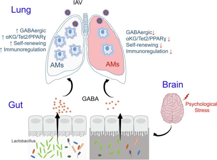
“Our study demonstrates that chronic psychological stress significantly increases host vulnerability to influenza A virus (IAV) infection characterized by a distorted gut microbiome and deregulated alveolar macrophages (AMs) response. We show that microbiome-derived γ-aminobutyric acid (GABA) functions as a tonic signal to support survival, self-renewing, and immunoregulation of AMs, and hence optimized pulmonary defensive response.

Chronic psychological stress causes gut microbiome dysbiosis and defective GABA generation, leading to loss of AMs homeostasis and aggravated viral pneumonia. The data indicate that:

  1. Microbial GABA is released in the circulation,
  2. Sensed by AMs via the GABAA receptor,
  3. Promoting cellular mitochondrial metabolism,
  4. For increased production of α-ketoglutarate (αKG),
  5. Which triggers Tet2-mediated DNA hydroxymethylation,
  6. To enable PPARγ-centered gene program,
  7. Supporting AMs homeostasis and function.

ga1_lrg

  • Re-localization of GABA-generating probiotics,
  • Supplementation of αKG, or
  • Adoptive transfer of GABA-conditioned macrophages,
  • Substantially rectifies stress-induced disruption inter-organ communication, and
  • Alleviates symptoms of viral pneumonia.

Our current study unveils an unappreciated regulatory circuitry that connects the brain, gut, and lung to mediate neurological modulation of host defensive response.”

https://www.sciencedirect.com/science/article/pii/S2090123223001716 “Gut microbial GABAergic signaling improves stress-associated innate immunity to respiratory viral infection”


consentofthegoverned

A biomarker for impaired cognitive function?

This 2023 rodent study investigated associations between a drug, a gut microbiota species, cognitive function, and proinflammatory cytokine interleukin-6:

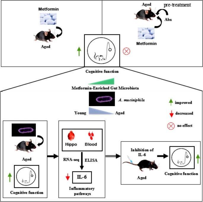
“We show that gut microbiota is altered by metformin, which is necessary for protection against ageing-associated cognitive function declines in aged mice.

  • Mice treated with antibiotics did not exhibit metformin-mediated cognitive function protection.
  • Treatment with Akkermansia muciniphila improved cognitive function in aged mice.
  • A. muciniphila decreased proinflammatory-associated pathways, particularly that of proinflammatory cytokine interleukin (IL)-6, in both peripheral blood and hippocampal profiles, which was correlated with cognitive function improvement.
  • An IL-6 antibody protected cognitive function, and an IL-6 recombinant protein abolished the protective effect of A. muciniphila on cognitive function in aged mice.

40168_2023_1567_Figa_HTML

A. muciniphila, which is mediated in gut microbiota by metformin, modulates inflammation-related pathways in the host and improves cognitive function in aged mice by reducing proinflammatory cytokine IL-6 both systemically and in the hippocampus. This is direct evidence to validate that gut microbiota mediate the effect of metformin on cognitive improvement.”

https://microbiomejournal.biomedcentral.com/articles/10.1186/s40168-023-01567-1Akkermansia muciniphila, which is enriched in the gut microbiota by metformin, improves cognitive function in aged mice by reducing the proinflammatory cytokine interleukin-6″


IL-6 may be useful with other biomarkers of impaired cognitive function. It’s too coarse to track improved cognitive function past a certain point, though. Maybe the current IL-6 blood test can be refined as high-specificity CRP and regular CRP blood tests were done?

We don’t need to take this drug or be concerned about this gut bacteria species in order to lower inflammation. Click the IL-6 link above and see blog posts such as Part 2 of Rejuvenation therapy and sulforaphane for other methods.

PXL_20230630_092833465