Maintaining your myelin, Part 1

Three papers on myelin and oligodendrocytes, starting with a 2023 review:

“Myelin is the spiral ensheathment of axons by a lipid and cholesterol-rich glial cell membrane that reduces capacitance and increases resistance of the axonal membrane. Axonal myelination speeds up nerve conduction velocity as a function of axon diameter.

While myelination proceeds rapidly after birth in the peripheral nervous system, central myelination is a spatially and temporally more regulated process. Ongoing myelination of the human brain has been documented at up to 40 years of age. This late myelination in the adult cortex is followed by exhaustion of oligodendrocyte precursor cells (OPC) with senescence and a gradual loss of myelin integrity in the aging brain.

The brain is well known for its high energy demands, specifically in gray matter areas. In white matter tracts, energy consumption is lower. Myelination poses a unique challenge for axonal energy generation where myelin sheaths cover more than 95% of the axonal surface areas.

Oligodendrocytes help support axonal integrity. Oligodendrocytes survive well in the absence of mitochondrial oxidative phosphorylation, and without signs of myelin loss, cell death, neurodegeneration or secondary inflammation.

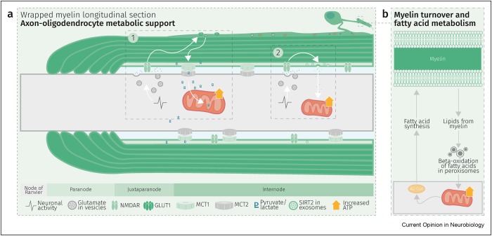
Glycolysis products of oligodendroglial origin are readily metabolized in axonal mitochondria. Oligodendroglial metabolic support is critical for larger and faster-spiking myelinated axons that also have a higher density of mitochondria. An essential requirement for the direct transfer of energy-rich metabolites from oligodendrocytes to the myelinated axonal compartment is ‘myelinic channels’ within the myelin sheath.

Interactions of oligodendrocytes and myelin with the underlying axon are complex and exceed the transfer of energy-rich metabolites. Continuous turnover of myelin membranes by lipid degradation and fatty acid beta-oxidation in mitochondria and peroxisomes leads to recycling of acetate residues by fatty acid synthesis and membrane biogenesis.
1-s2.0-S0959438823001071-gr2_lrg

In human multiple sclerosis (MS) and its animal model myelin oligodendrocyte glycoprotein-experimental autoimmune encephalomyelitis (MOG-EAE), acute inflammatory demyelination is followed by axonal degeneration in lesion sites that is mechanistically not fully understood. It is widely thought that demyelination and the lack of an axon-protective myelin sheath in the presence of numerous inflammatory mediators are the main causes of axon loss.

But unprotected axons improve rather than worsen the overall clinical phenotype of EAE mice which exhibited the same degree of autoimmunity. Thus, ‘bad myelin is worse than no myelin’ because MS-relevant myelin injuries perturb the integrity of myelinic channels and metabolic support.

Dysfunctional or injured oligodendrocytes that do not allow for compensation by any other cell types turn the affected myelin ensheathment into a burden of the underlying axonal energy metabolism, which causes irreversible axon loss. Any loss of myelin integrity, as seen acutely in demyelinating disorders or more gradually in the aging brain, becomes a risk factor for irreversible neurodegeneration.”

https://www.sciencedirect.com/science/article/pii/S0959438823001071 “Expanding the function of oligodendrocytes to brain energy metabolism”


A 2024 review focused on myelin and oligodendrocyte plasticity:

“This review summarizes our current understanding of how myelin is generated, how its function is dynamically regulated, and how oligodendrocytes support the long-term integrity of myelinated axons.

Apart from its unique ultrastructure, there are several other exceptional features of myelin. One is certainly its molecular composition. Another is its extraordinary stability. This was compellingly illustrated when 5000-year-old myelin with almost intact ultrastructure was dissected from a Tyrolean Ice Man.

Myelin is a stable system in contrast to most membranes. However, myelin is compartmentalized into structurally and biochemically distinct domains. Noncompacted regions are much more dynamic and metabolically active than tightly compacted regions that lack direct access to the membrane trafficking machinery of oligodendrocytes.

The underlying molecular basis for stability of myelin is likely its lipid composition with high levels of saturated, long chain fatty acids, together with an enrichment of glycosphingolipids (∼20% molar percentage of total lipids) and cholesterol (∼40% of molar percentage of total lipids). In addition, myelin comprises a high proportion of plasmalogens (ether lipids) with saturated long-chain fatty acids. In fact, ∼20% of the fatty acids in myelin have hydrocarbon chains longer than 18 carbon atoms (∼1% in the gray matter) and only ∼6% of the fatty acids are polyunsaturated (∼20% in gray matter).

With maturation of oligodendrocytes, the plasma membrane undergoes major transformations of its structure. Whereas OPCs are covered by a dense layer of large and negatively charged self-repulsive oligosaccharides, compacted myelin of fully matured oligodendrocytes lacks most of these glycoprotein and complex glycolipids.

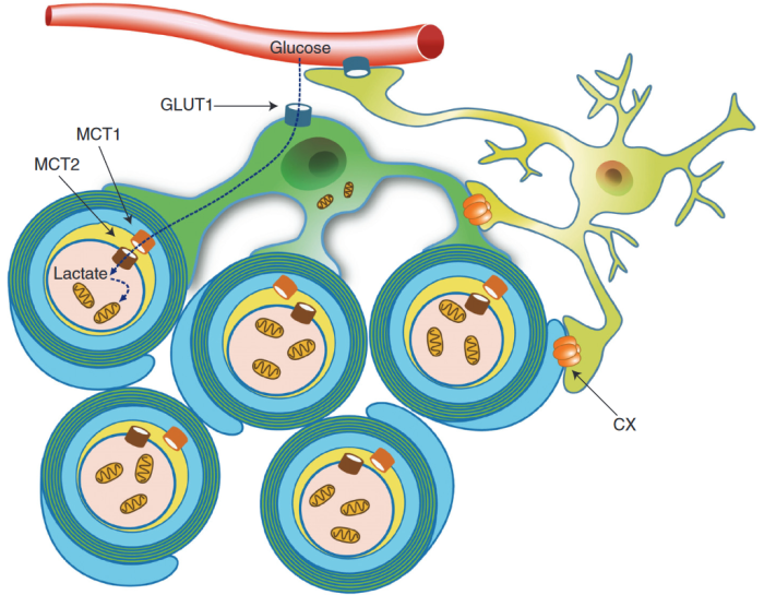
Schematic depiction of an oligodendrocyte that takes up blood-derived glucose and delivers glycolysis products (pyruvate/lactate) via monocarboxylate transporters (MCT1 and MCT2) to myelinated axons. Oligodendrocytes and myelin membranes are also coupled by gap junctions to astrocytes, and thus indirectly to the blood–brain barrier.
oligodendrocyte

Adaptive myelination refers to dynamic events in oligodendroglia driven by extrinsic factors such as experience or neuronal activity, which subsequently induces changes in circuit structure and function. Understanding how these adaptive changes in neuron-oligodendroglia interactions impact brain function remains a pressing question for the field.

Transient social isolation during adulthood results in chromatin and myelin changes, but does not induce consequent behavioral alterations. When mice undergo a social isolation paradigm during early life development, they similarly exhibit deficits in prefrontal cortex function and myelination, but these deficiencies do not recover with social reintroduction. This implicates a critical period for social deprivation effects on myelin dynamics. Experience-dependent changes in myelin dynamics may depend on not only the age, brain region, and cell type studied, but also the specific myelin structural change assessed.

Local synaptic neurotransmitter release along an axon not only affects the number of OPCs and oligodendrocytes associated with that axon and local synthesis of myelin proteins, but also drives preferential selection of active axons for myelination over the ensheathment of electrically silenced neighboring axons. Neuronal activity–induced plasticity may preferentially impact brain regions that remain incompletely myelinated compared to more fully myelinated tracts.

Whereas the myelin sheath has been regarded for a long time as an inert insulating structure, it has now become clear that myelin is metabolically active with cytoplasmic-rich pathways, myelinic channels, for movement of macromolecules into the periaxonal space. The myelin sheath and its subjacent axon need to be regarded as one functional unit, which are not only morphological but also metabolically coupled.”

https://cshperspectives.cshlp.org/content/early/2024/04/15/cshperspect.a041359 “Oligodendrocytes: Myelination, Plasticity, and Axonal Support” (not freely available) Thanks to Dr. Klaus-Armin Nave for providing a copy.


A 2024 rodent study investigated oligodendrocyte precursor cell transcriptional and epigenetic changes:

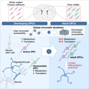
“We used single-cell RNA sequencing (scRNA-seq), single-cell ATAC sequencing (scATAC-seq), and single-cell spatial transcriptomics to characterize murine cortical OPCs throughout postnatal life. One group (active, or actOPCs) is metabolically active and enriched in white matter. The second (homeostatic, or hOPCs) is less active, enriched in gray matter, and predicted to derive from actOPCs. Relative to developing OPCs, both actOPCs and hOPCs are less active metabolically and have less open chromatin.

In adulthood, these two groups are transcriptionally but not epigenetically distinct, indicating that they may represent different states of the same OPC population. If that is the case, then one model is that the parenchymal environment maintains adult OPCs within an hOPC state, whereas those OPCs recruited into white matter or exposed to demyelinated axons may transition toward an actOPC state in preparation for making new oligodendrocytes. We do not yet know the functional ramifications of these differences, but this finding has clear implications for the development of therapeutic strategies for adult remyelination.
opcs

Another finding is that developing but not adult actOPC chromatin is preferentially open for binding motifs associated with neural stem cells, transit-amplifying precursors, and neurogenesis. Although this may simply reflect their origin as the immediate progeny of neonatal neural precursor cells, it may also explain why developing but not adult OPCs have the capacity to make neurons in culture.

If we could, at least in part, reverse the global chromatin shutdown that occurs between development and adulthood, then perhaps adult OPCs may reacquire the ability to make neurons or become better able to generate new oligodendrocytes for remyelination.”

https://www.cell.com/stem-cell-reports/fulltext/S2213-6711(24)00077-8 “Single-cell approaches define two groups of mammalian oligodendrocyte precursor cells and their evolution over developmental time”

Continued in Part 2.


PXL_20240414_103442372

What can be done today to fulfill early unmet needs?

Got agitated earlier this week watching Tucker Carlson’s freely-available interview with a maniac who thinks he’s graduated into a higher state by worshiping the Great AI (Artificial Intelligence, aka Automated Internet, inhabited solely by robots) which will dictate every aspect of what to do with his life. Nevermind that behind the Great AI curtain are the same people who have lied to billions of us, especially during every day of this decade.

Are his current set of beliefs better than previous ones he had of putting a chip into everybody’s brain? What’s wrong with getting to live your own life?

5000

What I saw expressed in the interview was an exhausting pursuit of substitutes for feeling loved. I doubt that many others saw the same, because feeling unloved is so devastating we’ll do anything to avoid it.

But re-experiencing early memories and feelings of unmet needs in a therapeutic setting is the way to keep them from subsequently running our lives. Otherwise, we’ll develop unfulfilling substitutes for what we missed, with misdirected ideas and beliefs accompanied by their unconscious act-outs.

While speaking with a mother who is doing a terrific job of meeting her six-month-old’s needs, I attempted to contrast this interview with the experiences she and her husband are giving their child. Maybe if they read this post, my poor explanation will become clearer.


Wild persimmon trees’ eclipse shadows

PXL_20240408_192336638

Sulforaphane’s effects on autism and liver disease

Here are two more papers that cited Precondition your defenses with broccoli sprouts, starting with a 2024 human / rodent study investigating gut microbiota / sulforaphane’s effects on autism:

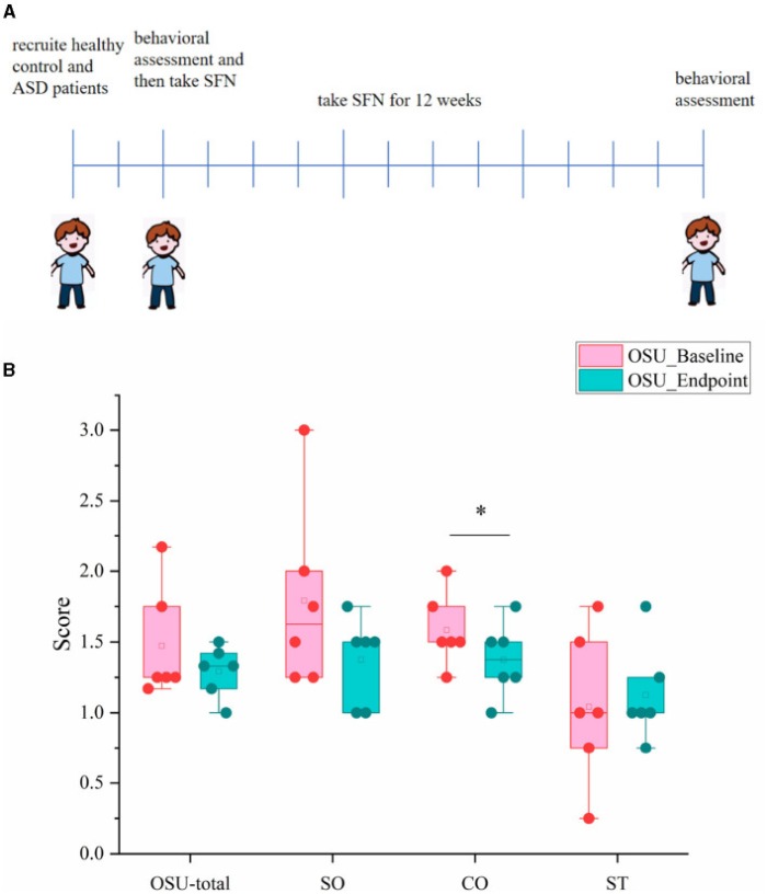
“Sulforaphane (SFN) has been found to alleviate complications linked with several diseases by regulating gut microbiota (GM), while the effect of GM on SFN for autism spectrum disorders (ASD) has not been studied. We evaluated therapeutic effects of SFN on maternal immune activation (MIA)-induced ASD-like rat model and pediatric autism patients aged 4–7 years.

OSU-SO for social interactive OSU behavioral subscores, OSU-CO for non-verbal communicative OSU behavioral [significant] subscores, and OSU-ST for repetitive or ritualistic OSU behavioral subscores:

fnut-10-1294057-g0005

Although gut microbiota composition was significantly altered in SFN-treated ASD-like rats, alteration of GM was not evident in ASD patients after 12 weeks of SFN treatment. Limitations in this study:

  1. Studies were conducted in male rats and boys only;
  2. The sample size of our clinical study is relatively small [6 SFN-treated boys] and needs to be further expanded in the future; and
  3. This study only uncovered a potential link between gut flora and the therapeutic effects of SFN on ASD.

SFN treatment alleviates social deficits in MIA-induced ASD-like rats and ASD patients, and improvements might be associated with gut microbiota.”

https://www.frontiersin.org/articles/10.3389/fnut.2023.1294057/full “Therapeutic efficacy of sulforaphane in autism spectrum disorders and its association with gut microbiota: animal model and human longitudinal studies”

The 2022 Efficacy of Sulforaphane in Treatment of Children with Autism Spectrum Disorder: A Randomized Double-Blind Placebo-Controlled Multi-center Trial (not freely available) was referenced for sulforaphane (actually, glucoraphanin with myrosinase enzyme) doses:

“Dosing was weight-based:

  • Two tablets/day for 10–29 lb;
  • Three tablets/day for 30–49 lb;
  • Four tablets/day for 50–69 lb.

An estimated delivery of approximately 24, 36, and 48 μmol of sulforaphane daily was expected in the respective SF dosage groups.”

Weights of the above μmol estimated dose amounts per https://pubchem.ncbi.nlm.nih.gov/compound/sulforaphane are 4.3, 6.4, and 8.5 mg, respectively. An average weight of a 4-year-old boy is 36 lbs / 16.3 kg, and a 7-year-old boy is 51.1 lbs / 23.2 kg.

This study’s maternal immune activation was done by injecting lipopolysaccharide into pregnant rats. Would injecting pregnant women with immune-activating substances have similar harmful effects on the fetus? We don’t have evidence because unbiased and unconflicted studies looking for such effects weren’t sponsored and/or published before immune-activating substances’ deployments.


A 2024 rodent study investigated sulforaphane’s effects on diabetic liver damage:

“We investigated whether sulforaphane, an Nrf2 activator and antioxidant, prevents diabetes-induced hepatic ferroptosis, and the mechanisms involved. Results showed that diabetes-induced inactivation of Nrf2 and decreased expression of its downstream antiferroptotic molecules critical for:

  • Antioxidative defense (catalase, superoxide dismutases, thioredoxin reductase);
  • Iron metabolism (ferritin heavy chain (FTH1), ferroportin 1);
  • Glutathione (GSH) synthesis (cystine-glutamate antiporter system, cystathionase, glutamate-cysteine ligase catalitic subunit, glutamate-cysteine ligase modifier subunit, glutathione synthetase); and
  • GSH recycling – glutathione reductase (GR)

were reversed/increased by sulforaphane treatment.

Diabetes-induced increases in serum glucose and triglyceride levels were also significantly reduced by sulforaphane. Taken together, our results demonstrate a potent effect of SFN in inhibiting ferroptotic death of hepatocytes under diabetic conditions in vivo, thereby alleviating liver injury.

This is the first study to demonstrate the protective role of SFN against ferroptosis in the liver of diabetic mice. This nominates sulforaphane as a promising phytopharmaceutical for the prevention/alleviation of ferroptosis in diabetes-related pathologies.”

https://iubmb.onlinelibrary.wiley.com/doi/10.1002/biof.2042 “Sulforaphane prevents diabetes-induced hepatic ferroptosis by activating Nrf2 signaling axis”

Improving peroxisomal function

A 2024 review provided details about “mysteries” in peroxisome research:

“Peroxisomes are key metabolic organelles with essential functions in cellular lipid metabolism (e.g., β-oxidation of fatty acids and synthesis of ether phospholipids, which contribute to myelin sheath formation), and metabolism of reactive oxygen species (ROS), particularly hydrogen peroxide. Loss of peroxisomal function causes severe metabolic disorders in humans.

Additional non-metabolic roles of peroxisomes have been revealed in cellular stress responses, regulation of cellular redox balance and healthy ageing, pathogen and antiviral defence, and as cellular signalling platforms. New findings also point to a role in regulation of immune responses.

In our previous reviews, we addressed the role of peroxisomes in the brain, in neurological disorders, in development of cancer, and in antiviral defence. To avoid repetition, we refer to those articles where appropriate, and to more specialised recent reviews on peroxisome biology.

418_2023_2259_Fig5

Proper functioning of peroxisomes in metabolism requires the concerted interaction with other subcellular organelles, including the endoplasmic reticulum (ER), mitochondria, lipid droplets, lysosomes, and the cytosol. A striking example of peroxisome-ER metabolic cooperation is de novo biosynthesis of ether phospholipids.

Metabolic activities of peroxisomes, such as ɑ- and β-oxidation of fatty acids, plasmalogen synthesis, and ROS/reactive nitrogen species metabolism, have been linked to numerous immune-related pathways. Roles for peroxisomes in immune and defence mechanisms have opened a new field of peroxisome research, and highlight once more how important peroxisomes are for human health and disease.

It is still not fully understood how peroxisomal functions and abundance are regulated, what kinases/phosphatases are involved, or how peroxisomes are linked to cellular signalling pathways and how they act as signalling platforms.”

https://link.springer.com/article/10.1007/s00418-023-02259-5 “The peroxisome: an update on mysteries 3.0”


Last Friday was Day 90 of a 90-day trial of plasmalogens coincident with improving peroxisomal function via resistance exercise and time-restricted eating. A sticking point has been leg resistance exercises. Ankle issues are interfering with progress, although beach walks aren’t similarly affected. I’m almost back to an upper body exercise routine of five years ago, and I’ve added a half-dozen abs exercises.

I’ll continue taking the two Prodrome plasmalogen precursor supplements (ProdromeGlia and ProdromeNeuro) and with efforts to improve peroxisomal function. Since achieving effective resistance exercise levels is taking longer than expected, and my crystal ball is out-of-commission, I don’t have a realistic end time estimate for stopping the supplements.

Building your plasmalogen savings account

A webinar from earlier this week with Dr. Goodenowe, a clinical trial facilitator, and a physician:

From the Q&A segment:

“Is there a particular age where it’s recommended to test for plasmalogen levels? And what levels would be considered normal?

That’s a good question. That actually raises this whole concept of optimal health and this concept of aging.

The best way to think about it – we talked about this paycheck-to-paycheck situation, where as long as our bills are paid every day, technically we think we’re normal. But we still feel this sense of health anxiety – if you will – like we just don’t know if my car breaks down, or my water heater breaks down, do I have enough money to pay these events in my life?

That’s what health feels like to a lot of people, because they’re just kind of getting by. From a health perspective, they’re considered normal, but they have no reserve capacity, and they have no vitality in terms of health.

Plasmalogens are a type of molecule that you build a savings account of, over years, over decades. Your heart builds them up, your brain builds them up, and you slowly accumulate them. Then when you get an oxidative stress like what’s happening now in today’s world with all the covid and myocarditis and brain fog – a lot of these things are being caused because that reserve of plasmalogens has been depleted.

We want plasmalogens for a longevity perspective. There are other situations that can have low plasmalogens, other things can really knock your plasmalogens down.

So you want to start early, you want to build a savings account, and you want to maintain it. Maintain health and function, and create a sustained surplus for optimal health, for optimal neuromuscular performance.”


PXL_20231207_185012059

Brain restoration with plasmalogens

In this 2023 presentation for a professional audience, Dr. Dayan Goodenowe showed an example of what could be done (in the form of what he personally did at ages 53-54) to restore and augment brain structure and function over a 17-month period by taking plasmalogens and supporting supplements:

https://drgoodenowe.com/recording-of-dr-goodenowes-presentation-from-the-peptide-world-congress-2023-is-now-available/

Follow the video along with its interactive transcript. Restorative / augmentative supplements included:

1. Nutritional Supplementation Strategy

Forms of MRI used to document brain structure and function changes were:

2. Advanced MRI Technologies

Brain volume decreases are the rule for humans beginning at age 40. Dr. Goodenowe documented brain volume increases, which aren’t supposed to happen, but did per the below slide of overall results:

3. Reversing Brain Shrinkage

“From a global cortical volume and thickness perspective, 17 months of high-dose plasmalogens reversed ~15 years of predicted brain deterioration.”


Specific increased adaptations in brain measurements over 17 months included:

  1. Cortical thickness .07/2.51 = +3%.
  2. White matter microstructure fractional anisotropy +8%.
  3. Nucleus accumbens volume +30%.
  4. Dopaminergic striatal terminal fields’ volume +18%.
  5. Cholinergic cortical terminal fields’ volume +10%.
  6. Occipital cortex volume +10%.
  7. Optic chiasm volume +225%.
  8. Nucleus basalis connectivity.
  9. Neurovascular coupling signal controlled by noradrenaline integrity.
  10. Amygdala volume +4% and its connectivity to the insula, indicating ongoing anxiety and emotional stress response.
  11. Parahippocampus volume +7%.
  12. Hippocampus fractional anisotropy +5%.

No changes:

  1. Amygdala connectivity to the ventral lateral prefrontal cortex, the same part of the brain that relates to placebo effect.
  2. Hippocampus connectivity.

Decreased adaptations in brain measurements included:

  1. White matter microstructure radial diffusivity -10%.
  2. Amygdala connectivity to the anterior cingulate cortex to suppress / ignore / deny anxiety response.
  3. Amygdala connectivity to the dorsal lateral prefrontal cortex.
  4. Entorhinal cortex volume -14%.
  5. Hippocampus volume -6%.
  6. Hippocampus mean diffusivity (white matter improved, with more and tighter myelin) -4%.

The other half of this video was a lively and wide-ranging Q&A session.


The referenced 2023 study of 653 adults followed over ten years showed what brain deterioration could be expected with no interventions. Consider these annual volume decrease rates to be a sample of a control group:

etable 3

https://jamanetwork.com/journals/jamanetworkopen/fullarticle/2806488 “Characterization of Brain Volume Changes in Aging Individuals With Normal Cognition Using Serial Magnetic Resonance Imaging”

Also see a different population’s brain shrinkage data in Prevent your brain from shrinking.


The daily plasmalogen precursor doses Dr. Goodenowe took were equivalent to 100 mg softgel/kg, double the maximum dose of 50 mg softgel/kg provided during the 2022 clinical trial of cognitively impaired old people referenced in Plasmalogens Parts 1, 2, and 3.

He mentions taking 5 ml in the morning and 5 ml at night because he used the Prodrome oil products. 1 ml of a Prodrome oil plasmalogen precursor product equals 900 mg of their softgel product.


“My brain is trying to minimize long-term effects of pain/stress by suppressing my memory of it. But this can only go on for so long before it becomes an entrenched state.

I have solved the sustenance side of the equation. I need to work harder to solve the environmental side.”

While I agree that we each have a responsibility to ourselves to create an environment that’s conducive to our health, the above phenomenon isn’t necessarily resolvable by changing an individual’s current environment. My understanding is that long-term effects of pain, stress, and related human experiences are usually symptoms of causes that started much earlier in our lives.

Adjusting one’s present environment may have immediate results, but probably won’t have much therapeutic impact on long-term issues. Early life memories and experiences are where we have to gradually go in order to stop being driven by what happened back then.

See Dr. Arthur Janov’s Primal Therapy for its principles and explanations. I started Primal Therapy at a similar age, 53, and continued for three years.

Plasmalogens, Part 3

The 2022 plasmalogen clinical trial mentioned in Parts 1 and 2 bypassed peroxisome metabolism of cognitively impaired people per discussion of the below diagram:

fcell-10-864842-g003

Increasing the body’s fasting state with time-restricted eating, and preventing muscle atrophy with resistance exercise, were offered as the two most important ways to improve peroxisomal function.

I didn’t find any relevant 2023 human studies (where I could access the full study) on different non-drug treatments that I was willing to do. A 2023 review outlined aspects of peroxisomes, to include a few older human studies:

“Peroxisomes are small, single-membrane-bound organelles, which are dynamic and ubiquitous. Peroxisomes directly interact with other organelles, such as endoplasmic reticulum, mitochondria, or lysosomes. Peroxisomes exert different functions in various cells through both catabolic and anabolic pathways.

The main functions of peroxisomes can be categorized as reactive oxygen species (ROS) metabolism, lipid metabolism, and ether-phospholipid biosynthesis. Peroxisomes also play important roles in inflammatory signaling and the innate immune response.”

1-s2.0-S2667325823001425-gr3_lrg

https://www.sciencedirect.com/science/article/pii/S2667325823001425 “Peroxisome and pexophagy in neurological diseases”


1. Since I haven’t recently tried the two main ways to improve peroxisomal function, I’ll give them a go over the next three months:

  • Expect to get my feeding timeframe to within eight hours. Don’t know about making it short like 6 hours, because my first meal of the day is 35 calories of microwaved cruciferous sprouts, then I wait an hour before eating anything else.
  • Resistance exercise progress should be measurable, as I recorded exercises during the first ten weeks of eating broccoli sprouts every day 3.5+ years ago.

2. Don’t know that I’ll recognize any cognitive improvements to the extent I did during Week 9.

  • I don’t have a young brain anymore, and I’m sure some decline could be measured in memory tests. But I’m not going to become a lab rat.
  • There’s an occasional annoyance that’s been going on for some time, especially when I’m distracted. It happens when I think of something to do, and it somehow becomes a short-term memory that I did it, instead of going into a Things To Do queue. It’s largely self-correcting. For example, regardless of what I paid, I’ll drive back to the grocery store self-checkout to retrieve a third bag that didn’t make it home. A pink-haired employee said young people leave their paid-for groceries behind all the time. It’s usually more of a reality disconnect for me than forgetfulness, because I have a memory that I performed the action. Definitely room for improvement.

3. Don’t know that I’d see biochemical changes such as some described in Part 1. Maybe I’ll move up an annual physical to compare it with the last one in May?

  • I already have very little oxidative stress, very little inflammation, low triglycerides, high HDL, and no major improvements are indicated on CBC / CMP / lipid panels.
  • Take supplements to ensure other things like acetylcholine neurotransmitter availability, one-carbon / methylation metabolism, vitamin / mineral adequacy.

4. I started the two Prodrome plasmalogen precursor supplements (ProdromeGlia and ProdromeNeuro) a week ago, and take their standard doses. My thought is that resultant plasmalogens won’t degrade very much if their primary use isn’t to immediately address oxidative stress and inflammation. That could give these extra plasmalogens a chance to make larger homeostatic contributions in myelin and membrane areas.

I don’t expect any particular effects to manifest. But I’m interested to see if these two areas would be affected:

  • My left ulnar nerve has been giving me problems for over five years, and several resistance exercises aggravate it. I’ve had two nerve continuity tests during that time to confirm. Numbness and pain are intermittent, though.
  • I still take acetaminophen several times a day for other pain.

None of the above treatments are specifically indicated. But if time-restricted feeding and/or extra plasmalogens have an effect on left ulnar or other pain, maybe I’ll be able to make better progress on resistance exercise.

Update #1 11/13/2023

Update #2 11/22/2023

Update #3 12/13/2023 comments

Update #4 1/30/2024

Update #5 3/31/2024

Plasmalogens, Part 1

The person who knows the most about this subject is Dayan Goodenowe, PhD. Some recent publications include:

https://www.frontiersin.org/articles/10.3389/fcell.2022.864842/full “Targeted Plasmalogen Supplementation: Effects on Blood Plasmalogens, Oxidative Stress Biomarkers, Cognition, and Mobility in Cognitively Impaired Persons”

https://www.frontiersin.org/articles/10.3389/fcell.2022.866156/full “Brain ethanolamine phospholipids, neuropathology and cognition: A comparative post-mortem analysis of structurally specific plasmalogen and phosphatidyl species”

plasmalogens and cognition


A sample of links freely available at https://drgoodenowe.com/.

1. Presentations to professional groups. Have your mouse ready to click the pause button.

https://drgoodenowe.com/dr-goodenowe-presents-at-the-iagg2023-in-yokohama-japan/ “A rare children’s disease that may be the key to reversing neurological decline in aging”

Includes videos of a treatment’s effects on a child.

https://neomarkgroup.wistia.com/medias/0qln0wy93t “The most influential biomarkers for aging and disease”

Despite the title, a considerable number of studies were presented on prenatal, infant, and early childhood development. He misspoke a few times, so read the slides.

Phenotype is reality. Genotype is possibility. Communications links between different fields are very poorly connected in science.

Peroxisomes are islands. They don’t have DNA like your mitochondria do. Peroxisomal transport issues are important things to understand.

All aging-related cross-sectional analyses are on the rate of decline. You’re declining from a previous well state. Age-matched controls are the most ridiculous thing to do.”


2. I’ll highlight the longest of several interviews because there was plenty of room to expand on points. Maybe the best detailed explanations came as responses to that interviewer challenging with contrasting AD, traumatic brain injury, and cholesterol paradigms. Its transcript is more accurate than a usual YouTube interpretation, but there are still mistakes such as “fossil lipid” vs. phospholipid.

https://www.betterhealthguy.com/episode186 “Plasmalogens with Dr. Dayan Goodenowe, PhD”

“Science is how do you push things to its failure, until you can’t fail it again. We’ve lost that. It’s become more hypothesis proving.

Plasmalogens levels go up for a different reason than people think. The reason why it peaks in our 40s and 50s is because we’ve been myelinating. The white matter of our brain is still increasing. It’s not because we’re making more plasmalogens. It’s because the lake, the reservoir, gets full. What you’re measuring in blood is overflow from the lake. The lower plasmalogens start trickling down in your blood, the bigger drain that’s occurring on that system.

Low plasmalogens don’t just predict dementia in the elderly population. It predicts the rate of decline of that dementia. It predicts the rate of death.

The biggest drivers of plasmalogen manufacturing and the biggest reasons why they decrease with age, or in other circumstances is two things. One, the failure to maintain a fasting state of the human body. The second one is muscle atrophy.

Amyloid has absolutely nothing to do with Alzheimer’s, or dementia. It’s just a bystander on the road watching an accident happen.

Age-related cognitive decline is clearly where plasmalogens have the greatest impact. You’re always going to have mixed pathologies in the brain.

Nutritional availability of plasmalogens is virtually non-existent. As soon as they hit the hydrochloric acid of your stomach, they’re gone. They don’t make it past the stomach, or the upper intestine.”


I came across Dr. Goodenowe’s work last month from clicking a comment on this blog that linked back to her blog. Always be curious.

Continued in Part 2.

Reversing biological age in rats

This 2023 rodent study wrapped together findings of the original study curated in A rejuvenation therapy and sulforaphane, and the second follow-on study mentioned in Signaling pathways and aging. I’ll start by highlighting specifics of the later study:

“Pronounced rejuvenation effects in male rats prompted us to conduct further confirmatory experiments. A particularly important consideration is the effectiveness of E5 with regards to sex, as sex-dependent rejuvenation by some interventions have previously been reported.

To assess E5’s applicability to both male and female Sprague Dawley rats, we studied 12 males (6 treated with E5, 6 with saline) and 12 females (6 treated with E5, 6 with saline). These rats were treated every 45 days with an injection of E5 or saline. Rats were monitored for 165 days, and blood was drawn at six time points: 0, 15, 30, 60, 150 and 165 days from the first injection.

We observed highly significant improvements in TNF alpha and IL-6 levels for both males and females in the blood of E5-injected rats over that of saline controls. We also observed a substantial improvement in grip strength.

Our study shows age reversal effects in both male and female rats, but E5 is more effective in males.”


Another experimental group was started with old rats of both sexes. Using the human / rat relative clock developed in the original study, a human equivalent age to these rats at 26 months old was ((112.7 weeks / 197.6 weeks maximum rat lifespan) x 122.5 years maximum human lifespan) = 69.8 years:

“To validate our epigenetic clock results, we conducted a second set of E5 experiments with Sprague Dawley rats of both sexes. When these rats turned 26 months old, half (9 rats) received the E5 treatment while the other half (8 rats) received only the control treatment (saline injection). We analyzed methylation data from two blood draws: blood draw before treatment (baseline) and a follow up sample (15 days after the E5/saline treatment).”

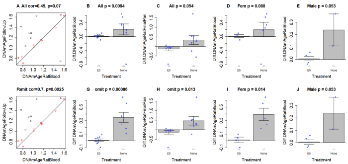
Treatment measurements were affected by one female control group outlier. Panels F through J were recalculated after removing the outlier to show significant effects in both sexes:

second follow-on results

“A) Final version of the rat clock for blood. Baseline measurement (x-axis) versus follow up measurement (15 days after treatment, y-axis). Points (rats) are colored by treatment: red=treated by E5, black=treated with saline only. Rotated grey numbers underneath each bar reports the group sizes. Each bar plot reports the mean value and one standard error.

B,D,E) Difference between follow up measurement and baseline measurement (y-axis) versus treatment status in B) all rats, D) female rats only, E) male rats only. C) is analogous to B) but uses the pan tissue clock for rats.

Panels in the second row (F,G,H,I,J) are analogous to those in the first row but the analysis omitted one control rat (corresponding to the black dot in the lower right of panel A).”

https://www.biorxiv.org/content/10.1101/2023.08.06.552148v1 “Reversal of Biological Age in Multiple Rat Organs by Young Porcine Plasma Fraction”


A description of how E5 plasma fraction was made starts on page 16 of the *.pdf file. The next E5 study will be done with dogs per July 2023 updates in blog post comments:

“On E5 our entire team is working hard towards the launch of an old Beagle dogs trial this month. We want to make them really young, healthy, happy, and jumping around like 1 and 2 year olds.

Primary endpoint is safety and toxicology to test various dose strengths and frequencies. Secondary endpoints are more than 20.

As you know, we like to test exhaustively to get a sharper perspective of what’s happening. In rat studies we tested 30 biomarkers, including functional. We are especially keen to check kidney markers.

There are two clocks for dogs we are interested in to get third party confirmation of age reversal. Horvath dog clock is ready and GlycanAge dog clock is under construction.

We are requesting all organizations that support pets and aging to financially support their project of building an accurate dog clock. Not only will it help veterinary aging research like ours, but also all the dog owners that may want to know how much improvement their dog received from treatment. Dr. Matt Kaeberlain is an advisor on their project.”

Neuritogenesis

Three 2023 papers on the initial stage of neuronal differentiation, starting with a rodent study of taurine’s effects:

“We aimed to assess the role of taurine (TAU) in axonal sprouting against cerebral ischemic injury, clarify the function of mitochondria in TAU-induced axonal sprouting, and further determine the underlying potential molecular mechanism.

experiment design

We determined that TAU improved motor function recovery and restored neurogenesis in ischemic stroke. This possibly occurred via improvements in mitochondrial function.

We investigated that the Sonic hedgehog (Shh) pathway exerted an important role in these effects. Our study findings highlighted the novel viewpoint that TAU promoted axonal sprouting by improving Shh-mediated mitochondrial function in cerebral ischemic stroke.”

https://www.scielo.br/j/acb/a/nxKvGXGk9g6gRkHxybMfbYJ/?lang=en “Taurine promotes axonal sprouting via Shh-mediated mitochondrial improvement in stroke”


A rodent study investigated effects of a soy isoflavone gut microbiota metabolite:

“Perinatally-infected adolescents living with HIV-1 (pALHIV) appear uniquely vulnerable to developing substance use disorders (SUD). Medium spiny neurons (MSNs) in the nucleus accumbens core (NAcc), an integrator of cortical and thalamic input, have been implicated as a key structural locus for the pathogenesis of SUD.

Treatment with estrogenic compounds (e.g., 17β-estradiol) induces prominent alterations to neuronal and dendritic spine structure in the NAcc supporting an innovative means to remodel neuronal circuitry. The carcinogenic nature of 17β-estradiol, however, limits its translational utility.

Plant-derived polycyclic phenols, or phytoestrogens, whose chemical structure resembles 17β-estradiol may afford an alternative strategy to target estrogen receptors. The phytoestrogen S-Equol (SE), permeates the blood-brain barrier, exhibits selective affinity for estrogen receptor β (ERβ), and serves as a neuroprotective and/or neurorestorative therapeutic for HIV-1-associated neurocognitive and affective alterations.

Beginning at approximately postnatal day (PD) 28, HIV-1 transgenic (Tg) animals were treated with a daily oral dose of 0.2 mg of SE. The SE dose of 0.2 mg was selected for two primary reasons, including:

  1. A dose-response experimental paradigm established 0.2 mg of SE as the most effective dose for mitigating neurocognitive deficits in sustained attention in the HIV-1 Tg rat; and
  2. The dose, which yielded a daily amount of 0.25–1.0 mg/kg/SE (i.e., approximately 2.5–10 mg in a 60 kg human), is translationally relevant (i.e., well below the daily isoflavone intake of most elderly Japanese.

Daily oral treatment continued through PD 90.

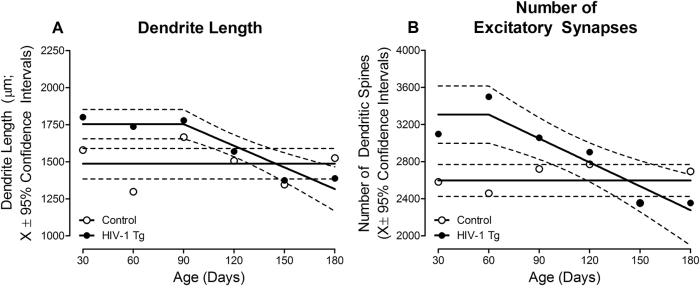
j_nipt-2023-0008_fig_002

HIV-1 Tg animals exhibited an initial increase in dendrite length (A) and the number of dendritic spines (B) early in development; parameters which subsequently decreased across time. In sharp contrast, dendrite length and the number of dendritic spines were stable across development in control animals.

Targeting these alterations with the selective ERβ agonist SE during the formative period induces long-term modifications to synaptodendritic structure, whereby MSNs in the NAcc in HIV-1 Tg animals treated with SE resemble control animals at PD 180.”

https://www.degruyter.com/document/doi/10.1515/nipt-2023-0008/html “Constitutive expression of HIV-1 viral proteins induces progressive synaptodendritic alterations in medium spiny neurons: implications for substance use disorders”


A rodent brain cell study investigated soy isoflavones’ effects on a different estrogen receptor:

“We evaluated effects of isoflavones using mouse primary cerebellar culture, astrocyte-enriched culture, Neuro-2A clonal cells, and co-culture with neurons and astrocytes. Soybean isoflavone-augmented estradiol mediated dendrite arborization in Purkinje cells.

These results indicate that ERα plays an essential role in isoflavone-induced neuritogenesis. However, G-protein-coupled ER (GPER1) signaling is also necessary for astrocyte proliferation and astrocyte–neuron communication, which may lead to isoflavone-induced neuritogenesis.

We highlight the novel possibility that isoflavones enhance dendritogenesis and neuritogenesis, indicating that they can be a useful supplementary compound during brain development or in the injured brain.”

https://www.mdpi.com/1422-0067/24/10/9011 “Isoflavones Mediate Dendritogenesis Mainly through Estrogen Receptor α”

Adverse Childhood Experiences, Part 2

A request was made to present studies that investigated epigenetic impacts of corporal punishments or physical trauma to children or adolescents. Here’s a follow-on of the 2015 Grokking an Adverse Childhood Experiences (ACE) score, since physical abuse is one factor of an ACE score.

1. The largest problem is that a person filling out an ACE questionnaire or Childhood Trauma Questionnaire can’t provide first-hand answers of their own experiences during womb life, infancy, and early childhood. These critical development periods are more impacted by adversity than are later life windows.

Human brains aren’t developed enough before age 3 to provide retrospective answers using cerebral memories. A self-reported ACE score can’t possibly address what happened during the times when we were most vulnerable to disrupted neurodevelopment. And good luck with parents providing factual histories of whether they physically or emotionally neglected, physically or emotionally abused, or otherwise adversely treated their fetus, infant, and young child.

2. Another problem is researchers can pretty much choose whatever questions they want as input criteria. I’ve seen pliable ACE scores developed from 5- to 25-item questionnaires.

Do these questionnaires cover all relevant adverse childhood experiences? For example, are researchers permitted to use as inputs societal-created adversities a child may have lived through such as the Khmer Rouge or Cultural Revolution? Studies are just starting to investigate adverse childhood experiences created by worldwide abuses of authority since 2020.

3. Other problems were discussed in a 2023 paper https://www.sciencedirect.com/science/article/abs/pii/S0145213423003162 “Adverse childhood experiences and adult outcomes using a causal framework perspective: Challenges and opportunities” (not freely available), two of which were:

  • Adding up ACE factors to a cumulative score ignores the impact of synergistic sets. For example, although both cumulative ACE scores are 2, a child who was physically and sexually abused would probably be more adversely affected than a child whose parents divorced or separated, and also had a family member incarcerated.
  • At any given time point, and especially with older people, there’s a potential selection bias against those most affected by adverse childhood experiences, such as those who died.

Using flawed, squishy, cumulative ACE scores as inputs, here are two 2023 studies that found epigenetic associations:

“We tested the following pre-registered hypotheses: Mothers’ adverse childhood experiences are correlated with DNA methylation (DNAm) in peripheral blood during pregnancy (hypothesis 1) and in cord blood samples from newborn infants (hypothesis 2), and women’s depression and anxiety symptoms during pregnancy mediate the association between mothers’ ACE exposure and prenatal/neonatal DNA methylation (hypothesis 3).

  1. Hypothesis 1: In 896 mother−infant pairs with available methylation and ACE exposure data, there were no significant associations between mothers’ ACE score and DNAm from antenatal peripheral blood, after controlling for covariates.
  2. Hypothesis 2: In infant cord blood, there were 5 CpG sites significantly differentially methylated in relation to mothers’ ACEs (false discovery rate < .05), but only in male offspring. Effect sizes were medium. CpG sites were in genes related to mitochondrial function and neuronal development in the cerebellum.
  3. Hypothesis 3: There was no mediation by maternal anxiety/depression symptoms found between mothers’ ACEs score and DNAm in the significant CpG sites in male cord blood.”

https://www.jaacap.org/article/S0890-8567(23)00313-1/fulltext “Epigenetic Intergenerational Transmission: Mothers’ Adverse Childhood Experiences and DNA Methylation”


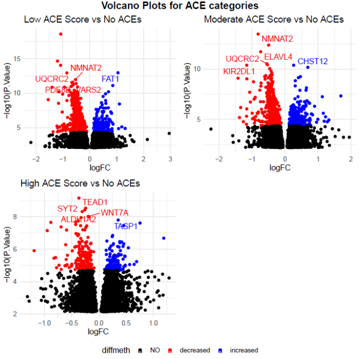
“In this study, the effect of cumulative ACEs experienced on human maternal DNAm was estimated while accounting for interaction with domains of ACEs in prenatal peripheral blood mononuclear cell samples. Intergenerational transmission of ACE-associated DNAm was explored used paired maternal and neonatal cord blood samples. Replication in buccal samples was also explored.

We used a four-level categorical indicator variable for ACEs exposure: none (0 ACEs), low (1–3 ACEs), moderate (4–6 ACEs), and high (> 6 ACEs). 🙄

125a4c3cacfe4b922e5b864c

https://www.researchsquare.com/article/rs-2977515/v1 “Effect of Parental Adverse Childhood Experiences on Intergenerational DNA Methylation Signatures”

Transgenerational transmission of stress

This 2023 rodent study found that effects of stress during mid-late gestation were epigenetically transmitted to the first, second, and third female generations:

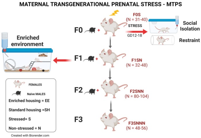
“We investigated effects of gestational chronic variable stress (CVS) in rats using restraint and social isolation stress in the parental F0 generation. Only the F0 pregnant dams were subjected to stress.

When a pregnant female experiences adversity, impacts of that stress affect exposed somatic tissues (F0 generation), the fetuses (F1 generation), and the fetuses’ germline (F2 generation). A true transgenerational inheritance arises when germline epimutations are transmitted to unexposed F3 offspring.

A subset of F1 rats was housed in an enriched environment (EE) to mitigate adverse effects of CVS. F2 offspring reared in EE had increased birth weights, but their uterine gene expression patterns remained comparable to those of stressed animals.

ijms-24-03734-g001

We provide evidence that psychological and psychosocial CVS alters inflammatory status and endocrine markers in uteri of adult dams through transgenerational programming of the female germline. EE therapy in prenatally stressed F1 offspring had no beneficial effects on uterine expression of inflammatory and endocrine markers for them or their future offspring.”

https://www.mdpi.com/1422-0067/24/4/3734 “Environmental Enrichment Promotes Transgenerational Programming of Uterine Inflammatory and Stress Markers Comparable to Gestational Chronic Variable Stress”


PXL_20230611_100728709

Amphibian epigenetic clocks

This 2023 study of two frog species expanded one of the cited studies in Epigenetic clocks so far in 2022 to include post-embryonic epigenetic clock measurements:

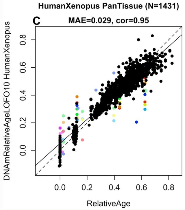
“We generated DNA methylation data from African clawed frogs (Xenopus laevis) and Western clawed frogs (Xenopus tropicalis) and built multiple epigenetic clocks. Dual species clocks were developed that apply to both humans and frogs (human-clawed frog clocks), supporting that epigenetic aging processes are evolutionary conserved outside mammals.

The two species underlying our Xenopus clocks have markedly different maximum lifespans (30.3 for X. laevis and 16 for X. tropicalis), and average ages of sexual maturity (1 year for laevis and 0.375 for tropicalis). When building our Xenopus clocks, we addressed this fact in two ways:

  • In our pan-clock, we used a log-linear transformation of age that effectively normalizes ages with respect to age at sexual maturity.
  • In our relative pan-clock, we instead estimate relative age (chronological age divided by maximum lifespan), which normalizes ages with respect to maximum lifespan.

We also created dual-species clocks, referred to as human-clawed frog clocks, for estimates of chronological age and relative age. Relative age is the ratio of chronological age to maximum lifespan, and takes on values between 0 and 1. Maximum lifespan observed for humans was 122.5 years.

The relative age clock allows for alignment and biologically meaningful comparison between species with different lifespans.

relative age

Previous studies in humans showed that a hallmark of age-related CpGs is their association with target sites of Polycomb repressive complex 2 (PRC2), which gain methylation with age. This feature is fully recapitulated in Xenopus, and physiological significance of this association is an important open question.

PRC2 plays a prominent role during embryonic development and consequently, many aging-clock-associated genes relate to developmental processes. Given its evolutionary conservation from frogs to humans, methylation status of PRC2 targets supports some critical causal relationship to systemic aging.

Since the association with PRC2 with aging stems from analyses of adult postmitotic cells, and of different tissue origin rather than from embryonic cells, it is tempting to speculate that adult methylation status will get important input during embryonic development, the very phase when PRC2 target gene expression is prominent.

Genes associated with both positive and negative age-related CpGs relate to neural processes, although in somewhat opposite direction. While DNAm increase is linked to neural developmental genes, DNAm decrease links to synaptic transmission, roughly corresponding to processes of immature vs. mature neuronal cells, respectively. This leads to the counter-intuitive suggestion that studying Xenopus neural development may yield new insights into biological aging.”

https://link.springer.com/article/10.1007/s11357-023-00840-3 “DNA methylation clocks for clawed frogs reveal evolutionary conservation of epigenetic aging”


I’ve seen dual-species epigenetic clocks – introduced in A rejuvenation therapy and sulforaphane – referenced elsewhere, most recently in Selective Breeding for High Intrinsic Exercise Capacity Slows Pan-Tissue Epigenetic Aging in Rats. These clocks still aren’t in wide use by researchers, though. Don’t know what it will take to persuade researchers to use dual-species relative age clocks in their model organism studies so that they can justifiably invoke human applicability.

PXL_20230601_182718629

Nrf2 Week #2: Neurons

To follow the Nrf2 Week #1 suggestion that Nrf2 target neurological disorders, this 2023 cell study investigated Nrf2 expression in neurons:

“Oxidative metabolism is inextricably linked to production of reactive oxygen species (ROS), which have the potential to damage all classes of macromolecules. Yet ROS are not invariably detrimental. Several properties make ROS useful signaling molecules, including their potential for rapid modification of proteins and close ties to cellular metabolism.

We used multiple single cell genomic datasets to explore Nrf2 expression and regulation in hundreds of neuronal and non-neuronal cell types in mouse and human. With few exceptions, Nrf2 is expressed at far lower levels in neurons than in non-neuronal support cells in both species.

This pattern is maintained in multiple disease states, and the chromatin accessibility landscape at the Nrf2 locus parallels these expression differences. These results imply that Nrf2 activity is limited in almost all neurons of the mouse and human central nervous system (CNS).

nrf2 expression

We separated cell types into neuron or non-neuronal ‘support’ cell categories. The general ‘support’ term is not meant to minimize the functional relevance of non-neuronal cells in the CNS, but is an umbrella term meant to cover everything from glial cell types (astrocytes, microglia, oligodendrocytes) to endothelial cells.

It is not clear why an important, near ubiquitous cytoprotective transcription factor like Nrf2 remains off in mature neurons, especially considering oxidative stress is a driver of many diseases. The simplest explanation is that Nrf2 activity also disrupts normal function of mature neurons.

ROS play a key role in controlling synaptic plasticity in mature neurons. These activity-dependent changes in synaptic transmission, which are important for learning and memory, are disrupted by antioxidants.

A subset of important Nrf2-targeted antioxidant genes (e.g., Slc3a2, Slc7a11, Nqo1, Prdx1) are also low in neurons. So it is likely that these and/or other Nrf2 targets must remain low or non-ROS-responsive in mature neurons. Future work exploring why this expression pattern persists in mature neurons will inform our models on roles of antioxidant genes in normal neuronal physiology and in neurological disorders.

https://www.biorxiv.org/content/10.1101/2023.05.09.540014v1.full “Limited Expression of Nrf2 in Neurons Across the Central Nervous System”


PXL_20230520_182827767

Minds of their own

It’s the weekend, so it’s time for: Running errands? Watching sports? Other conditioned behavior?

Or maybe broadening our cognitive ability with Dr. Michael Levin’s follow-ups to his 2021 Basal cognition paper and 2020 Electroceuticals presentation with a 2022 paper and presentation starting around the 13:30 mark:

Michael Levin - Cell Intelligence in Physiological and Morphological Spaces

“A homeostatic feedback is usually thought of as a single variable such as temperature or pH. The set point has been found to be a large-scale geometry, a descriptor of a complex data structure.”


His 2022 paper Technological Approach to Mind Everywhere: An Experimentally-Grounded Framework for Understanding Diverse Bodies and Minds:

“It is proposed that the traditional problem-solving behavior we see in standard animals in 3D space is just a variant of evolutionarily more ancient capacity to solve problems in metabolic, physiological, transcriptional, and morphogenetic spaces (as one possible sequential timeline along which evolution pivoted some of the same strategies to solve problems in new spaces).

Developmental bioelectricity works alongside other modalities such as gene-regulatory networks, biomechanics, and biochemical systems. Developmental bioelectricity provides a bridge between the early problem-solving of body anatomy and the more recent complexity of behavioral sophistication via brains.

This unification of two disciplines suggests a number of hypotheses about the evolutionary path that pivoted morphogenetic control mechanisms into cognitive capacities of behavior, and sheds light on how Selves arise and expand.

While being very careful with powerful advances, it must also be kept in mind that existing balance was not achieved by optimizing happiness or any other quality commensurate with modern values. It is the result of dynamical systems properties shaped by meanderings of the evolutionary process and the harsh process of selection for survival capacity.”


PXL_20220904_102050409