Neuritogenesis

Three 2023 papers on the initial stage of neuronal differentiation, starting with a rodent study of taurine’s effects:

“We aimed to assess the role of taurine (TAU) in axonal sprouting against cerebral ischemic injury, clarify the function of mitochondria in TAU-induced axonal sprouting, and further determine the underlying potential molecular mechanism.

experiment design

We determined that TAU improved motor function recovery and restored neurogenesis in ischemic stroke. This possibly occurred via improvements in mitochondrial function.

We investigated that the Sonic hedgehog (Shh) pathway exerted an important role in these effects. Our study findings highlighted the novel viewpoint that TAU promoted axonal sprouting by improving Shh-mediated mitochondrial function in cerebral ischemic stroke.”

https://www.scielo.br/j/acb/a/nxKvGXGk9g6gRkHxybMfbYJ/?lang=en “Taurine promotes axonal sprouting via Shh-mediated mitochondrial improvement in stroke”


A rodent study investigated effects of a soy isoflavone gut microbiota metabolite:

“Perinatally-infected adolescents living with HIV-1 (pALHIV) appear uniquely vulnerable to developing substance use disorders (SUD). Medium spiny neurons (MSNs) in the nucleus accumbens core (NAcc), an integrator of cortical and thalamic input, have been implicated as a key structural locus for the pathogenesis of SUD.

Treatment with estrogenic compounds (e.g., 17β-estradiol) induces prominent alterations to neuronal and dendritic spine structure in the NAcc supporting an innovative means to remodel neuronal circuitry. The carcinogenic nature of 17β-estradiol, however, limits its translational utility.

Plant-derived polycyclic phenols, or phytoestrogens, whose chemical structure resembles 17β-estradiol may afford an alternative strategy to target estrogen receptors. The phytoestrogen S-Equol (SE), permeates the blood-brain barrier, exhibits selective affinity for estrogen receptor β (ERβ), and serves as a neuroprotective and/or neurorestorative therapeutic for HIV-1-associated neurocognitive and affective alterations.

Beginning at approximately postnatal day (PD) 28, HIV-1 transgenic (Tg) animals were treated with a daily oral dose of 0.2 mg of SE. The SE dose of 0.2 mg was selected for two primary reasons, including:

  1. A dose-response experimental paradigm established 0.2 mg of SE as the most effective dose for mitigating neurocognitive deficits in sustained attention in the HIV-1 Tg rat; and
  2. The dose, which yielded a daily amount of 0.25–1.0 mg/kg/SE (i.e., approximately 2.5–10 mg in a 60 kg human), is translationally relevant (i.e., well below the daily isoflavone intake of most elderly Japanese.

Daily oral treatment continued through PD 90.

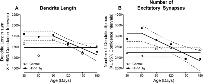
j_nipt-2023-0008_fig_002

HIV-1 Tg animals exhibited an initial increase in dendrite length (A) and the number of dendritic spines (B) early in development; parameters which subsequently decreased across time. In sharp contrast, dendrite length and the number of dendritic spines were stable across development in control animals.

Targeting these alterations with the selective ERβ agonist SE during the formative period induces long-term modifications to synaptodendritic structure, whereby MSNs in the NAcc in HIV-1 Tg animals treated with SE resemble control animals at PD 180.”

https://www.degruyter.com/document/doi/10.1515/nipt-2023-0008/html “Constitutive expression of HIV-1 viral proteins induces progressive synaptodendritic alterations in medium spiny neurons: implications for substance use disorders”


A rodent brain cell study investigated soy isoflavones’ effects on a different estrogen receptor:

“We evaluated effects of isoflavones using mouse primary cerebellar culture, astrocyte-enriched culture, Neuro-2A clonal cells, and co-culture with neurons and astrocytes. Soybean isoflavone-augmented estradiol mediated dendrite arborization in Purkinje cells.

These results indicate that ERα plays an essential role in isoflavone-induced neuritogenesis. However, G-protein-coupled ER (GPER1) signaling is also necessary for astrocyte proliferation and astrocyte–neuron communication, which may lead to isoflavone-induced neuritogenesis.

We highlight the novel possibility that isoflavones enhance dendritogenesis and neuritogenesis, indicating that they can be a useful supplementary compound during brain development or in the injured brain.”

https://www.mdpi.com/1422-0067/24/10/9011 “Isoflavones Mediate Dendritogenesis Mainly through Estrogen Receptor α”

A blood plasma aging clock, Part 2

Quite a few people recently looked at Part 1 which curated “Undulating changes in human plasma proteome across lifespan are linked to disease” in December 2019. Let’s start with a 2023 human study coauthored by Part 1’s lead researcher:

“The aim of this study is to identify a set of proteins in human plasma associated with aging by integration of data of four independent, large-scaled datasets. We identified a set of 273 plasma proteins significantly associated with aging (aging proteins, APs) across these cohorts consisting of healthy individuals and individuals with comorbidities and highlight their biological functions.

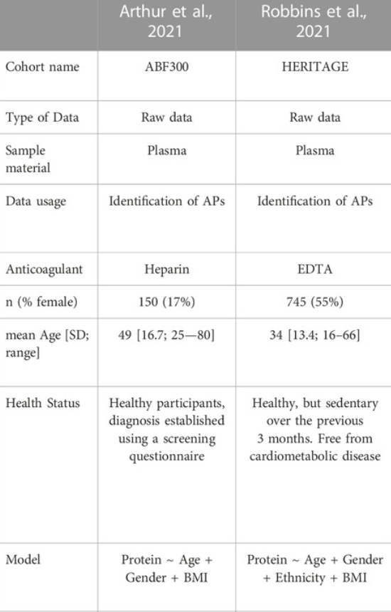
arthur and robbins cohorts

Although these presented proteins may be different compared to other presented proteomic clocks [like Part 1’s], this can be explained due to a variety of factors. Across studies there may be several technical factors, such as used anti-coagulants, and biological differences, such as different age ranges, ethnicity and corrections for BMI, which may influence the plasma proteome in the cohorts. To overcome these differences, we focused on the overlap between the different studies as they also present several of these confounding factors.

We show that individuals presenting accelerated or decelerated aging based on their plasma proteome, respectively have a more aged or younger systemic environment. These results provide novel insights in understanding the aging process and its underlying mechanisms and highlight potential modulators contributing to healthy aging.”

https://www.frontiersin.org/articles/10.3389/fragi.2023.1112109/full “Markers of aging: Unsupervised integrated analyses of the human plasma proteome”


A 2023 human study cited the above study and found:

“Our cross-sectional study of adults adherent and non-adherent to recommended lifestyle habits established strong group differences for 39 proteins primarily related to innate immunity and lipoprotein metabolism. Many of these protein differences were best explained by group contrasts in adiposity and visceral fat. The relatively small number of upregulated and downregulated proteins associated with good lifestyle habits should facilitate development of a targeted lifestyle proteomic panel that can be used in future studies to determine efficacy of various prevention and treatment strategies.”

https://www.researchsquare.com/article/rs-3097901/v1 “Adherence to Lifestyle Recommendations Linked to Innate Immunity and Lipoprotein Metabolism: A Cross-Sectional Comparison Using Untargeted Proteomics”


A 2023 human study from Google-owned Calico:

“In most cases, direction of effects between cause-specific and all-cause mortality was concordant, but all-cause mortality association was not statistically significant. Neither do we have insight into conditional causal effects of these proteins nor interaction effects between them.”

https://www.researchsquare.com/article/rs-2626017/v1 “Plasma Proteomic Determinants of Common Causes of Mortality”

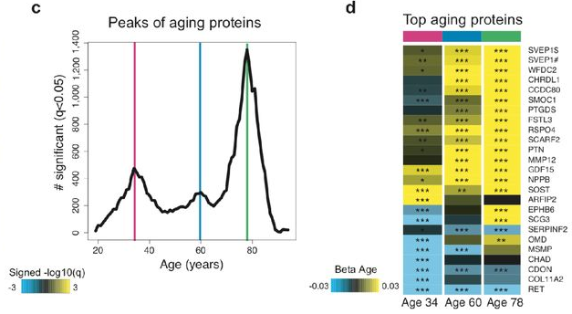
“Undulating” in Part 1 described plasma proteins changing over time with peaks at ages 34, 60, and 78. Those peaks don’t provide a base for linearly extrapolating all-cause mortality.

peaks


A 2023 rodent study did a touch better with one of Part 1’s 46 proteins of a conserved aging signature that changed in the same direction with mice and humans, although it didn’t fully investigate protein expression over time.

“Interactions between CHRDL1 levels, age, and plasma lipids that might affect cardiometabolic health should be further investigated.”

https://www.mdpi.com/2073-4409/12/4/624 “Chordin-like 1, a Novel Adipokine, Markedly Promotes Adipogenesis and Lipid Accumulation”

Sulforaphane, TFEB, and ADH1

Looked for a recent follow-on study of the 2021 Precondition your defenses with broccoli sprouts, specifically:

“NFE2L2/NRF2 is a target gene of TFEB (transcription factor EB), a master regulator of autophagic and lysosomal functions, which we show here to be potently activated by sulforaphane.”

Some interesting papers cited it, but no studies continued its sulforaphane/TFEB line of inquiry. A 2022 review made a good point when citing this study for TFEB, but didn’t tie it in with sulforaphane:

“TFEB is translocated into the nucleus with a moderate increase of ROS through a Ca2+-dependent, but mTOR (mechanistic target of rapamycin kinase)-independent mechanism. Essential genes involved in lysosome biogenesis and autophagosome are activated, which are crucial for removal of damaged mitochondria.

https://www.ncbi.nlm.nih.gov/pmc/articles/PMC9730074/ “Phytochemicals and modulation of exercise-induced oxidative stress: a novel overview of antioxidants”


A search of TFEB brought up a 2023 nematode study:

“We searched for effectors acting downstream of the transcription factor EB (TFEB), known as HLH-30 in C. elegans, because TFEB/HLH-30 is necessary across anti-aging interventions. Its overexpression is sufficient to extend C. elegans lifespan, and reduce biomarkers of aging in mammals including humans.

While investigating the potential role of autophagy in hlh-30 dependent longevity of the mxl-3 C. elegans mutant, we found that the current model has exceptions. Contrary to expectation, we found that autophagy is not activated in the mxl-3 mutant, and that neither autophagy nor lysosomal activity are required for the longevity phenotype observed in these mutant animals. mxl-3 longevity is hlh-30-dependent but autophagy-independent.

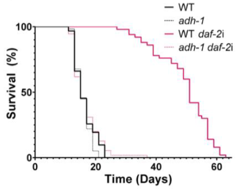
Instead, we found the gene encoding Alcohol DeHydrogenase ADH-1 induced in mxl-3 and other hlh-30-dependent anti-aging interventions. adh-1 is induced in an hlh-30-dependent manner in longevity models caloric restriction (eat-2), insulin insensitivity (daf-2), and mTOR inhibition (let-363 RNAi).

insulin insensitivity longevity model

We present an alcohol-dehydrogenase-mediated anti-aging response (AMAR) that is essential for C. elegans longevity driven by HLH-30 overexpression, caloric restriction, mTOR inhibition, and insulin-signaling deficiency. Overexpression of ADH-1 is sufficient to activate AMAR, which extends healthspan and lifespan by reducing levels of glycerol, an age-associated and aging-promoting alcohol.”

https://www.cell.com/current-biology/fulltext/S0960-9822(23)00128-8 “Increased alcohol dehydrogenase 1 activity promotes longevity” (not freely available) Thanks to Dr. Eyleen O’Rourke for providing a copy.


A 2022 human study found that chronic ADH1 activation occurs in liver disease:

“Activity of total ADH, ADH isoenzymes and aldehyde dehydrogenase (ALDH) was evaluated in the blood serum of patients with primary biliary cholangitis (PBC), a chronic autoimmune disease of the liver. An increase in class I ADH and total ADH activity indicates that the isoenzyme class I ADH is released by compromised liver cells and can be useful diagnostic markers of PBC.”

https://link.springer.com/article/10.1007/s00005-022-00667-4 “An Assessment of the Serum Activity of ADH and ALDH in Patients with Primary Biliary Cholangitis”

Chronically activating any of the body’s systems points to a problem. There’s has to be a balance.


A 2022 rodent study investigated ADH1 activation and MEK1/2 inhibitors for beneficial effects:

“Alcohol is mainly catabolized by class I alcohol dehydrogenase (ADH1) in liver. ADH deficiency can aggravate ethanol-induced tissue injury.

Extracellular signal-regulated kinases 1/2 (ERK1/2) is involved in alcohol metabolism. However, the relationship between ERK1/2 and ADH1 remains unclear.

Mitogen-activated protein kinases 1/2 (MEK1/2) is required to phosphorylate and activate ERK1/2. Protein expression of phosphorylated ERK1/2 in liver is inversely associated with ethanol-induced liver injury and hepatocytes apoptosis, suggesting inhibition of ERK1/2 may protect hepatocytes from ethanol-induced cytotoxicity. We hypothesize that inhibition of ERK1/2 by MEK1/2 inhibitors may protect hepatocytes from ethanol cytotoxicity by activating ADH1 expression.

Results showed MEK1/2 inhibitors significantly increased ADH1 protein expression by inducing its transcription activity. Our findings revealed inhibition of ERK1/2 can significantly increase ADH1 expression, indicating MEK1/2 inhibitors may possess potential application in alcohol-related diseases.”

https://link.springer.com/article/10.1007/s11033-022-07361-w “MEK1/2 inhibitors induce class I alcohol dehydrogenase (ADH1) expression by regulating farnesoid X receptor in hepatic cell lines and C57BL/6J mouse” (not freely available)

Chronically inhibiting any of the body’s systems also points to a problem.


A 2022 rodent study investigated TFEB activation and MEK1/2 inhibitors for beneficial effects:

“Inhibiting MEK/ERK signaling using a clinically available MEK1/2 inhibitor induces protection of neurons through autophagic lysosomal activation mediated by transcription factor EB (TFEB) in a model of AD.”

https://www.nature.com/articles/s41380-022-01713-5 “MEK1/2 inhibition rescues neurodegeneration by TFEB-mediated activation of autophagic lysosomal function in a model of Alzheimer’s Disease”


PXL_20230720_102047833

Adverse Childhood Experiences, Part 2

A request was made to present studies that investigated epigenetic impacts of corporal punishments or physical trauma to children or adolescents. Here’s a follow-on of the 2015 Grokking an Adverse Childhood Experiences (ACE) score, since physical abuse is one factor of an ACE score.

1. The largest problem is that a person filling out an ACE questionnaire or Childhood Trauma Questionnaire can’t provide first-hand answers of their own experiences during womb life, infancy, and early childhood. These critical development periods are more impacted by adversity than are later life windows.

Human brains aren’t developed enough before age 3 to provide retrospective answers using cerebral memories. A self-reported ACE score can’t possibly address what happened during the times when we were most vulnerable to disrupted neurodevelopment. And good luck with parents providing factual histories of whether they physically or emotionally neglected, physically or emotionally abused, or otherwise adversely treated their fetus, infant, and young child.

2. Another problem is researchers can pretty much choose whatever questions they want as input criteria. I’ve seen pliable ACE scores developed from 5- to 25-item questionnaires.

Do these questionnaires cover all relevant adverse childhood experiences? For example, are researchers permitted to use as inputs societal-created adversities a child may have lived through such as the Khmer Rouge or Cultural Revolution? Studies are just starting to investigate adverse childhood experiences created by worldwide abuses of authority since 2020.

3. Other problems were discussed in a 2023 paper https://www.sciencedirect.com/science/article/abs/pii/S0145213423003162 “Adverse childhood experiences and adult outcomes using a causal framework perspective: Challenges and opportunities” (not freely available), two of which were:

  • Adding up ACE factors to a cumulative score ignores the impact of synergistic sets. For example, although both cumulative ACE scores are 2, a child who was physically and sexually abused would probably be more adversely affected than a child whose parents divorced or separated, and also had a family member incarcerated.
  • At any given time point, and especially with older people, there’s a potential selection bias against those most affected by adverse childhood experiences, such as those who died.

Using flawed, squishy, cumulative ACE scores as inputs, here are two 2023 studies that found epigenetic associations:

“We tested the following pre-registered hypotheses: Mothers’ adverse childhood experiences are correlated with DNA methylation (DNAm) in peripheral blood during pregnancy (hypothesis 1) and in cord blood samples from newborn infants (hypothesis 2), and women’s depression and anxiety symptoms during pregnancy mediate the association between mothers’ ACE exposure and prenatal/neonatal DNA methylation (hypothesis 3).

  1. Hypothesis 1: In 896 mother−infant pairs with available methylation and ACE exposure data, there were no significant associations between mothers’ ACE score and DNAm from antenatal peripheral blood, after controlling for covariates.
  2. Hypothesis 2: In infant cord blood, there were 5 CpG sites significantly differentially methylated in relation to mothers’ ACEs (false discovery rate < .05), but only in male offspring. Effect sizes were medium. CpG sites were in genes related to mitochondrial function and neuronal development in the cerebellum.
  3. Hypothesis 3: There was no mediation by maternal anxiety/depression symptoms found between mothers’ ACEs score and DNAm in the significant CpG sites in male cord blood.”

https://www.jaacap.org/article/S0890-8567(23)00313-1/fulltext “Epigenetic Intergenerational Transmission: Mothers’ Adverse Childhood Experiences and DNA Methylation”


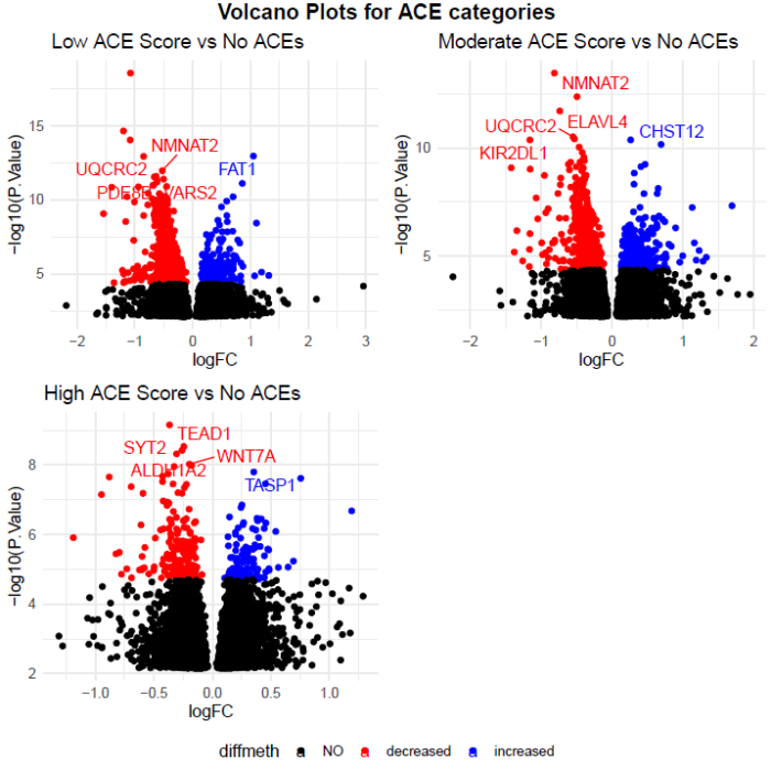
“In this study, the effect of cumulative ACEs experienced on human maternal DNAm was estimated while accounting for interaction with domains of ACEs in prenatal peripheral blood mononuclear cell samples. Intergenerational transmission of ACE-associated DNAm was explored used paired maternal and neonatal cord blood samples. Replication in buccal samples was also explored.

We used a four-level categorical indicator variable for ACEs exposure: none (0 ACEs), low (1–3 ACEs), moderate (4–6 ACEs), and high (> 6 ACEs). 🙄

125a4c3cacfe4b922e5b864c

https://www.researchsquare.com/article/rs-2977515/v1 “Effect of Parental Adverse Childhood Experiences on Intergenerational DNA Methylation Signatures”

Natural ways to modify GDF11

Three 2023 studies to follow up mention of GDF11 in the Brain endothelial cells post. Two are selected for non-pharmaceutical interventions people can do on their own. Let’s start with a human exercise study:

“We explored the exercise-related regulation of Growth Differentiation Factor 11 (GDF11) in cerebrospinal fluid (CSF) and blood. Samples of serum, plasma, and CSF were obtained before and 60 min after acute exercise (90 min run) from twenty healthy young individuals. Additional serum and plasma samples were collected immediately after run. GDF11 protein content, body composition, physical fitness, and cognitive functions were evaluated.

Controversies regarding the role of GDF11 in aging originate mainly from the absence of a reliable, validated and widely accepted method of GDF11 detection. To support the reliability of our findings as well as to distinguish GDF11 from its close homologue GDF8, we determined GDF11 in CSF, serum, and plasma, by immunoblotting, using two different GDF11-specific antibodies, as well as GDF11/GDF8 non-specific antibody. These antibodies have been previously successfully used by others. Reliability of our findings is further supported by correlations between GDF11 in serum and plasma, as well as between GDF11 and serum GDF11/GDF8.

We report an association between levels of GDF11 and adiponectin in CSF as well as in serum after acute endurance exercise. These observations support potentially synergic effects of GDF11 and adiponectin on the brain. The experimental design we implement seems to represent a reliable model to study regulation of bioactive molecules, potential mediators of neuroprotective effects of exercise in the human brain.

We show for the first time a direct link between endurance exercise and GDF11 levels in human cerebrospinal fluid. This study provided the first albeit indirect (correlative) evidence on the putative role of GDF11 in promoting healthy aging in humans, by demonstrating a tight relationship between serum GDF11 and peak power output. We extend this observation by showing that the level of physical fitness is an important determinant of regulation of GDF11 by acute exercise.

In this work, we confirm in a bigger cohort our previous finding that blood-brain barrier permeability, as assessed by CSF/serum albumin ratio, is decreased after an acute bout of endurance exercise. We observed a modest positive correlation between CSF/serum albumin ratio and CSF/serum GDF11/GDF8 ratio, with a trend also for GDF11. However, exercise-induced changes of CSF/serum albumin ratio and that of GDF11 or GDF11/GDF8 did not correlate, indicating that there are other factors that could modulate levels of this growth factor rather than blood-brain barrier permeability.”

https://www.frontiersin.org/articles/10.3389/fendo.2023.1137048/full “Acute endurance exercise modulates growth differentiation factor 11 in cerebrospinal fluid of healthy young adults”


Next is a rodent study of intermittent fasting before and after cerebral ischemia:

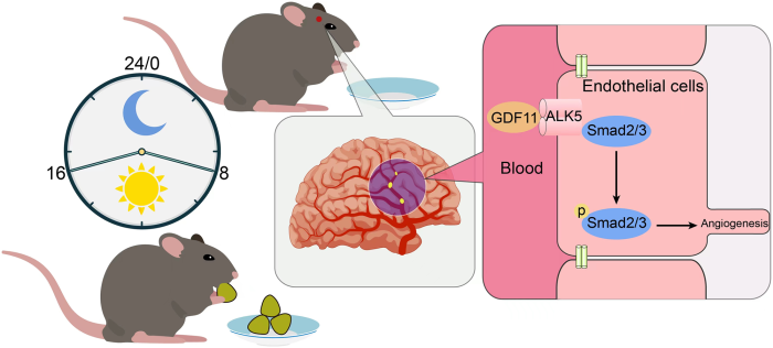
“The present study focused on the cerebral angiogenesis effect of intermittent fasting (IF) on ischemic rats. Rats were fed within strict time periods for 8 h out of every 24 h, with free access to food between 0800 and 1600 h.

In the first step, we designed different time schedules (10 d, 1 month, and 3 months) of IF before middle cerebral artery occlusion (MCAO). We monitored whether IF accelerated neurobehavioral recovery and induced expression of endothelial cells after MCAO. Then we explored whether GDF11 and downstream signals mediated angiogenesis in the peri-infarct area.

journal.pone.0282338.g006

We found that 3 months (p < 0.01) and 1 month (p < 0.05) of IF conditioning, respectively, markedly increased GDF11-positive cells in the peri-infarct area 3 d after MCAO compared with ad libitum dietary regimen. There were no significant differences between the cerebral ischemia (CI) + ad libitum group and the CI + IF 10-day group.

We also assayed plasma expression pattern of GDF11 protein. Plasma level of GDF11 protein was significantly upregulated in the IF dietary groups compared with the ad libitum dietary group 3 d after MCAO, which was consistent with the brain level. However, short-term CI + IF 10-day group results were not statistically different from CI + ad libitum group.

Taken together, our results strongly indicated that pretreatment of long-term IF might promote circulation of GDF11 and cerebral GDF11 protein during the post-ischemic, recovery period. Preoperative long-term IF might be beneficial for inducing cerebral angiogenesis in acute cerebral infarction.

These findings suggested that the longer the period of IF before MCAO, the better the protective effects after surgery.”

https://journals.plos.org/plosone/article?id=10.1371/journal.pone.0282338 “Long-term intermittent fasting improves neurological function by promoting angiogenesis after cerebral ischemia via growth differentiation factor 11 signaling activation”

Per Week 28 of Changing to a youthful phenotype with broccoli sprouts, using species maximum lifespan to estimate a human-equivalent multiplication factor that can be applied to a rat post-development time period is 122.5 years / 3.8 years = 32.2. Applying it to this study’s findings:

  • 10 rat days (322 human days) of intermittent fasting provided little protection from cerebral ischemia;
  • 1 rat month (32.2 human months) of intermittent fasting had better protection; and
  • 3 rat months (a little over 8 human years) of intermittent fasting had even stronger protection.

Is it worth the hassle of intermittently fasting every day for years to prevent a future stroke, or better recover from one, or keep other subclinical / clinical diseases from accelerating, or keep aging from accelerating? This study also cited more immediate benefits of intermittent fasting.


Might be too late for a gradual approach for people who are already diseased or close, though, like subjects in this human study:

“We aimed to explore the correlation among serum GDF11, the severity of coronary artery lesions, and the prognosis of patients with ST-segment elevation myocardial infarction (STEMI). A total of 367 patients were enrolled and divided into control (n = 172) and STEMI (n = 195) groups. Control group fulfilled the following criteria:

  1. Presented with typical chest tightness, chest pain, or other discomfort symptoms on admission;
  2. Electrocardiogram examination suggested ST-T changes;
  3. Levels of myocardial injury markers did not suggest abnormalities; and
  4. The diagnosis of unstable angina was considered clinically valid.

14 variables that were significant in univariate logistic regression analysis were included in the subsequent multivariate logistic regression analysis. Multivariate analysis indicated that smoking, diabetes, C-reactive protein, homocysteine, and lipoprotein (a) were positively correlated with STEMI occurrence, whereas serum GDF11 and the Apolipoprotein A1-to-Apolipoprotein B ratio were negatively correlated with STEMI occurrence.

Serum GDF11 was negatively correlated with severity of coronary lesions, and was also an independent prognostic indicator of major adverse cardiovascular events in patients with STEMI.”

https://link.springer.com/article/10.1007/s12265-023-10358-w “Correlation Between GDF11 Serum Levels, Severity of Coronary Artery Lesions, and the Prognosis of Patients with ST-segment Elevation Myocardial Infarction” (not freely available)

Brain endothelial cells

Six 2023 papers on the subject, starting with a rodent study:

“One of the primary discoveries of our study is that the endothelial cell (EC) transcriptome is dynamically regulated by both aging and heterochronic parabiosis. We found that ECs, when compared with other brain cell types, exhibited one of the highest fractions of aging-related genes that were rescued after heterochronic parabiosis in the old brain, and similarly, the highest fraction of aging-related genes that were disrupted after heterochronic parabiosis in the young brain. This finding supports our previous research that vasculature is strongly affected by aging and disease, and is capable of regrowth after heterochronic parabiosis or systemic GDF11 treatment.

parabiosis

We observed that a subset of ECs was classified as mitogenic. It is reasonable to speculate that the growth of these cells, which is probably prevented or suspended by the inflammatory environment of the aged brain, may be among the cell populations that respond to these interventions.

Although proteostasis in brain ECs has not been thoroughly investigated, they are apparently long-lived cells and, like neurons, might therefore accumulate protein aggregates with age, potentially compromising their function. ECs become senescent with age, but parabiosis may reverse that phenotype as well.

These findings underline the strong susceptibility and malleability of ECs, which are directly exposed to secreted factors in both brain parenchyma and blood, to adapt to changes in their microenvironment. ECs, despite comprising <5% of the total number of brain cells, are a promising and accessible target for treatment of aging and its associated diseases.”

https://www.nature.com/articles/s43587-023-00373-6 “Heterochronic parabiosis reprograms the mouse brain transcriptome by shifting aging signatures in multiple cell types”


A review elaborated on endothelial cell senescence:

“ECs form highly dynamic and differentiated monolayers arranged in a vascular network. Within brain tissue, the ECs of arteries, capillaries, and veins present different molecular characteristics. The main functions of ECs as a major cellular component of the blood-brain barrier (BBB) are to express cell membrane transport proteins, produce inflammatory mediators, deliver nutrients to brain tissue, and prevent drugs and toxins from entering the central nervous system.

ECs are the first echelons of cells affected at the onset of senescence due to their special structural position in the vascular network. Senescent ECs produce reactive oxygen species (ROS), which directly inhibit smooth muscle potassium channels and cause vasoconstriction.

The vascular endothelium is in a constant process of damage and repair, and once damage occurs, ECs replenish themselves to remove damaged cells. EC senescence makes the endothelium less capable of self-repair. With the decline in endothelial function, excess accumulated senescent cells express senescence-associated secretory phenotypes (SASPs), which result in senescence of adjacent cells, and eventually degeneration of vascular function.”

https://www.aginganddisease.org/EN/10.14336/AD.2023.0226-1 “Endothelial Senescence in Neurological Diseases”


A human study investigated above-mentioned differences in brain endothelial cells:

“We performed single nucleus RNAseq on tissue from 32 Alzheimer’s Disease (AD) and non-AD donors each with five cortical regions: entorhinal cortex, inferior temporal gyrus, prefrontal cortex, visual association cortex, and primary visual cortex. Analysis of 51,586 endothelial cells revealed unique gene expression patterns across the five regions in non-AD donors.

Visual cortex areas, which are affected late in AD progression and experience less neurodegeneration, expressed more genes related to vasculogenesis and angiogenesis. Highly vulnerable areas such as the entorhinal cortex expressed more oxidative stress-related genes in normal aged brain, suggesting endothelial dysfunction in this region even in the absence of severe AD pathology.

The present work shows that senescence-related gene signatures are increased across several brain regions, and confirms these changes in endothelial cells in the absence of other vascular cell types. While endothelial cells are not typically associated with protein aggregation, upregulated protein folding pathways suggest that proteostatic stress is a key pathway in this cell type.”

https://www.biorxiv.org/content/10.1101/2023.02.16.528825v1.full “Endothelial Cells are Heterogeneous in Different Brain Regions and are Dramatically Altered in Alzheimer’s Disease”


A human cell study abstract on above-mentioned blood-brain barrier endothelial cells:

“The BBB is a semi-permeable and protective barrier of the brain, primarily composed of endothelial cells interconnected by tight junction proteins, that regulates movement of ions and molecules between blood and neural matter. In pathological conditions such as traumatic brain injury (TBI), disruption of the BBB contributes to leakage of solutes and fluids into brain parenchyma, resulting in onset of cerebral edema and elevation of intracranial pressure.

The objective of this study was to determine upstream regulators of NLRP3 signaling and BBB hyperpermeability, particularly to determine if extracellular adenosine triphosphate (ATP) via P2X7R, a purinergic receptor, promotes NLRP3 inflammasome activation. Extracellular ATP is a major contributor of secondary injuries following TBI.

Our results suggest that extracellular ATP promotes NLRP3 inflammasome activation. Subsequent caspase-1 and MMP-9-mediated tight junction disorganization are major pathways that lead to BBB dysfunction and hyperpermeability following conditions such as TBI.”

https://journals.physiology.org/doi/abs/10.1152/physiol.2023.38.S1.5732827 “Regulation of Blood-Brain Barrier Endothelial Cell Hyperpermeability by NLRP3 Inflammasome Inhibition”


A human study further investigated effects of traumatic brain injury on brain endothelial cells:

“We previously demonstrated that extracellular vesicles (EVs) released from injured brains led to endothelial barrier disruption and vascular leakage. Here, we enriched plasma EVs from TBI patients (TEVs), detected high mobility group box 1 (HMGB1) exposure to 50.33 ± 10.17% of TEVs, and found the number of HMGB1+TEVs correlated with injury severity. We then investigated for the first time the impact of TEVs on endothelial function using adoptive transfer models.

HMGB1 is secreted by activated cells or passively released by necrotic or injured cells. After post-translational modifications, it interacts with receptors such as toll-like receptors (TLRs; e.g., TLRs 2, 4, and 9) and the receptor for advanced glycation end products (RAGE) to trigger multiple signaling pathways and mediate inflammatory and immune responses. Extracellular HMGB1 promotes endothelial dysfunction, leukocyte activation and recruitment, as well as thrombosis.

These results suggest that circulating EVs isolated from patients with TBI alone are sufficient to induce endothelial dysfunction. They contribute to secondary brain injury that are dependent on immunologically active HMGB1 exposed on their surface. This finding provided new insight for development of potential therapeutic targets and diagnostic biomarkers for TBI.”

https://www.sciencedirect.com/science/article/pii/S1043661823001470 “Circulating extracellular vesicles from patients with traumatic brain injury induce cerebrovascular endothelial dysfunction”


To wrap up, eat mushrooms to protect your brain endothelial cells!

“Natural compound ergothioneine (ET), which is synthesised by certain fungi and bacteria, has considerable cytoprotective potential. We previously demonstrated anti-inflammatory effects of ET on 7-ketocholesterol (7KC)-induced endothelial injury in human blood-brain barrier endothelial cells (hCMEC/D3). 7KC is an oxidised form of cholesterol present in atheromatous plaques and sera of patients with hypercholesterolaemia and diabetes mellitus. The aim of this study was to elucidate the protective effect of ET on 7KC-induced mitochondrial damage.

Protective effects of ET were diminished when endothelial cells were coincubated with verapamil hydrochloride (VHCL), a nonspecific inhibitor of the ET transporter OCTN1 (SLC22A4). This outcome demonstrates that ET-mediated protection against 7KC-induced mitochondrial damage occurred intracellularly and not through direct interaction with 7KC.

OCTN1 mRNA expression itself was significantly increased in endothelial cells after 7KC treatment, consistent with the notion that stress and injury may increase ET uptake. Our results indicate that ET can protect against 7KC-induced mitochondrial injury in brain endothelial cells.”

https://www.mdpi.com/1422-0067/24/6/5498 “Protective Effect of Ergothioneine against 7-Ketocholesterol-Induced Mitochondrial Damage in hCMEC/D3 Human Brain Endothelial Cells”

A flawed broccoli sprouts clinical trial

This 2023 human study investigated commercially available broccoli sprouts’ effects on platelets. I’ll provide details of some procedures, but not of findings, as there were several issues:

“Administration of intervention (sulforaphane/placebo) was followed in 90 min by administration of standardized caloric challenge PhenFlex. Urine samples were classified into three groups: (A) baseline, green line, (B) after intervention or placebo, blue lines, and (C) after PhenFlex challenge, red lines. Samples were divided into 5 timepoints: (0) baseline, (1) <60 min after intervention or placebo, (2) >60 min after intervention or placebo, (3) <60 min after PhenFlex challenge, and (4) >60 min after PhenFlex challenge.

fnut-10-1204561-g001

Shortly (maximum of 3 min) before administration, sprouts were cut approximately 1 cm below the leaves, weighed, and mashed with a small amount of tap water (approximately 13°C) in a kitchen blender for 30s at room temperature. Subsequently, tap water was added to a total amount of 250 mL and participants were instructed to drink the entire mixture.

Commercially available pea sprouts (Affilla Cress®) were used as placebo in this study since pea sprouts do not contain glucoraphanin/sulforaphane. Affilla Cress (16 g) was prepared and administered in a similar fashion. Blinding of participants was ensured by the even appearance of both drinks and the use of nasal plugs during consumption of the investigational products. 🙂

Ninety minutes after administration of investigational products, participants were asked to drink PhenFlex, a high-fat, high-glucose, high-caloric product. PhenFlex mixtures were freshly prepared, and participants were instructed to consume the drink within 5 min.”

https://www.frontiersin.org/articles/10.3389/fnut.2023.1204561/full “The beneficial effect of sulforaphane on platelet responsiveness during caloric load: a single-intake, double-blind, placebo-controlled, crossover trial in healthy participants”


Two main issues were:

1. It was stated throughout that sulforaphane did or could do this and that. None of this was supported by sulforaphane intake measurements / estimates, although measuring equipment was available.

Researchers couldn’t assume that blending 16 grams of broccoli sprouts of unknown age creates x amount of sulforaphane. 3-day-old broccoli sprouts have the optimal yields measured 6 broccoli varieties’ sulforaphane content over 3, 5, and 7-day ages, and published 15 different answers.

Sulforaphane and two metabolites’ urinary output was measured. Supposing that only output measurements were adequate leads to the second main issue.

2. Genes were asserted for certain effects. Plausible alternate explanations such as individual differences in gut microbiota composition, excretion, and metabolism weren’t explored.

These researchers knew or should have known about the 2016 https://onlinelibrary.wiley.com/doi/abs/10.1002/mnfr.201600766 “Stabilized Sulforaphane for Clinical Use: Phytochemical Delivery Efficiency” (not freely available). That study measured two known sulforaphane inputs, and in ten people each, blood plasma and urinary outputs.

The first sulforaphane input had sulforaphane bioavailability from 19.5% to 86.9% of dose. The second input ranged from 48% to 96% of dose. Widely different responses to sulforaphane intake prompted those researchers to state:

“These differences in SF bioavailability may be due to differences in gut microbial metabolism, in the levels of drug metabolizing enzymes (e.g. well-known polymorphisms of glutathione S-transferases that catalyze the conjugation of SF with glutathione), and in excretion kinetics. Innate metabolic differences must not be discounted when assessing the metabolism of SF.”

PXL_20230705_093411243

The brain-gut-lung circuit

This 2023 rodent study investigated mechanisms of improving stress-worsened respiratory viral infection:

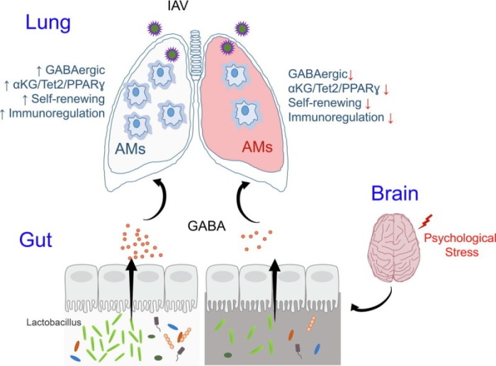
“Our study demonstrates that chronic psychological stress significantly increases host vulnerability to influenza A virus (IAV) infection characterized by a distorted gut microbiome and deregulated alveolar macrophages (AMs) response. We show that microbiome-derived γ-aminobutyric acid (GABA) functions as a tonic signal to support survival, self-renewing, and immunoregulation of AMs, and hence optimized pulmonary defensive response.

Chronic psychological stress causes gut microbiome dysbiosis and defective GABA generation, leading to loss of AMs homeostasis and aggravated viral pneumonia. The data indicate that:

  1. Microbial GABA is released in the circulation,
  2. Sensed by AMs via the GABAA receptor,
  3. Promoting cellular mitochondrial metabolism,
  4. For increased production of α-ketoglutarate (αKG),
  5. Which triggers Tet2-mediated DNA hydroxymethylation,
  6. To enable PPARγ-centered gene program,
  7. Supporting AMs homeostasis and function.

ga1_lrg

  • Re-localization of GABA-generating probiotics,
  • Supplementation of αKG, or
  • Adoptive transfer of GABA-conditioned macrophages,
  • Substantially rectifies stress-induced disruption inter-organ communication, and
  • Alleviates symptoms of viral pneumonia.

Our current study unveils an unappreciated regulatory circuitry that connects the brain, gut, and lung to mediate neurological modulation of host defensive response.”

https://www.sciencedirect.com/science/article/pii/S2090123223001716 “Gut microbial GABAergic signaling improves stress-associated innate immunity to respiratory viral infection”


consentofthegoverned

Impact of processing and storage methods on nutritional values of a dozen fruits and vegetables

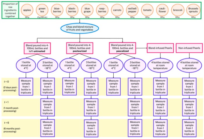
This 2023 study investigated a dozen fruits and vegetables processed with three methods and freezer times for impacts on their sixteen main nutrients. I’ll focus on sulforaphane:

“This paper compares how different processing methods (pasteurization vs. high hydrostatic pressure processing or pascalization) affect phytochemical concentrations of a complex mixture of fruits and vegetables, and investigates how these methods influence their stability during freezing and over time in frozen storage. Phytochemicals tested were vitamin C, quercetin-3-glucoside, delphinidin-3-glucoside, cyanidin-3-glucoside, peonidin-3-glucoside, catechin, epigallocatechin-3-gallate, epicatechin, epicatechin gallate, chlorogenic acid, sulforaphane, resveratrol, lycopene, lutein, alpha-carotene, and beta-carotene.

After freezing to −18 °C, one bottle from each condition was immediately removed from the freezer and thawed at 4 °C, which took about two days. Measurements at t = 0 for the fresh and frozen condition were technically made two days after processing.

antioxidants-12-01252-g001

antioxidants-12-01252-g010

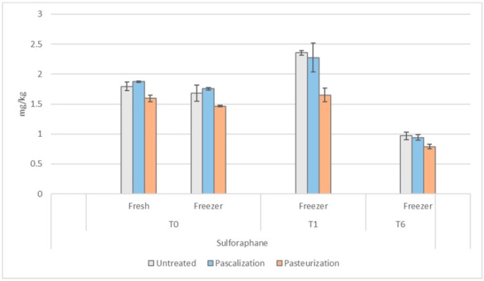
The effect of immediate freezing and thawing on broccoli, cauliflower, and Brussels sprouts sulforaphane levels was consistent despite the processing method (−6% for pascalized and untreated samples, and −8% for pasteurized) at t = 0. Pasteurized samples at t = 0 were 11% lower in sulforaphane than untreated in fresh samples and 13% lower in frozen.

At one month in the freezer, levels of sulforaphane increased in each processing method from t = 0:

  • Untreated by +18%;
  • Pascalized by +57%; and
  • Pasteurized by +94%.

At six months in the freezer, sulforaphane levels in all samples decreased below their t = 0 levels:

  • Untreated by -31%;
  • Pascalized by -35%; and
  • Pasteurized by -35%.

Optimal processing method seems to vary based on the phytochemical of interest. These impacts should be considered to produce foods aimed at preventing chronic disease development.'”

https://www.mdpi.com/2076-3921/12/6/1252 “Impact of Processing Method and Storage Time on Phytochemical Concentrations in an Antioxidant-Rich Food Mixture”


Untreated samples’ sulforaphane took a hit in this study from fresh levels to initial freezing at -18°C then thawing for two days at 4°C. Untreated levels recovered after a month to be more than their two-day levels, but lowered again after six months.

My refrigerator / freezer has one control for both compartments. Pretty sure the freezer can’t get to 0°F / -18°C without ruining refrigerator fruits and vegetables.

In any event, a (1 – .06) x 1.18 = +11% sulforaphane gain after a month isn’t worth my effort. We can increase sulforaphane more than 1100% by microwaving broccoli sprouts in a 1000W microwave on full power for 35 seconds to 60°C (140°F) per Week 6 of Changing an inflammatory phenotype with broccoli sprouts.

PXL_20230610_093652335