People will forgive you for being wrong, but they will never forgive you for being right – especially if events prove you right while proving them wrong. Thomas Sowell
“The contribution of transfer RNA (tRNA)-specific modifications to aging remains largely unexplored. We systematically profile tRNA modifications across multiple organs, species, and senescence models, and identify mannosyl-queuosine (manQ) as the first tRNA-specific modification that consistently declines with age.
Across species, queuine supplementation extends lifespan and enhances healthspan. In naturally aging mice, long-term oral administration beginning at 16-months-old (human equivalent 50 years) extends mean lifespan by 15.3%, reduces DNA methylation age, improves cognitive and motor performance, strengthens antioxidant defenses, remodels the gut microbiota, and alleviates inflammation and metabolic dysfunction without detectable toxicity.
These findings establish tRNA epitranscriptomic remodeling as a previously unrecognized layer of aging regulation, and identify restoration of manQ through queuine supplementation as a multi-system strategy to delay aging.
manQ hypomodification is selective rather than reflecting global tRNA depletion. Aging preferentially reduces the manQ-containing tRNAAsp fragment while leaving the corresponding unmodified tRNAAsp fragment, and other queuosine-modified tRNAs, relatively unchanged.
This pattern supports a regulated defect in modification homeostasis rather than a generalized change in transcript abundance. Such specificity argues that manQ loss is not merely a passive consequence of tissue degeneration, but instead represents a conserved, biologically meaningful aging-associated event with mechanistic impact.
Because proteostasis intersects with multiple canonical hallmarks (e.g. mitochondrial dysfunction, impaired stress resilience, and altered intercellular communication), translation-coupled proteome destabilization offers a unifying explanation for how a single tRNA modification defect can elicit multi-system consequences. In this view, manQ decline is not merely one of many molecular changes observed in aging, but rather a proximate determinant capable of amplifying downstream hallmarks through a common axis of proteome quality control.
Our findings further suggest that manQ depletion may engage self-reinforcing feedback loops that accelerate aging trajectories. This architecture offers a conceptual framework in which aging progressively erodes ‘epitranscriptomic integrity’ at the tRNA level, pushing translation toward an error-prone regime that accelerates proteostatic collapse and functional decline.
A distinctive implication of this work is that queuine introduces a microbiota-host epitranscriptomic axis into aging biology. Queuine is produced by gut microbiota and cannot be synthesized de novo by mammals. These findings expand the conceptual scope of geroscience by placing a microbiota-derived nutrient upstream of translational quality control.
Queuine supplementation offers a distinct therapeutic logic: rather than modulating a single signaling cascade, it restores a tRNA modification state that governs translational fidelity – an upstream determinant of proteome quality that can, in principle, influence multiple downstream hallmarks concurrently. These findings highlight an intervention paradigm centered on restoring molecular fidelity, rather than suppressing a single downstream phenotype, as a strategy to delay systemic aging.”
I switch things around pretty often, but I haven’t said much about diet and supplement changes since this time last year. Here’s what I’ve done in terms of changes that I’ve since abandoned or reduced, followed by additions or increases that I’ve kept.
Abandoned and reduced items
1. I stopped using Avena sativa oats to grow 3-day-old oats sprouts. I again ran into the same situation where I got < 10% yield.
The first time this happened in 2023, I related to the Montana farmer that degraded seed vitality was probably caused by the way that Amazon handled their oat products. I’m the customer, though, and I won’t make it my problem if the vendor can’t meet expectations.
I switched to sprouting Avena nuda oats based on Sprouting hulless oats. I’ll note that this Illinois farmer doesn’t let Amazon handle their organic Avena nuda oats, and they add on post office shipping costs. They don’t recommend sprouting, probably because of liability, although I’ve had a 91% germination rate over three days. I might have ordered Avena sativa oats directly from the Montana farmer bypassing Amazon if they were also organic.
2. I stopped taking alpha ketoglutarate. In my view, increasing tricarboxylic acid (TCA) cycle intermediate metabolites such as alpha ketoglutarate and CoQ10 should not be the primary way to improve mitochondrial electron transport chain function.
Instead of biochemical considerations, focus on photon modulation, which precedes biochemical reactions. Which means mitochondrial studies should be controlled for light exposures, and very few of them do that, although it’s the way nature works.
This past winter I increased indoor non-LED light exposure within a circadian rhythm framework. I’ll switch back to walking the beach at sunrise from being out in mid-day sun after it gets a little bit warmer.
3. I’ve taken creatine on and off during the past year. There’s a bit of literature on its use for improving methyltransferase system components like homocysteine.
Stopping creatine fits one of the overall patterns that studies demonstrate – people who are initially deficient in the studied item get a benefit, while people who are initially sufficient don’t benefit from treatment. I’ve always tested mid-range for homocysteine, which is desirable.
4. I had some cocoa powder lying around for a year or two, and I used it this past winter to improve the taste of coffee I bought on sale. Cocoa flavanols are supposed to improve various health measures. But I haven’t been provided access to the most recent human studies, so I won’t repeat their results without reading their details.
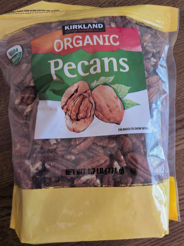
For me, though, the dryness of a chewed pecan bolus creates a swallowing problem that walnuts don’t have. YMMV.
6. I stopped taking 2 g magnesium L-threonate. I’ve always tested high for magnesium without using a specific supplement.
7. I reduced D3 by 25 mcg to a daily 2400 IU. Winter is over.
New and increased items
1. I curated five 2025 ergothioneine studies in Human studies of ergothioneine after stopping mushroom intake via AGE-less chicken soup. I wasn’t thrilled that none of them investigated long-term effects of persistent plasma ergothioneine levels.
This year I decided to start taking the higher 25 mg dose of the first study once a week. That should produce some benefits at a lower ergothioneine blood level than daily doses produce. I’ll check periodically for 2026 research.
2. The only paper I’ve curated on deuterium (heavy hydrogen) is Taurine and mitochondrial health. I started using Icelandic glacier water to make coffee and tea, and for just drinking.
It isn’t advertised as deuterium-depleted water, and it isn’t manufactured as such. But I think any glacier water contains less deuterium than local water. I use local filtered water for sprouting and cooking.
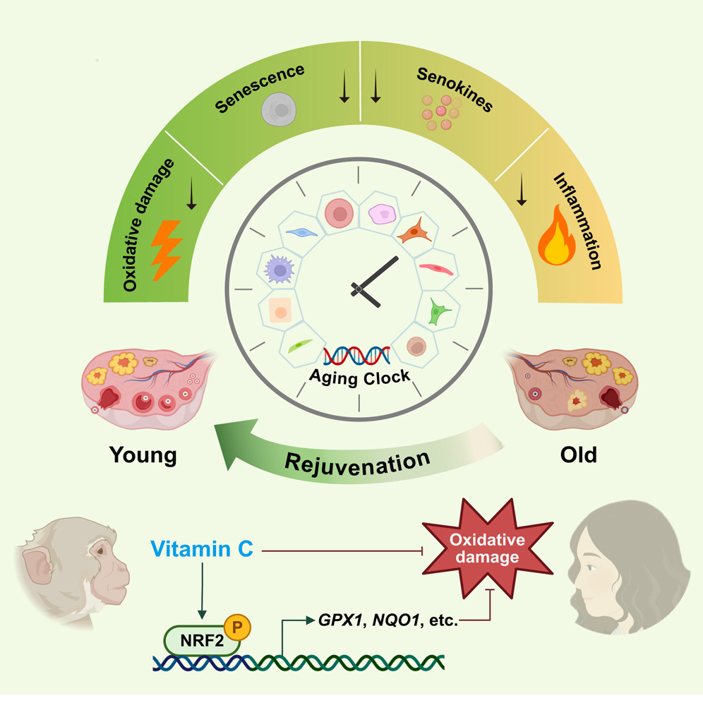
3. Per The return of the free radical theory of aging I started taking extra vitamin C separately from other supplements in the form of Now brand liposomal 1 gram twice daily this past winter. That study found vitamin C to be an anti-aging compound for primates.
“VC slowed aging in various ovarian cell types. Moreover, VC protected human ovarian endothelial and stromal cells (SCs) from aging partially via NRF2 activation. This study establishes a proof-of-concept for delaying primate ovarian aging with a single compound, and provides important insights into preventing and treating degenerative diseases related to ovarian aging.”
4. I restarted taking inulin last year, about 3 grams (a heaping teaspoon) daily after posting Inulin vs. FOS. My 2.5 year-old grandchild takes a level teaspoon daily, as inulin’s beneficial effects aren’t just for old people.
5. I started taking 12 mg astaxanthin twice in the morning. I use Nrf2 activators in the morning because Nrf2 is especially involved in the circadian cycle, as noted in papers such as Broccoli sprouts activate the AMPK pathway, Part 4.
6. I increased daily raw egg consumption from 3 eggs a day to 3 eggs twice daily.
7. This year, Ovega 3 algae oil DHA 420 mg/EPA 140 mg became no longer available AFAIK. I substituted Vegan Omega 3 algae oil DHA 300 mg/EPA 150 mg in the morning and Sports Research Omega 3 fish oil DHA 310 mg/EPA 690 mg in the afternoon.
8. I picked up this Korean seaweed in a 10-pack at Costco. The label doesn’t say what its iodine content is. I eat it as a snack whenever I get a salt craving, maybe once a week.
A 2026 rodent study investigated sulforaphane’s ability to affect ALS-like symptoms:
“The objective of this study was to evaluate neuroprotective efficacy and safety of sulforaphane (SUFP) in a methylmercury (MMHg⁺)-induced preclinical rat model of amyotrophic lateral sclerosis (ALS). ALS is characterized by progressive motor neuron degeneration and muscle wasting, leading to impairments in gait, swallowing, salivation, and routine motor activities.
64 animals were classified into eight groups: 1st: normal control, 2nd: vehicle control; 3rd: SUFP perse (4 mg/kg, i.p.), 4th: MMHg + (5 mg/kg, p.o.), 5th: MMHg + 5 + SUFP (2 mg/kg, i.p.), 6th: MMHg+ 5 + SUFP (4 mg/kg, i.p.), 7th: MMHg+ 5 + omaveloxolone (OVX) (30 mg/kg, i.p.), and 8th: MMHg + 5 + dimethyl fumarate (DIMT) (50 mg/kg, i.p.). Neurotoxin MMHg + was orally administered at 5 mg/kg for the first 21 days. For the next 22 days, SUFP, OVX, and DIMT were administered intraperitoneally (i.p.).
SUFP modulates neurotransmitter levels such as acetylcholine (A), dopamine (B), GABA (C), glutamate (D), and serotonin (E).
SUFP4 exerted broad neuroprotective effects in ALS pathology by restoring antioxidant proteins (Nrf2, HO-1, SIRT1), suppressing apoptotic (Bax, caspase-3, Bcl-2) and inflammatory markers (TNF-α, IL-1β), and enhancing the anti-inflammatory cytokine IL-10. It also downregulated stress-related signaling pathways (PI3K/Akt, p75NTRECD, MAPKs) associated with neurodegeneration. These molecular effects translated into meaningful functional recovery, as evidenced by improvements in grip strength, locomotor performance, spatial memory, and depressive-like behavior.
Histopathological evaluation demonstrated attenuation of demyelination and preservation of neuronal architecture including the cerebral cortex, hippocampus, striatum, midbrain, and cerebellum. Beyond central neuroprotection, SUFP exerted systemic benefits by normalizing hepatic enzymes, improving skeletal muscle integrity, restoring redox balance, stabilizing neurofilament and myelin-associated proteins, and correcting hematological alterations.
Despite limitations related to study duration and animal sex, this work strongly positions SUFP as a promising, multi-target therapeutic candidate for ALS with both neural and systemic protective efficacy.”
https://link.springer.com/article/10.1007/s12035-026-05683-5 “Sulforaphane-Mediated Multitarget Therapeutic Effects in Methylmercury-Induced ALS-Like Pathology: Comparative Analysis and Multifaceted Approach to Neuroprotection and Systemic Recovery” (not freely available) Thanks to Dr. Sidharth Mehan for providing a copy.
Unlike A Nrf2 treatment for ALS?, this study didn’t present evidence that its treatment compound was effective for preventing ALS. For one thing, currently-known disease factors involving heat shock proteins and associated genes, some of which are Nrf2 targets, weren’t investigated.
Two Nrf2 activators were used in both studies as comparators of Nrf2 activation effects. Neither omaveloxolone nor dimethyl fumarate are ALS causal treatments, though, and have undesirable side effects.
A human equivalent of this study’s higher sulforaphane dose is ((4 mg x .162) x 70 kg) = 45 mg. 45 mg of sulforaphane might be too much to consistently take at one time because of unpalatability. But I documented taking an estimated 52 mg for a year during 2020-2021 by eating microwaved 3-day-old broccoli sprouts twice a day.
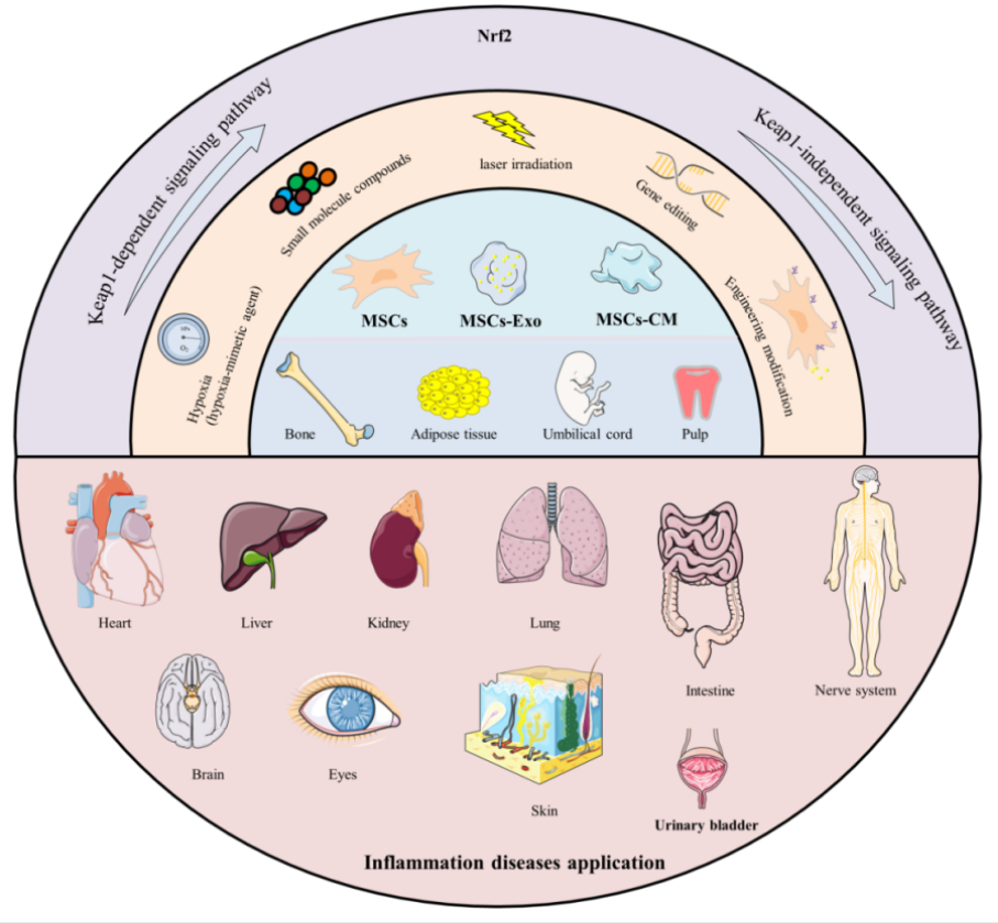
A 2026 review subject was mechanisms and therapeutic potential for Nrf2 activators in combination with mesenchymal stem cells:
“Mesenchymal stromal/stem cells (MSCs) are multipotent stem cells that can be isolated from various tissues – such as bone marrow (BM), umbilical cord (UC), adipose tissue (AD), dental pulp (DP), hair follicle (HF), and placenta – and differentiated into multiple lineages under appropriate conditions. Their functional repertoire includes immunomodulation, homing, and differentiation, which collectively help establish a balanced inflammatory and regenerative niche within damaged tissues during severe inflammation. MSCs-derived extracellular vesicles (MSCs-EVs) and conditioned medium (MSCs-CM) play remarkable roles, exhibiting potent anti-inflammatory and antioxidant properties that offer novel therapeutic alternatives for inflammatory diseases.
Therapeutic capacity of MSCs in inflammatory conditions is increasingly attributed to their potent paracrine activity rather than solely to their differentiation potential. A key mechanism underlying this paracrine effect is activation of the Nrf2 antioxidant pathway.
MSCs and their secreted products including exosomes (Exos), EVs, and CM, activate Nrf2 through multi-dimensional/target mechanisms, thereby enhancing cellular antioxidant defenses, modulating immune responses, and promoting tissue repair. It is noteworthy that therapeutic efficacy of MSCs and their derivatives can be enhanced through external modulation, including pretreatment with natural compounds.
Preconditioning refers to brief treatment of MSCs or their derivatives with physical, chemical, or biological factors prior to application, aiming to enhance their ability to counteract oxidative stress and improve their therapeutic efficacy. Flavonoids precondition and prime MSCs via the direct Keap1-Nrf2 pathway or indirect PI3K-Akt pathway, which enhances cellular resilience to adverse conditions by reducing apoptosis and promoting survival. Primed MSCs, in turn, remodel the microenvironment through an altered secretory profile, releasing bioactive factors that create more favorable conditions for their own persistence.
The core logic of these strategies lies in simulating or inducing adaptive stress, such as employing specific chemical molecules or drug stimuli, or utilizing physical / microenvironmental preconditioning to mimic specific physical conditions of the in vivo injury environment. The most straightforward strategy is overexpression of Nrf2 or its key downstream effector molecules.
The majority of existing studies remain at the level of observing correlations with Nrf2 upregulation, and there is still a lack of precise causal validation regarding key upstream signals – such as specific cytokines, miRNAs, or proteins – through which MSCs or derivatives initiate Nrf2 activation. Mechanistic insights are predominantly derived from in vivo or rodent (mouse/rat) model experiments, with a notable absence of clinical validation, insufficient long-term safety and pharmacokinetic data, and a lack of standardization in administration routes and dosages, all of which hinder clinical translation.
The essential role of the Nrf2 pathway has not been rigorously confirmed, as most studies have not employed reverse genetic validation using Nrf2-knockout animals or specific inhibitors. Consequently, it remains unclear whether therapeutic effects are necessarily and exclusively dependent on Nrf2, and potential synergistic contributions from other pathways may have been overlooked.
Most natural flavonoids face challenges such as low oral bioavailability, rapid metabolism, and poor targeting. Numerous challenges remain to be addressed in order to translate these promising preclinical findings into clinical practice. Future research should focus on the following aspects:
Elucidating precise upstream molecular mechanisms by which MSCs activate Nrf2;
Employing more clinically relevant chronic disorder models;
Systematically evaluating long-term safety, optimal delivery strategies (including dosage and route of administration), and immunogenicity of MSCs-based therapies;
Validating selection criteria (optimal source), quality control, batch-to-batch consistency of MSCs, and addressing regulatory and ethical barriers to clinical translation; and
Integrating molecular docking, ADMET (Absorption, Distribution, Metabolism, Excretion, Toxicity) prediction, and in vitro and in vivo validation to further elucidate regulatory effects of flavonoids and enhance understanding of their mechanisms of action.”
This paper was overly long at 127 pages, so I focused on the later sections. None of these treatments are currently ready for clinical trials.
I also didn’t mention specific flavonoids as Nrf2 activators. It’s beyond a reviewer’s task to rank Nrf2 activators, and a study’s researchers seldom address why they used a poorly-activating flavonoid instead of a higher-ranked natural plant compound such as sulforaphane.
A 2026 paper provided details of a 2020-2023 human trial of broccoli sprouts:
“In a 42-month randomized, double-blind, placebo-controlled trial, 26 participants aged 63–90 years with memory impairment were randomly assigned to receive either 30 mg/day of glucoraphanin (GLR) or placebo. The primary outcome was the change in Memory Performance Index (MPI) scores from the mild cognitive impairment (MCI) screen. This study evaluated the long-term efficacy of GLR supplementation on cognitive function in older adults at an elevated risk for Alzheimer’s disease (AD), including those with MCI.
Participants were instructed to take three capsules of either the GLR or placebo supplements daily for 42 months. The GLR supplement contained 30 mg of GLR purified from broccoli sprouts, along with 120 mg of mustard powder per three capsules. Mustard powder was included as a source of exogenous active myrosinase to enhance the enzymatic conversion of GLR to sulforaphane. The placebo supplement contained 0 mg of GLR.
No significant group difference was observed in the initial 6 months. A marginal difference in favor of GLR appeared in the later phase (30 and 42 months), including the 42-month endpoint.
The GLR group demonstrated superior performance on immediate recall and delayed free recall tests. MCI participants showed a greater MPI improvement with GLR.
Long-term GLR supplementation may help preserve cognitive function in individuals at elevated risk for AD, particularly those with MCI. Larger trials are warranted to confirm efficacy and clarify underlying mechanisms.”
This study was funded by the supplement manufacturer. There was no explanation of what the supplement’s “purified from broccoli sprouts” entails. Also, I didn’t mention results of voluntary group exercise because there was a long gap in the participants’ data due to government response to covid.
For comparison of this study’s 30 mg glucoraphanin dose, Our model clinical trial for Changing to a youthful phenotype with broccoli sprouts provided 30 grams of fresh broccoli sprouts that contained an estimated 51 mg of glucoraphanin for ten weeks. That study’s corresponding coauthor said of their 30 gram broccoli sprouts dose in Understanding a clinical trial’s broccoli sprout amount that “When we carried out tests with consumers, previous to the bioavailability studies, higher amounts per day were not easy to consume and to get eaten by participants.” There was no rationale provided for this study’s 30 mg dose other than citing two previous human studies that also used a 30 mg glucoraphanin dose.
For comparison of this study’s 120 mg mustard seed powder dose, my daily cruciferous food intake since five and a half years ago includes sprouted yellow mustard seeds started with 3.5 grams of seeds, along with sprouted broccoli and red cabbage started with 3.6 grams of each vegetable’s seeds, all sprouted for three days. I haven’t seen studies that show sprouting has effects on myrosinase enzyme activity.
This study cited Does sulforaphane reach the colon? which used 2% mustard seed powder to create sulforaphane from glucoraphanin. This study’s 120 mg mustard seed powder / 30 mg glucoraphanin is a lot more than 2%. My daily sprout intake started from 3.5 g mustard seeds / (3.6 g broccoli seeds +3.6 g red cabbage seeds) is also a lot more than 2%.
I’ve changed some items along the way, switching supplier from True Leaf to Johnny’s for organic broccoli seeds, and from non-organic Red Acre red cabbage seeds to True Leaf organic red cabbage seeds. I recently had to find another supplier of organic yellow mustard seeds when Naturevibe stopped carrying that product. I tried Food to Live, but their yellow mustard seeds when sprouted aren’t mild. I’ll next try Frontier Co-op to see if those are mild as advertised.
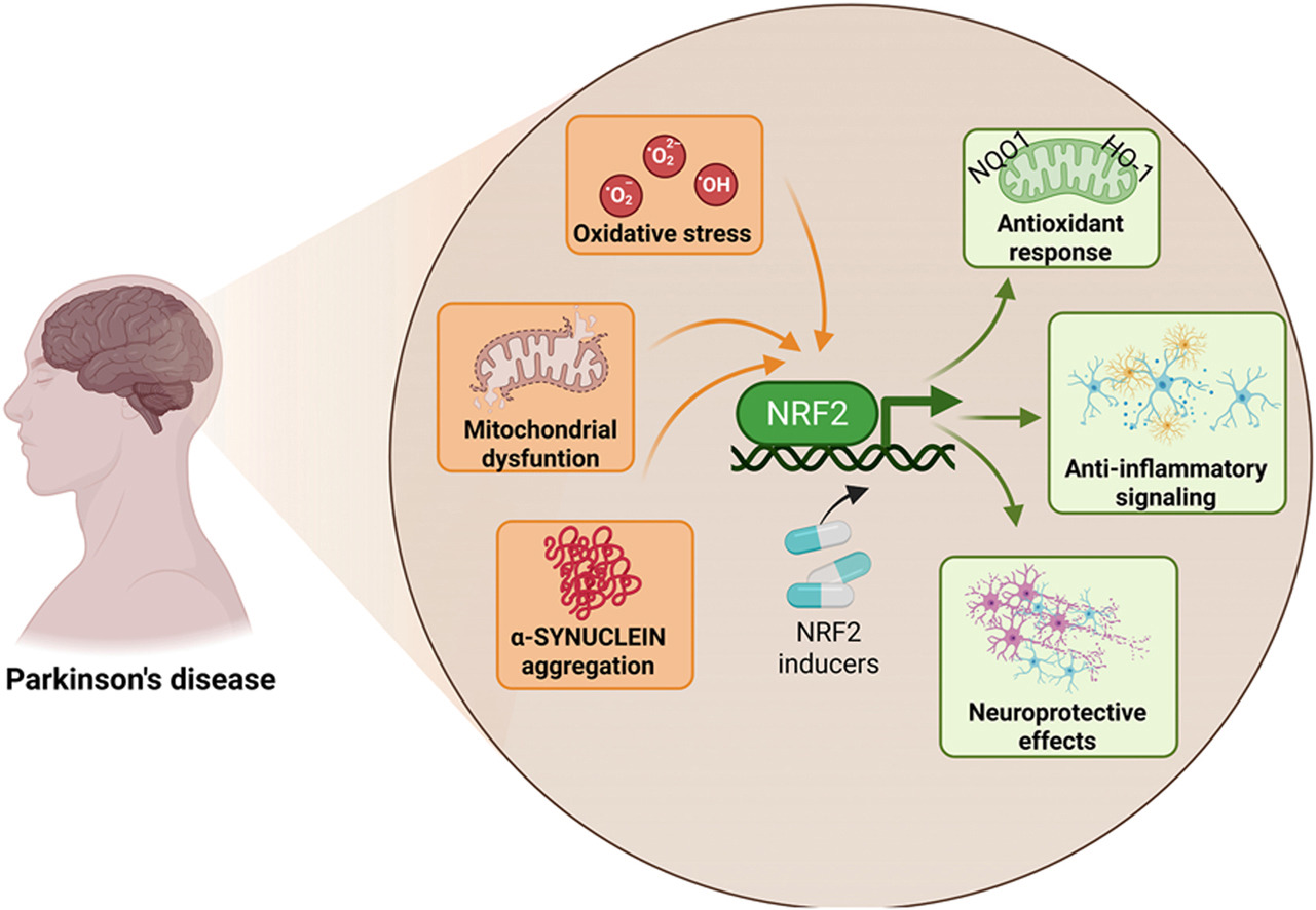
Starting this blog’s twelfth year by curating a poorly-done 2026 review of Nrf2 and its capability to change a person’s development of Parkinson’s disease. I’ll emphasize precedent conditions that if not effectively dealt with in youth, can’t prevent PD from occurring at some later life stage.
“This review explicitly examines how age-associated decline in NRF2 responsiveness intersects with redox imbalance, mitochondrial dysfunction, proteostatic failure, and neuroinflammation, core mechanisms shared between aging and PD. PD unfolds through a complex interplay of cellular stress and immune responses. Oxidative stress, mitochondrial dysfunction, and chronic neuroinflammation converge to damage dopaminergic neurons, with microglia playing a central role in amplifying this injury.
NRF2 emerges as a key regulator of antioxidant defenses, inflammatory balance, and mitochondrial protection, offering a promising target for clinical intervention. NRF2 activity favors the anti-inflammatory microglial over the pro-inflammatory phenotype. Decline in NRF2 inducibility with age impairs microglial clearance, promotes neuroinflammation, and reduces antioxidant defenses, while NRF2 activation restores protective functions and offers a promising therapeutic target.
Strategies aimed at restoring or enhancing NRF2 activity hold significant promise as disease-modifying interventions, not only to slow PD progression but also to promote resilience against the broader spectrum of age-associated neurodegenerative and inflammatory conditions.”
This review only gave lip service to PD progression outside of the brain, as if the importance of prodromal factors to a person’s neurodegeneration such as dysfunction in gut, eyes, skin, and olfactory systems can be minimized. But failure to recognize early what will doom a person to be unable to recover health in later decades is disingenuous. Since these reviewers omitted early interventions into PD prodromal factors, the best they came up with was interventions to “slow PD progression.”
Maybe these reviewers felt it would be outside the scope of this review to discuss early non-brain PD factors for more than one sentence? However, while PD is defined by striatal brain neurons, Nrf2 activity is much less in brain and central nervous system neurons than elsewhere in the body per Nrf2 Week #2: Neurons.
I disagree with these reviewers’ self-imposed emphasis on aging. Repeating ‘age-associated’ numerous times seemed as if they wanted to influence the reader into thinking age in and of itself was a cause for PD, rather than an imputed mathematical correlation. Their emphasis led to dumb mentions such as senolytics for no apparent reason than senescence is a ‘hallmark of aging’, and to meaningless ‘diseasome of aging’ characterizations, and to ignoring the existence of early non-age-associated PD diagnoses in 20- and 30-year-olds.
Whatever it takes to get published, I’d guess. Or maybe it’s that the number of omissions and useless points a review paper makes increases with the number of reviewers and their sponsors’ agendas.
For example, why was it permissible to dedicate lip service to ‘exposome’ factors like microplastics, environmental pollution, and viruses, but it’s still not permitted in 2026 to discuss research into the impacts on vascular disease and neurodegeneration of lipid nanoparticles and DNA contamination in what a large number of humans were exposed to by injected pharmaceuticals starting in late 2020? Not to mention two studies published in 2024 of over 2.5 million people whose incidences of neurologic issues, mild cognitive impairment, and Alzheimer’s disease rapidly increased after ‘vaccination’?
I’ve mentioned in this blog many times how it’s every human’s choice whether or not we take responsibility for our own one precious life. I suggest, if it’s not too late, do that for your children’s lives, too.
Here are five 2025 human ergothioneine studies, starting with a clinical trial of healthy older adults:
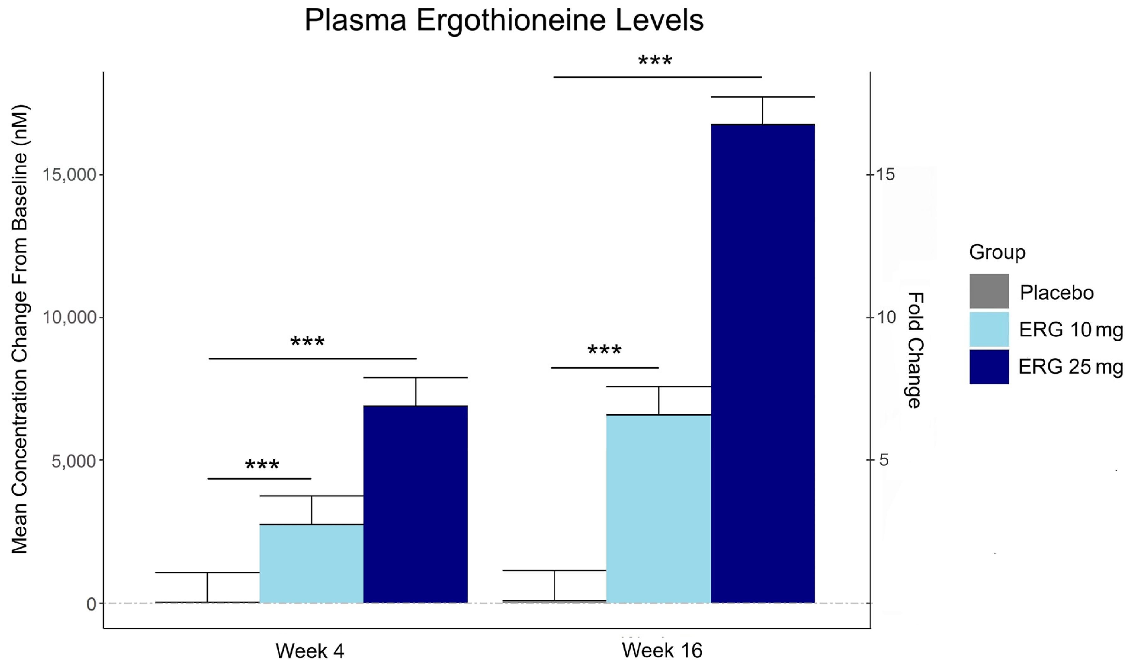
“In this 16-week randomized, double-blind, placebo-controlled trial, 147 adults aged 55–79 with subjective memory complaints received ergothioneine (10 mg or 25 mg/day ErgoActive®) or placebo. Across all the groups, approximately 73% of participants in each group were female, with a median age of 69 years.
The primary outcome was the change in composite memory. Secondary outcomes included other cognitive domains, subjective memory and sleep quality, and blood biomarkers. At baseline, participants showed slightly above-average cognitive function (neurocognitive index median = 105), with plasma ergothioneine levels of median = 1154 nM.
Although not synthesized in the human body, ergothioneine is efficiently absorbed via the OCTN1 transporter (also known as the ergothioneine transporter, or ETT), which is expressed in many tissues, including the intestine, red blood cells, kidneys, bone marrow, immune cells, skin, and brain. This transporter enables ergothioneine to accumulate in high concentrations in organs vulnerable to oxidative stress and inflammation. Ergothioneine has multiple cellular protective functions, including scavenging reactive oxygen species, chelating redox-active metals, suppressing pro-inflammatory signaling, and protecting mitochondrial function.
Plasma ergothioneine increased by ~3- and ~6-fold for 10 mg, and ~6- and ~16-fold for 25 mg, at weeks 4 and 16, respectively.
While the primary outcome, composite memory, showed early improvement in the 25 mg group compared to baseline, this effect was not sustained and did not differ from placebo. Reaction time showed a significant treatment-by-time interaction favoring ergothioneine, yet the between-group differences were not significant, suggesting that any potential benefits were modest and require validation in larger or longer studies.
Other cognitive effects observed were primarily within-group and not consistently dose-responsive, highlighting the challenge of detecting objective cognitive changes over a relatively short study duration in high-functioning healthy populations. However, positive effects of ergothioneine supplementation were observed on subjective measures of prospective memory and sleep initiation that were not seen in the placebo group.
This trial adds to the growing body of evidence supporting the favorable safety profile of ergothioneine. No adverse events attributable to ergothioneine were reported. Additionally, we observed potential hepatoprotective effects, with significant reductions in the plasma AST and ALT levels, particularly among males in the ERG 25 mg group.”
https://www.mdpi.com/1661-3821/5/3/15 “The Effect of Ergothioneine Supplementation on Cognitive Function, Memory, and Sleep in Older Adults with Subjective Memory Complaints: A Randomized Placebo-Controlled Trial”
The third graphic for Ergothioneine dosing, Part 2 showed a human study where a 25 mg dosing stopped after Day 7, but the plasma ergothioneine level stayed significantly higher than baseline through Day 35.
The second graphic for Ergothioneine dosing, Part 2 was a male mouse experiment where plasma ergothioneine levels of a human equivalent 22 mg to 28 mg daily dose kept rising through 92 weeks.
This trial couldn’t explain the desirability of a 25 mg daily dose that was likely (per the second and third graphics for Ergothioneine dosing, Part 2) to sustain the subjects’ increased plasma ergothioneine levels well after the trial ended at Week 16. What effects can be expected from a sustained plasma ergothioneine level that’s 16 times higher than the subjects’ initial levels? Were these 16-fold sustained plasma ergothioneine levels better or worse than the 6-fold increases in the 10 mg group, both of which were likely to continue past the trial’s end?
A representative of the trial’s sponsoring company talked a little more about the trial in this interview:
Another clinical trial investigated ergothioneine’s effects on skin:
“We conducted an 8-week, randomized, double-blind, placebo-controlled clinical trial to evaluate effects of daily oral supplementation with 30 mg of ergothioneine (DR.ERGO®) on skin parameters in healthy adult women aged 35–59 years who reported subjective signs of skin aging. Objective measurements including melanin and erythema indices, skin glossiness, elasticity, and wrinkle and pigmentation counts were used to comprehensively evaluate changes in skin condition.
The OCTN1 transporter is preferentially expressed in basal and granular epidermal layers, where cellular renewal and barrier maintenance are most active. Once internalized, ergothioneine localizes to mitochondria, where it directly scavenges reactive oxygen species (ROS) and protects mitochondrial DNA from UV- and inflammation-induced damage.
At the signaling level, ergothioneine activates key protective pathways such as the Nrf2/ARE axis, enhancing expression of antioxidant enzymes including HO-1, NQO1, and γ-GCLC. These enzymes contribute to redox homeostasis and glutathione regeneration, reinforcing cellular defense systems against photoaging and environmental insult.
In parallel, ergothioneine modulates the PI3K/Akt/Nrf2 and SIRT1/Nrf2 pathways, which are implicated in collagen preservation, inflammation resolution, and mitochondrial maintenance. These pathways converge to reduce matrix metalloproteinase (MMP) activity, enhance collagen synthesis, and suppress pro-inflammatory cytokines (TNF-α, IL-6, IL-1β), all of which are central to maintaining skin structure and function.
Compared to placebo, the DR.ERGO® ergothioneine group showed significantly greater improvements in melanin and erythema reduction, skin glossiness, elasticity, and wrinkle and spot reduction. No adverse events were reported.
These findings corroborate and extend previous clinical evidence from (Hanayama et al., 2024), who investigated an ergothioneine-rich mushroom extract (Pleurotus sp., 25 mg ergothioneine/day) in a 12-week randomized double-blind trial, and (Chunyue Zhang, 2023), who examined pure ergothioneine supplementation (25 mg/day) in a 4-week open-label study. We contextualized our results within this existing literature by comparing key outcomes.
Several limitations should be acknowledged:
The study cohort consisted solely of Japanese women aged 35–59 years, which may limit generalizability across sexes, ethnicities, and age groups.
The 8-week intervention period, while sufficient to detect short-term effects, does not allow conclusions about the sustainability of benefits or the risk of relapse upon discontinuation.
The placebo group also showed modest improvements in self-perception, highlighting the well-documented placebo response in beauty and wellness studies.
This study focused on a single daily dosage (30 mg/day) without evaluating dose–response relationships, and hydration-specific endpoints such as corneometry or transepidermal water loss (TEWL) were not included.”
Two clinical trials investigated ergothioneine’s effects on sleep quality:
“A four-week administration of 20 mg/day ergothioneine (EGT), a strong antioxidant, improves sleep quality; however, its effect at lower doses remains unclear. This study estimated the lower effective doses of EGT using a physiologically based pharmacokinetic (PBPK) model in two clinical trials.
In Study 1, participants received 5 or 10 mg/day of EGT for 8 weeks, and their plasma and blood EGT concentrations were measured. An optimized PBPK model incorporating absorption, distribution, and excretion was assembled. Our results showed that 8 mg/day of EGT for 16 weeks was optimal for attaining an effective plasma EGT concentration.
In Study 2, a randomized, double-blind, placebo-controlled study, participants received 8 mg/day EGT or a placebo for 16 weeks. The subjective sleep quality was significantly improved in the EGT group than in the placebo group.
In mammals, EGT is not generated in the body but is acquired from the diet via the carnitine/organic cation transporter OCTN1/SLC22A4. Its plasma concentration after oral administration is quite stable and gradually increases after repeated dosing on a multi-day basis.
Blood concentrations of EGT increase after Day 8 when EGT intake is interrupted, and they continue to increase until Day 35. The delayed increase in EGT concentration in the blood, compared with that in the plasma, can be interpreted as its efficient uptake by undifferentiated blood cells, which express high levels of OCTN1/SLC22A4 in the bone marrow, and subsequent differentiation to mature blood cells that enter the circulation. This may imply the nonlinear absorption, distribution, and excretion of EGT owing to saturation of the transporter at higher concentrations, potentially leading to difficulty in model construction.
This is the first study to propose a strategy to estimate lower effective doses based on the PBPK model.”
The bolded section above referenced a 2016 study / third graphic for Ergothioneine dosing, Part 2, where a 25 mg dosing stopped after Day 7, but the plasma ergothioneine level stayed high through Day 35. I didn’t see that the referenced 2004 and 2010 studies addressed this 2016 finding.
I also didn’t see that this study’s mathematical model accounted for saturation of the OCTN1 transporter or other effects, such as a very small ergothioneine clearance rate. Okay, lower the ergothioneine dose, and achieve a lower persistent plasma ergothioneine level, to what benefit?
“The present study demonstrated that OCTN1 is associated with myeloid cells rather than lymphoid cells, and especially with erythroid-lineage cells at the transition stage from immature erythroid cells to peripheral mature erythrocytes.”
Persistent high ergothioneine levels aren’t costless. Skewing bone marrow stem cells and progenitor cells toward a myeloid lineage is done at the expense of lymphocytes, T cells, B cells, and other lymphoid lineages.
Where are the studies that examine these tradeoffs? Subjective sleep quality in this study and sleep initiation in the first study above aren’t sufficiently explanatory.
A study investigated associations of plasma ergothioneine levels and cognitive changes in older adults over a two-year period:
“Observational studies have found that lower plasma levels of ergothioneine (ET) are significantly associated with higher risks of neurodegenerative diseases. However, several knowledge gaps remain:
Most of the above studies were based on cross-sectional study design, and potential reverse causation cannot be excluded. It has been suggested that plasma ET declines concomitantly with the deterioration of cognitive function.
Since the impact of a single dietary factor on health is mild, it is prone to be affected by the baseline characteristics of subjects (such as sex, educational level, disease status and gene polymorphism). However, no study has systematically evaluated potential effect modifiers on the association between ET levels and cognitive function.
The dose-response distribution between ET and cognitive function remains undetermined.
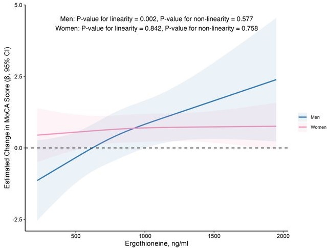
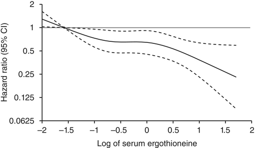
In this prospective cohort study of 1,131 community-dwelling older adults (mean age 69 years), higher baseline plasma ET levels were significantly associated with slower cognitive decline, as assessed by Montreal Cognitive Assessment (MoCA) scores, during a 2-year follow-up period.
When the plasma concentration of ET exceeds 1,000 ng/mL, the decline in cognitive function significantly slows down. However, this association has only been observed in men.
Domain-specific analysis found that the observed ET-MoCA association was mainly driven by the temporary slowdown in the decline of visuospatial/executive and delayed recall. Impaired delayed recall represents one of the earliest and most sensitive cognitive markers of dementia progression, predictive of conversion from MCI to dementia. The preferential preservation of this function by ET suggests targeted neuroprotective effects within the hippocampus.
Visual inspection of the spline curves revealed a potential plateauing effect at ET concentrations ≥1,000 ng/mL in the total population.
Baseline ET concentrations differed between men and women. Most men (81.5%) had concentrations below 1,000 ng/mL (median 754.2, IQR 592.0–937.9 ng/mL). Women exhibited substantially higher median plasma ET concentrations than men, with 35.7% of women exceeded 1,000 ng/mL (median 890.1, IQR 709.7–1,095.6 ng/mL).
Our study included only participants with normal cognitive function, and the results remained robust even after excluding those with baseline cognitive function at the lower end of the normal range. Collectively, our findings support that low ET intake occurs prior to cognitive decline.
Our findings indicate that higher plasma ET levels are significantly associated with slower cognitive decline independent of confounders in non-demented community-dwelling elderly participants, with such association observed in men but not women. Dose-response curves indicated plateauing effects above 1000 ng/mL.”
The average age of this study and the first trial above were both 69 years. Since the first trial’s participants showed slightly above-average cognitive function (neurocognitive index median = 105), with plasma ergothioneine levels of median = 1154 nM at baseline, and this study showed plateauing effects above 1000 ng/mL, I wonder how raising plasma ergothioneine levels above 1000 ng/mL could possibly show a net benefit for older people? What are the trade-offs for older people between potentially increasing slightly above-average cognitive function with ergothioneine and its other effects from saturating their OCTN1 transporter?
This study is at its preprint stage. I’m interested to see if its peer review prompts these researchers to also investigate the common finding that people who are most deficient at baseline have the greatest improvements. If so, would these sex-specific associations still hold?
Wrapping up with a study that investigated associations of serum ergothioneine levels with the risk of developing dementia:
“1344 Japanese community-residents aged 65 years and over, comprising 765 women and 579 men, without dementia at baseline were followed prospectively for a median of 11.2 years.
During follow-up, 273 participants developed all-cause dementia. Among them, 201 had Alzheimer’s disease (AD) and 72 had non-Alzheimer’s disease (non-AD) dementia.
Age- and sex-adjusted hazard ratios (HRs) for all-cause dementia, AD, and non-AD dementia decreased progressively across increasing quartiles of serum ergothioneine. These associations remained significant after adjustment for a wide range of cardiovascular, lifestyle, and dietary factors, including daily vegetable intake.
In subgroup analysis, association between serum ergothioneine levels and the risk of dementia tended to be weaker in older participants and in women:
In older individuals, cumulative burden of multiple risk factors such as hypertension, diabetes mellitus, and smoking may contribute to both neurodegenerative and vascular pathology, potentially diminishing the relative influence of ergothioneine.
In women, postmenopausal hormonal changes, particularly the decline in estrogen, have been associated with increased oxidative stress and a higher vulnerability to neurodegenerative changes.
Several limitations should be noted:
Since serum ergothioneine levels and other risk factors were measured only at baseline, we could not evaluate the changes of serum ergothioneine levels during the follow-up period. Lifestyle modifications during follow-up could have influenced serum ergothioneine levels and other risk factors. In addition, serum ergothioneine level was measured only once, and from a sample.
We cannot rule out residual confounding factors, such as other nutrients in mushrooms and socioeconomic status.
There is a possibility that dementia cases at the prodromal stage were included among participants with low serum ergothioneine levels at baseline.
We are unable to specify which mushroom varieties were predominantly consumed by participants in the town of Hisayama.
Given the limited discriminative ability of serum ergothioneine and potential degradation due to long-term sample storage, we were unable to explore a clinically meaningful threshold value of serum ergothioneine.
Generalizability of findings was limited because participants of this study were recruited from one town in Japan.
These findings suggest that the potential benefit of ergothioneine may be attenuated in individuals with pre-existing, multifactorial risk profiles for dementia.
Our findings showed that higher serum ergothioneine levels were associated with a lower risk of developing all-cause dementia, AD, and non-AD dementia in an older Japanese population. Since ergothioneine cannot be synthesized in the human body, a diet rich in ergothioneine may be beneficial in reducing the risk of dementia.”
For five years I got most of my estimated 7 mg daily ergothioneine intake from mushrooms in AGE-less chicken vegetable soup per Ergothioneine dosing. The soup was always boring, but I got too bored this year and stopped making it. I haven’t replaced mushroom intake with supplements.
I still don’t eat fried or baked foods, preferring sous vide and braising cooking methods to avoid exogenous advanced glycation end products. I avoid buying foods that evoke a hyperglycemic response or otherwise form excessive endogenous AGEs per All about AGEs.
Here are two 2025 papers, starting with a rodent study that investigated interactions between the Nrf2 and kynurenine pathways:
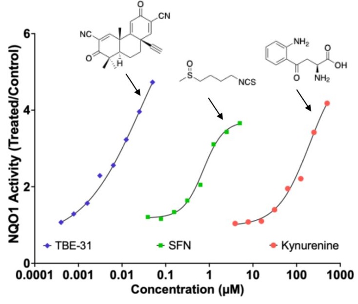
“Exposure to the tryptophan metabolite kynurenine and its electrophilic derivative kynurenine-carboxyketoalkene (Kyn-CKA) leads to an increase in the abundance of transcription factor Nrf2 and induction of Nrf2-target genes. The Keap1/Nrf2 system is the main orchestrator of cellular defence against environmental stress, most notably oxidative and inflammatory stress.
Nrf2 can be activated pharmacologically by small molecules, the majority of which are electrophiles and oxidants that modify specific cysteine-based sensors in Keap1. C151 in Keap1 is the target of the isothiocyanate sulforaphane, a classical Nrf2 activator that has been employed in ∼90 clinical trials, as well as for the two Nrf2 activators that are clinically in use: dimethyl fumarate, for relapsing remitting multiple sclerosis, and omaveloxolone, for Friedreich’s ataxia.
Kynurenine is an endogenous metabolite derived from the essential amino acid tryptophan. Kynurenine and its metabolites, such as the electrophilic kynurenine-carboxyketoalkene (Kyn-CKA), have been demonstrated to activate Nrf2 in other pathologies, including sickle cell disease, attenuating inflammation. Moreover, identification of the gene encoding the kynurenine-metabolising enzyme kynureninase as a gene transcriptionally upregulated by Nrf2, provides a plausible negative feedback regulatory mechanism.
Because kynurenine is not electrophilic, whereas its metabolite Kyn-CKA is, we considered the possibility that Kyn-CKA is the actual Nrf2 activator. Using biochemical and cell-based assays, we found that Kyn-CKA reacts with C151 in the BTB domain of Keap1 and increases the thermostability of Keap1, indicating target engagement. Consequently, Nrf2 accumulates and induces transcription of antioxidant/electrophile-responsive element (ARE/EpRE)-driven genes.
These findings demonstrate that Kyn-CKA targets C151 in Keap1 to derepress Nrf2, and reveal that Nrf2 is a main contributor to the anti-inflammatory activity of Kyn-CKA in macrophages.”
A review subject was targeting nicotinamide adenine dinucleotide, oxidized form (NAD+) for clinical use:
“Mammalian NAD+ biosynthesis includes four known pathways, primarily occurring in cytoplasm:
(a) the NRH pathway;
(b) the salvage pathway;
(c) the Preiss–Handler pathway; and
(d) the kynurenine pathway.
The de novo kynurenine pathway metabolizes tryptophan (Trp) to NAD+, producing various intermediates that serve as biomarkers for different diseases. These intermediates show alterations in various pathological conditions.
While kynurenine and its metabolic derivatives are intermediates in the de novo NAD+ biosynthesis pathway, these are also produced independently in various physiological contexts, particularly in immune cells, where they act as immunomodulatory compounds.”
This second paper above showed a graphic of the Nrf2 and kynurenine pathways together in a diagram showing relationships between NAD+ augmentation and the hallmarks of aging, but didn’t elaborate other than labeling their box Dysbiosis. So how these two pathways interact is better outlined in the first paper above with explaining how a kynurenine-metabolizing enzyme is one of the hundreds of Nrf2 target genes, creating a natural feedback loop between Nrf2 activation and the kynurenine pathway.
These reviewers also lumped SIRT1 in their Dysbiosis box, and into several other boxes, probably due to the penultimate coauthor’s influence:
However, repeating something over and over doesn’t make it scientifically valid regardless of the number of citations. Or, as a 2022 review Sirtuins are not conserved longevity genes concluded:
“A global pursuit of longevity phenotypes was driven by a mixture of framing bias, confirmation bias, and hype. Review articles that propagate these biases are so rampant that few investigators have considered how weak the case ever was for sirtuins as longevity genes.
Acknowledging that a few positive associations between sirtuins and longevity have been identified after thousands of person-years and billions of dollars of effort, we review the data and suggest rejection of the notions that sirtuins (i) have any specific connection to lifespan in animals and (ii) are primary mediators of the beneficial effects of NAD repletion.”
Continuing Plasmalogens Week with three 2025 papers, starting with a rodent study of genetically deleting a plasmalogen catabolizing enzyme:
“In this study, we investigated the impact of global and tissue-specific loss-of-function of a plasmalogen catabolizing enzyme, lysoplasmalogenase (TMEM86B), on circulatory and tissue lipidomes. Mice with homozygous global inactivation of Tmem86b (Tmem86b KO mice) were viable and did not display any marked phenotypic abnormalities.
Tmem86b KO mice demonstrated significantly elevated levels of plasmalogens alkenyl phosphatidylethanolamine (PE(P)) and alkenyl phosphatidylcholine (PC(P)), as well as lysoplasmalogens, in the plasma, liver, and natural killer cells compared to their wild-type counterparts. The endogenous alkenyl chain composition of plasmalogens remained unaltered in Tmem86b KO mice. Consistent with the global knockout findings, hepatocyte-specific Tmem86b knockout mice also exhibited increased plasmalogen levels in the plasma and liver compared to their floxed control counterparts.
Plasmalogens may be synthesized locally within various tissues, with each organ possessing the necessary enzymatic machinery to regulate its own plasmalogen levels. Plasmalogens are important structural constituents of the biological membranes of animals and certain anaerobic bacteria, and have several well-described functions, including regulating membrane dynamics and vesicular cholesterol transport and homeostasis.
One of the most interesting features of plasmalogens is their endogenous antioxidant activity, which is mostly due to the vinyl ether bond, which can scavenge reactive oxygen species and thereby protect other biomolecules from oxidative damage.
They increase the gene expression of multiple antioxidant enzymes to protect against chemically induced cytotoxicity and lipid peroxidation in cultured hepatocytes.
Plasmalogen derivatives such as polyunsaturated fatty acids (AA or DHA) and lysoplasmalogens can act as lipid mediators for multiple cellular signaling activities.
Plasmalogens are important for phagocytosis of macrophages, lipid droplet formation, and development and function of neuromuscular junctions.
They play vital roles in mediating immune responses, and mitochondrial fission to regulate adipose tissue thermogenesis, and protecting neuronal cells against cell death and inflammation.
All of these are suggestive of a critical role played by plasmalogens in maintaining cellular homeostasis.
While plasmalogen anabolism is well defined, its catabolism has been less studied. During catabolism, plasmalogens are deacylated by the action of a calcium-independent phospholipase A2 enzyme (iPLA2) to produce lysoplasmalogens. However, cytochrome C has also been shown to act as a plasmalogenase under certain circumstances.
The amount of lysoplasmalogens in cells is tightly regulated either by reacylation into plasmalogens through a coenzyme A-independent transacylase, or by degradation into fatty aldehydes and glycerophospholipids by an alkenyl ether hydrolase commonly known as lysoplasmalogenase. Lysoplasmalogenase is a microsomal transmembrane enzyme highly specific for lysoplasmalogens, and has no activity against plasmalogens.
While research on the distinct biological functions of lysoplasmalogens and plasmalogens is lacking, some reports indicate potential toxic effects of lysoplasmalogens. Degradation products of lysoplasmalogens, such as fatty aldehydes, are highly reactive electrophilic compounds that can form toxic adducts with cellular proteins and lipids. These interactions can lead to cellular dysfunction and contribute to various pathological conditions. Their accumulation in ischemic/reperfused tissues has been associated with cellular damage.
However, we observed that the amount of lysoplasmalogens as a proportion of total plasmalogens in the liver of Tmem86b KO mice was only ∼3.5%, indicating that elevated lysoplasmalogens are rapidly converted into plasmalogens within the liver. In adipose tissue-specific Tmem86a KO mice, which also exhibited higher lysoplasmalogens, no toxic effects were observed. Instead, these mice showed elevated mitochondrial oxidative metabolism and energy expenditure, offering protection from high-fat diet-induced metabolic dysfunction. These findings suggest that any potential toxic effects of lysoplasmalogens are largely mitigated by their rapid reacylation into plasmalogens.
This study enhances our understanding of regulatory mechanisms governing plasmalogen metabolism, and highlights the potential of targeting Tmem86b to therapeutically raise plasmalogen levels.”
An independent researcher published a commentary on the above study:
“While the biosynthesis of this particular lipid subclass, starting in the peroxisomes and ending at the endoplasmic reticulum, has been the subject of extensive research, the degradation pathway of these compounds remains to be further elucidated. Plasmalogen breakdown is a complex process involving enzymatic hydrolysis, oxidative cleavage, and possibly also a recycling mechanism.
A fundamental unresolved question in the field of plasmalogen catabolism is which of the two possible reaction routes is actually the more important one. Either 1) directly via plasmalogenase or 2) via a deacylation step by a plasmalogen-specific phospholipase A2 (cPLA2, PLA2G4A), yielding a lysoplasmalogen as the first degradation product, and subsequent hydrolysis of the ether bond by a lysoplasmalogenase such as TMEM86A and TMEM86B. It is also unclear how these pathways interact or compensate for each other, how they are regulated, and whether they are tissue- or cell type–specific.
To make the story even more complex, a CoA-independent transacylase activity was described that reacylates lysoplasmalogen intermediates back to plasmalogens by transferring polyunsaturated fatty acids to the vacant sn-2 position of ether lysophospholipids. But no gene for this enzyme has so far been identified.
Why is plasmalogen breakdown so important? Disturbances in plasmalogen metabolism are associated with several human disorders. Neurodegenerative diseases such as Alzheimer’s disease, Parkinson’s disease, and multiple sclerosis have been shown to be associated with reduced levels of plasmalogens.
Unfortunately, it is still too early to draw conclusions about the individual roles of TMEM86A and TMEM86B, as their cellular localisation and function are not sufficiently studied, and reliable antibodies for these proteins are not yet available. Localization of the two TMEM86 homologs overlaps to some extent, as shown, for example, by their gene expression in small intestine. However, whether one isoform is able to compensate for a deficiency in the other is uncertain, and was not found in small intestine of Tmem86b knockout mice [in the above study].
In contrast to the two proteins TMEM86A and TMEM86B, cytochrome c is much better studied. It is associated with the inner mitochondrial membrane, and can be released into the cytosol during apoptosis. It has a wide tissue distribution with most abundant gene expression levels in the digestive tract and heart.“
The statement “no gene for this enzyme has so far been identified” revealed a paradigm. But maybe what’s being observed evolved before genes?
One example of this principle is from the 1966 https://www.science.org/doi/10.1126/science.152.3720.363 “Evolution of the Structure of Ferredoxin Based on Living Relics of Primitive Amino Acid Sequences” which provided evidence pointing to heme protein evolution beginning before gene evolution. Its abstract included this statement:
“We explain the persistence of living relics of this primordial structure by invoking a conservative principle in evolutionary biochemistry: The processes of natural selection severely inhibit any change in a well-adapted system on which several other essential components depend.”
Maybe the process of reassembling plasmalogen breakdown products back into plasmalogens without involving a specific gene likewise became essential?
A role of plasmalogens in diabetic kidney disease was found in a third study that investigated a genetic rodent model of diabetes:
“Diabetic nephropathy (DN) represents a frequent cardiovascular complication of diabetes, affecting about 20–50% of individuals with the disease. Globally, it constitutes a primary etiology for end-stage kidney disease (ESKD) and chronic kidney disease (CKD), while also serving as a significant independent risk factor for cardiovascular morbidity and mortality.
Although intensive management strategies targeting blood pressure and glucose levels demonstrably attenuate the risk of DN development, they do not confer complete protection. This residual risk strongly implicates pathogenic factors beyond impaired glucose metabolism and hemodynamic alterations in DN pathogenesis.
In the present study, we employed the db/db mice as the DN model. When compared to other diabetes models, such as those induced by streptozotocin (STZ) or high-fat diet combined with STZ, the db/db model more accurately recapitulates the pathological features of human type 2 diabetes mellitus (T2DM). It also possesses a stable genetic background, making it particularly well-suited for the investigation of diabetes complications.
Transcriptomics revealed extensive dysregulation of metabolic and lipid regulatory pathways in db/db. Lipidomics uncovered pronounced abnormalities in cardiolipin species composition and plasmalogen profiles. Transcriptome-lipidome integration demonstrated impaired phosphatidylcholine (PC) biosynthesis, mechanistically linked to dysregulation of choline phosphotransferase 1 (chpt1), which correlated significantly with compromised tissue regeneration capacity.
Volcano plot analysis delineated specific lipid alterations, particularly in plasmalogen species in plasmalogen lipids. Plasmenylcholines (plas-PC) and plasmenylethanolamine (plas-PE) containing n-3 polyunsaturated fatty acids (PUFAs) were significantly decreased in the kidneys of db/db mice. Conversely, plas-PCs and plas-PEs esterified with n-6 PUFAs showed substantial accumulation in diabetic kidneys.
In conclusion, the highly sensitive and extensively targeted UHPLC-MS/MS methodology enabled a more in-depth characterization of renal metabolic and lipid perturbations in db/db mice. These alterations principally reflect the sustained inflammatory milieu and compromised antioxidant defenses characteristic of DN renal tissues.”
Continuing Part 1 with three 2025 papers, starting with a rodent study of dietary mussel plasmalogens’ effects on atherosclerosis:
“The purpose of this study was to clarify the underlying mechanisms of Mytilus edulis-derived plasmalogens (Pls) against atherosclerosis (AS) in ApoE−/− mice induced by a high-fat diet (HFD), through a comprehensive analysis of hepatic metabolomics and aortic transcriptomics data. Besides Pls role as the storage pool of n-3 PUFAs, the structural feature of vinyl ether bond at sn-1 position confers multiple advantages upon Pls compared to their diacyl counterparts, including enhanced antioxidant capacity, increased membrane fluidity, as well as improved stability and stability of biomembranes.
The C57BL/6 mouse strain is susceptible to high-fat diet (HFD)-induced AS lesions, and ApoE knockout accelerates AS development. Molecular mechanisms by which Pls ameliorate AS were investigated through a comprehensive analysis of hepatic metabolomics and aortic transcriptome profiles, focusing on changes in gene related to the p38 mitogen-activated protein kinase (MAPK) signaling pathway and the downstream inflammatory response.
The concentration of Pls in mussel tissues is 32 μgmg−1 (dry weight), and the obtained Pls contains 49.53% of phosphatidylethanolamine-Pls, 35.87% of phosphatidylcholine-Pls, and 14.60% of phosphatidylserine-Pls. The main fatty acid compositions of Pls are presented in Supplementary Table 1, which indicates that EPA accounts for 45.82% and the n-3/n-6 ratio is 3.84.
Pls inhibited aortic lipid accumulation, prevented thickening of the aortic wall, and suppressed collagen accumulation at the aortic-heart junction. Pls inhibited HFD-induced loosening of hepatocyte arrangement, vacuolization, and accumulation of lipid droplets.
Although several key components of MAPK signaling pathway were suppressed at both the transcriptional and protein levels in Pls-treated mice, no significant changes in phosphorylated p38 protein were observed among the experimental groups in our study. Further research is needed to elucidate the overall inhibitory mechanism of Pls on p38 protein and the MAPK signaling pathway.”
A rodent / human cell study investigated effects of plasmalogens in innate immune system macrophages on atherosclerosis:
“We demonstrate that simultaneous inactivation of two key enzymes involved in macrophage polyunsaturated fatty acid (PUFA) metabolism—ELOVL5, which elongates long-chain PUFAs, and LPCAT3, which incorporates them into phospholipids—disrupts membrane organization by promoting the formation of cholesterol-enriched domains. This increases macrophage sensitivity to cytotoxic oxysterols and leads to more vulnerable atherosclerotic plaques with enlarged necrotic cores in a mouse model of atherosclerosis.
We identified ELOVL5 as one elongase facilitating the conversion of C20 to C22 PUFA. In humans, analysis of 187 carotid plaques reveals a positive correlation between LPCAT3/ELOVL5-generated phospholipids—including arachidonate (C20:4 n-6)-containing ether lipids—and more stable plaque profiles. Additionally, Mendelian randomization analysis supports a causal relationship between LPCAT3 expression and reduced risk of ischemic stroke.
Potentially beneficial effects we observed in mice and in human atheroma plaques were mainly associated with PLs enriched in omega-6, particularly in AA. Although omega-6 FAs are often considered as pro-inflammatory, their role is undergoing reconsideration, with markers linked to the intake of omega-6 appearing beneficial in the context of cardiovascular diseases. In this context, it is worth to note that AA-containing plasmalogens have been previously identified as markers of healthy obesity.
Our findings uncover a regulatory circuit essential for PUFA-containing phospholipid generation in macrophages, positioning PUFA-containing ether lipids as promising biomarkers and therapeutic targets.”
A human study included plasmalogens in investigating associations among people with mental illness and their lipid profiles:
“Plasma lipidomic profiles of 623 individuals (188 schizophrenia (SCZ), 243 bipolar disorder (BD), 192 healthy controls) belonging to the PsyCourse Study were assessed using liquid chromatography and untargeted mass spectrometry. Exact etiology of these major mental health disorders is yet unknown and while their symptoms overlap, their diagnostic criteria are based on clinical evaluations of symptoms without objective markers.
Cognitive dysfunction is among the most disabling symptoms of SCZ and BD, and is difficult to treat with the commonly used pharmacologic regimes. Consequently, it has important impacts on long-term functional outcomes.
We aimed to answer the question, whether specific lipid species or classes were associated with differential performance across various cognitive domains, including psychomotor and processing speed, executive function, short-term and working memory and crystalized intelligence and whether these associations were affected by diagnoses.
Lipids belonging to the phosphatidylethanolamine plasmalogen (PE-P) class emerged as the main lipid class associated negatively with DG-SYM test performance, representative of processing and psychomotor speed. Our findings showed that higher levels of PE-P 42:5, PE-P 40:4, PE-P 40:5, and ceramide 38:1 in plasma samples of our study are significantly associated with poorer DG-SYM test performance. The DG-SYM test mainly measures processing speed, the amount of time required to complete a series of cognitive tasks. Enrichment analysis also showed significant associations between other lipid classes and various cognitive tests.
Our findings suggest a link between lipids and cognitive performance independent of mental health disorders. Independent replication is warranted to better understand if phosphatidylethanolamines could represent an actionable pharmacologic target to tackle cognitive dysfunction, an important unmet clinical need that affects long-term functional outcomes in individuals with severe mental health disorders.”
It was apparently beyond these researchers’ expertise to offer informed discussion on this study’s associative link between enrichment of these three phosphatidyl ethanolamine plasmalogens and cognitive dysfunction. Grok countered that their depletion was associated with neurodegenerative diseases (Alzheimer’s, Parkinson’s, multiple sclerosis), cardiovascular risk / oxidized-LDL burden, and chronic fatigue / post-viral syndromes.
Continuing Plasmalogens Week with three 2025 papers, starting with a human study that included plasmalogen biomarkers of non-communicable disease fatigue symptoms:
“This study explored the biological mechanisms underlying fatigue in patients with NCDs using a multi-omics approach. Our findings indicate that distinct metabolic pathways, salivary microbiota, and genetic factors may contribute to different dimensions of fatigue, including general, physical, and mental fatigue.
General fatigue is associated with unsaturated fatty acid biosynthesis, indicating its role in lipid metabolism.
Physical fatigue was associated with plasmalogen synthesis, mitochondrial beta-oxidation of long-chain fatty acids, and selenoamino acid metabolism, suggesting a potential contribution of impaired energy production.
Mental fatigue is associated with homocysteine degradation and catecholamine biosynthesis, which may influence cognitive fatigue.
This exploratory study suggests that fatigue in patients with NCDs may involve disruptions in lipid metabolism, neurotransmitter pathways, microbial composition, and genetic variations. Blood-based biomarkers showed better predictive potential for physical fatigue, whereas salivary-based models were more indicative of mental fatigue.
Although our findings support the role of lipid metabolism, the contribution of plasmalogen synthesis remains underexplored. Further studies are needed to validate these findings and understand their mechanisms of action.”
A human study of metabolic dysfunction-associated steatotic liver disease (MASLD) included investigating plasmalogens:
“In this study, we applied untargeted metabolomic profiling to serum samples from individuals with and without MASLD, classified by the Fatty Liver Index, with the goal of identifying characteristic metabolic signatures and pathways that may underlie disease presence and progression. Individuals in the MASLD group displayed significantly higher levels of ALT, AST, ALP, and GGT, reflecting ongoing hepatic injury, cholestasis, and oxidative stress. However, albumin and bilirubin levels remained within normal limits, indicating early to intermediate disease stages rather than advanced fibrosis or cirrhosis.
A consistent and highly significant lipidomic pattern in the MASLD group is the depletion of plasmalogens and sphingomyelins. Depletion of these lipid classes was identified as a hallmark of insulin resistance as defined by the triglyceride-glucose index. In contrast, phosphatidylcholine, phosphatidylethanolamine, and phosphatidylinositol species were elevated in MASLD, pointing toward broader lipid remodeling events.
Reduced plasmalogen and sphingomyelin levels positions their depletion as a core feature of metabolic dysfunction. Plasmalogens are ether phospholipids with strong antioxidant capacity, and their reduction suggests a loss of protective buffering against oxidative stress, one of the main drivers of MASLD progression. Similarly, sphingomyelin depletion implicates altered membrane dynamics and signaling disturbances, further contributing to metabolic dysfunction.
Depletion of plasmalogens 1-(1-enyl-palmitoyl)-2-oleoyl-GPC (P-16:0/18:1), 1-(1-enyl-palmitoyl)-2-linoleoyl-GPC (P-16:0/18:2), 1-(1-enyl-palmitoyl)-2-palmitoyl-GPC (P-16:0/16:0), 1-(1-enyl-palmitoyl)-2-palmitoleoyl-GPC (P-16:0/16:1), 1-(1-enyl-palmitoyl)-2-oleoyl-GPE (P-16:0/18:1), 1-(1-enyl-palmitoyl)-2-linoleoyl-GPE (P-16:0/18:2), and disruption of the glutamate–gamma-glutamyl pathway stand out as central features of metabolic dysfunction in MASLD, with clear potential to inform biomarker discovery, disease classification, and the design of targeted therapeutic strategies.”
https://www.mdpi.com/2218-1989/15/11/687 “Metabolomic Signatures of MASLD Identified by the Fatty Liver Index Reveal Gamma-Glutamyl Cycle Disruption and Lipid Remodeling”
A rodent study investigated dietary sea squirt (AM) plasmalogen ethanolamine (PlsEtn) extract’s and dietary pig liver (PL) phosphatidyl ethanolamine (PtdEtn) extract’s effects on acetaminophen liver injury:
“We investigated dietary effects of PlsEtn from ascidian on chronic hepatic injury in acetaminophen (APAP)-treated mice. Five-week-old male mice were divided into four groups (n = 12), which were treated with experimental diets for two weeks and then the respective APAP-containing diet for five weeks.
Ingested PlsEtn is digested into lysoPlsEtn and free fatty acid in the small intestine. PlsEtn digests are absorbed and are subsequently resynthesized into PlsEtn preferentially with PUFA.
Acetaminophen is a frequently used analgesic and antipyretic. Approximately 90% of APAP is metabolized by UDP-glucuronosyltransferase and sulfotransferase into glucuronic acid and sulfate conjugates, respectively.
5–9% of APAP is metabolized into the highly reactive intermediate N-acetyl-p-benzoquinone imine (NAPQI). This metabolite is considered a pivotal molecule in APAP-induced hepatotoxicity and is conjugated by glutathione (GSH). Excessive NAPQI levels deplete GSH and covalently bind to cellular proteins, resulting in organelle dysfunction, such as mitochondria dysfunction. These impairments induce oxidative stress, cell malfunctions, and subsequently, cell death, such as ferroptosis and apoptosis.
Mice were treated with continuous APAP consumption to induce oxidative stress and impaired lipid metabolism in the liver. Effects of diets were evaluated based on levels of malondialdehyde (MDA), a marker of lipid oxidation, on fatty acid content, and on expression of apoptosis-related proteins in the liver.
The PlsEtn-rich diet effectively suppressed APAP-induced decrease in body and liver weights of mice. However, this suppressive effect was not observed in mice fed a PtdEtn-rich diet. APAP administration decreased the total fatty acid content in the liver, whereas a PlsEtn-rich diet alleviated this decrease and increased the hepatic content of docosahexaenoic acid (DHA).
Owing to the alkenyl linkage, which exhibits antioxidant properties, PlsEtn was expected to markedly suppress hepatic lipid oxidation. However, its suppressive effect was the same extent as that by PtdEtn. Both PlsEtn and PtdEtn contain an ethanolamine base in their structures, and free ethanolamine and its metabolite choline suppress lipid peroxidation. Dietary PlsEtn and PtdEtn may be metabolized into free ethanolamine and its further metabolites, which may alleviate APAP-induced hepatic lipid oxidation.
Dietary ethanolamine glycerophospholipids (EtnGpls) rich in PlsEtn or PtdEtn suppressed APAP-induced lipid oxidation in the liver. Protein expression results revealed that dietary EtnGpls reduced expression of certain apoptosis-related proteins compared to the APAP group. This reduction was more effective in mice fed the PlsEtn-rich diet than in those on the PtdEtn-rich diet.”
https://www.mdpi.com/2076-3417/15/11/5968 “Dietary Ethanolamine Plasmalogen from Ascidian Alleviates Chronic Hepatic Injury in Mice Treated with Continuous Acetaminophen”
This study neither demonstrated nor provided citations for its dietary plasmalogen recycling statements.
Three more plasmalogen health and disease papers are curated in Part 2.
Continuing Plasmalogens Week with two 2025 papers, starting with a simulated in vitro model of how humans digest mussel plasmalogens:
“Plasmalogens (Pls) have promising therapeutic potential in the treatment of neurological disorders, but their distribution, compositional intricacies, and structural alterations during the digestive process are unclear. This study aimed to address this gap by isolating Pls-enriched fractions from mussel (Mytilus edulis) and simulating their digestion in vitro across the mouth, stomach, and intestine phases.
Comparison between Pls and normal phospholipids, sharing identical fatty acyl compositions, illuminated a heightened susceptibility of Pls to catabolism during stomach digestion, which is mainly attributed to the hydrolysis reaction of Pls sensitive to acidic conditions. Phospholipid digestion commenced during the gastric phase and continued with notable catabolism in the intestinal phase, resulting in the release of substantial amounts of free fatty acids (FFAs) and lysophospholipids (LPs), which subsequently formed lipid droplets of larger sizes. Larger droplets delay intestinal absorption, extending the window period for Pls hydrolysis by pancreatic lipase.
The digestive behaviour of Pls with different polar head groups indicated that pancreatic lipase appears to digest phosphatidylethanolamine plasmalogen (PlsPE) to a greater extent than phosphatidylcholine plasmalogen (PlsPC). 41 PlsPE and 14 PlsPC were observed, suggesting that Pls may be more readily digested in the gastrointestinal tract compared to conventional phospholipids.
Generally, lipids are first absorbed by intestinal epithelial cells and undergo lipid remodeling before being transported into lymphatic fluid and then entering the bloodstream. During lipid absorption, PE can be partially converted into PC for lipid remodeling. Since in vitro digestion models cannot fully simulate the intestinal microenvironment (such as microbial metabolism and intestinal epithelial absorption), animal experiments are required to verify the actual bioavailability of PlsPE and PlsPC.”
A review highlighted nutritional implications of changes in plasmalogen chemistry:
“Plasmalogens vary quantitatively in biological systems due to biosynthesis, degradation, remodeling, and certain external stressors. Not only concentrations, but also the composition of molecular species within the plasmalogen pool changes. These shifts often involve the shortening of sn-2 fatty acyl chains, the loss of PUFAs such as DHA and EPA, and the accumulation of oxidized, truncated, or degraded species, as a result of radical-mediated oxidation and/or enzymatic degradation.
The possible increase in lysophospholipids (typically LPE and LPC, corresponding to PlsEtn and PlsCho, respectively) may be attributed to the loss of intact plasmalogens during degradation, especially in the sn-1 position. Lysoplasmalogens can be re-acylated to regenerate the original plasmalogens or create new plasmalogen species with different sn-2 fatty acyl compositions.
These molecular-level transitions highlight the complexity of plasmalogen dynamics and emphasize the need for quantitative, species-specific analysis. Variations are influenced by physiological conditions, pathological states, and nutritional supplementation.
Plasmalogens are primarily those derived from animal products, such as fish, meat, and dairy products, as well as certain marine foods. Microorganism-derived plasmalogens are attracting researchers’ attention, representing a new way of effectively utilizing bacterial resources as a ‘food’ source. Compounds provided can be plasmalogens (either PlsCho and PlsEtn, extracted from natural sources or synthesized) or plasmalogen precursors (e.g., alkylglycerols).”
A challenge researchers haven’t satisfactorily addressed yet is the question of whether beneficial oral intake of plasmalogens can be mechanistically attributed to specific plasmalogen breakdown products or to intact plasmalogens. This review introduced two other mechanistic uncertainties in that 1) absorbed and digested breakdown products can be recycled back into plasmalogens, and 2) gut microbiota can also produce plasmalogens. I’ve read papers that speculated but didn’t demonstrate that either of these factors contributed to their results.
This review cited Dr. Goodenowe’s plasmalogen precursor clinical trial mentioned in Plasmalogens Parts 1, 2, and 3. The first paper above, and most of the papers in Plasmalogen Week cited his other research.
Continuing Plasmalogens Week with two 2025 papers, starting with a rodent study of plasmalogens’ effects on mitigating cognitive decline:
“We evaluated beneficial effects of plasmalogens (PLS), phosphatidylcholine (PC), and phosphatidylserine (PS) on age-associated cognitive decline. We established a mouse model of aging-associated cognitive impairment using the subcutaneous injection of d-galactose (D-gal) at a dosage of 400 mg/kg/day.
We randomly divided six-week-old female mice into nine groups: control, model, high-dose PLS (0.3 mg/kg/day), low-dose PLS (0.09 mg/kg/day), high-dose PC (200 mg/kg/day), low-dose PC (50 mg/kg/day), high-dose PS (200 mg/kg/day), low-dose PS (50 mg/kg/day), AMC-Plas (120 mg/kg/day; and functional component PLS (0.252 mg/kg/day).
We administered PLS, PC, and PS separately by oral gavage once daily. We extracted PLS from scallops according to the literature. AMC-Plas is a commercially available health supplement known for its neuroprotective properties and memory-enhancing effects. In this study, we included AMC-Plas as a positive control group to evaluate the effects of different phospholipids.
Synaptophysin (SYP), synapsin-1 (SYN-1), postsynaptic density protein 95 (PSD-95), and brain-derived neurotrophic factor (BDNF) play important roles in synapse formation and synaptic plasticity. Synaptic function alterations or losses are key pathological mechanisms that underlie development of cognitive impairment. Therapeutic strategies that attempt to restore synaptic function or promote synaptic remodeling are considered to be increasingly promising strategies to mitigate cognitive decline.
Results showed that:
PLS improved spatial memory performance by 44% and object recognition by 80% in D-galactose-induced cognitively impaired mice.
PLS significantly decreased glial fibrillary acidic protein (GFAP)-positive cells (an indicator of astrocyte activation) in the dentate gyrus (DG) of the hippocampus, an important result because the DG is a crucial neurogenesis region.
PLS alleviated neuronal damage and protected against synaptic injury, verified by a 228% increase in PSD-95 expression in the hippocampus.
PLS showed a more prominent role for the mitigation of age-related cognitive impairment compared with PC and PS.
In conclusion, the evaluation of PLS using both behavioral and neuropathological assessments in cognitively impaired mice highlighted its exceptional efficacy compared with other phospholipids. PLS at a remarkably low effective dose significantly ameliorated cognitive deficits in cognitively impaired mice. This result further emphasized its potential relevance in neurodegenerative disease research.
We found that PLS alleviated cognitive impairment potentially by improving synaptic function; however, the molecular mechanisms that underlie its effects on synaptic function warrant further investigation.”
There was no disclosed chemical analysis of the PLS scallop extract’s plasmalogen types or other contents. Despite its name, I didn’t see that the AMC-Plas product contained plasmalogens or plasmalogen precursors.
A fruit fly study investigated plasmalogen effects on mitochondria during aging:
“We identify plasmalogens—endogenous ether-linked phospholipids—as key regulators of age-associated mitochondrial fission in Drosophila melanogaster. Loss of Kua (also known as plasmanylethanolamine desaturase (PEDS) / TMEM189 in mammals), the enzyme essential for plasmalogen biosynthesis, leads to inhibition of mitochondrial fission and impaired recruitment of the fission protein Drp1, similar to what is observed during aging.
Mitochondrial dynamics, comprising balanced cycles of fission and fusion, are essential for preserving organelle quality, metabolic flexibility, and cellular homeostasis throughout life. Aging disrupts this balance, with multiple studies reporting a decline in mitochondrial fission that contributes to the accumulation of enlarged and dysfunctional mitochondria.
These morphological changes are linked to impaired mitophagy, altered energy production, and tissue dysfunction. Midlife induction of Drp1—the dynamin-related GTPase that drives mitochondrial division—has been shown to reverse age-related mitochondrial defects and prolong lifespan in Drosophila.
To determine whether plasmalogen biosynthesis is essential for mitochondrial fission, we used KuaMI04999, a hypomorphic allele. Western blot analysis revealed significantly reduced Kua protein levels in KuaMI04999/+ heterozygotes compared to wild-type controls.
Our findings reveal a previously unrecognized lipid-based mechanism that controls mitochondrial fission during aging and position plasmalogens as key effectors linking membrane composition to mitochondrial homeostasis. It is not merely expression or stability of Drp1 that is affected, but rather its recruitment to the mitochondrial surface, which is a critical activation step for fission.
While our study highlights the requirement of plasmalogen biosynthesis for Drp1 recruitment, further work is needed to understand how plasmalogens mechanistically facilitate this interaction.”
It’s been a while since I curated plasmalogen papers. Let’s start out a week’s worth of 2025 papers with a review of plasmalogens as biomarkers:
“Reduced levels of plasmalogens in circulation or in cell membranes are associated with rare peroxisomal disorders, systemic disease, neurological impairment, cancer, and diseases of the heart, kidney, and liver. Roles for plasmalogens have been identified in lipid rafts, myelin, chlorolipids, bromolipids, hemostasis, cholesterol metabolism, and redox responses.
Plasmalogens account for approximately 5-20% of the phospholipids in mammalian cell membranes. Circulating choline and ethanolamine are incorporated into lipid membranes through the synthesis of plasmalogens. These lipids are formed through a separate multistep process involving precursors in the cytoplasm, peroxisome, and endoplasmic reticulum.
Cytochrome c (cyt-c) typically serves as an electron carrier in the mitochondrial membrane, but under oxidative stress, cyt-c undergoes a conformational alteration conferring peroxidase activity that cleaves the vinyl-ether linkage in plasmalogens. Plasmalogens may act as precursors to platelet-activating factor (PAF), and PAF can be enzymatically converted to plasmalogens. PAF is a potent pro-inflammatory mediator in cancer, cardiovascular, neurological, chronic and infectious disease, suggesting that increased PAF levels may inversely correspond to lower ethanolamine plasmalogen levels identified in human diseases.
Plasmalogens are abundant in myelin, and crucial to the function of central nervous system oligodendrocytes and peripheral nervous system Schwann cells in supporting neuronal action potential.
Catabolism of plasmalogens occurs in response to oxidative stress and activation of TLRs, which promote pro-inflammatory responses during disease progression. Release of fatty acids (e.g., arachidonic acid, eicosapentaenoic acid, docosahexaenoic acid) during plasmalogen catabolism can either exacerbate or resolve pro-inflammatory and thrombotic responses depending on the type of fatty acid released and mediator produced.
Continued research of the types of plasmalogens and plasmalogen precursors and their natural or synthetic sources, the frequency and amount of plasmalogens administered, the route of administration, and the timing of treatment is needed.”
A second review highlighted various strategies for regulating plasmalogen levels:
“Plasmalogens serve as significant structural components of cellular membranes, particularly enriched in tissues with high membrane trafficking. Plasmalogens are recognized as major reservoirs for polyunsaturated fatty acids (PUFAs), notably docosahexaenoic acid (DHA) and arachidonic acid (AA). Incorporation of these PUFAs influences membrane physical properties, including fluidity and the propensity to form non-lamellar structures.
Effective delivery of plasmalogens or their precursors faces significant hurdles, including chemical instability (especially oxidation of the vinyl-ether bond), low oral bioavailability, and challenges in crossing biological barriers like the blood–brain barrier (BBB). Exploration of plasmalogen-based nanoparticles is currently quite limited.”
A 2025 rodent in vivo / human cell ex vivo study investigated effects of a Nrf2 activator on ALS rodent models and ALS human nervous system cells:
“M102 is a central nervous system (CNS) penetrant small molecule electrophile which activates in vivo the NF-E2 p45-related factor 2-antioxidant response element (NRF2-ARE) pathway, as well as transcription of heat-shock element (HSE) associated genes. Apart from the recent promising emergence of tofersen as a disease modifying therapy for the 2% of ALS patients who harbor mutations in the SOD1 gene, other approved drugs have only marginal effects on life expectancy (riluzole) or indices of disease progression (edaravone).
Data from disease model systems and from human biosamples provide strong evidence for a role of redox imbalance, inflammation, mitochondrial dysfunction, and altered proteostasis, including autophagy and mitophagy, as four key drivers in the pathobiology of ALS. We demonstrate that M102 is a dual activator of NRF2 and HSF1 transcription factor pathways, two upstream master regulators of neuroprotective mechanisms, with the potential to modulate all four of these key drivers of neurodegeneration and with excellent penetration across the blood brain barrier.
Stress response of the KEAP1-Nrf2-ARE system is stronger in astrocytes compared to neurons. A body of evidence from in vitro and in vivo model systems and from post-mortem CNS tissue from ALS patients has indicated that the NRF2 response is impaired in ALS, and has also been shown to decline with age.
HSF1 is a stress-inducible transcription factor that is the key driver for expression of multiple heat shock proteins which act as chaperones responsible for correct folding of newly synthesized proteins, refolding of denatured proteins, and prevention of aggregation of misfolded proteins. However, to date, many small molecule activators of HSF-1 have shown undesirable properties e.g. by acting as Hsp90 inhibitors or by exerting direct proteotoxic effects.
M102 (S-apomorphine hydrochloride hemihydrate) is a proprietary new chemical entity (NCE) and the S-enantiomer of the marketed R-apomorphine (Apokyn®; pure R-enantiomer). The R-enantiomer is a dopamine agonist administered subcutaneously for management of advanced Parkinson’s disease. M102 is a very weak dopamine antagonist and does not show the adverse effects associated with dopamine agonism.
M102 treatment rescues motor neuron (MN) survival in co-cultures with C9, SOD1 and sporadic ALS patient-derived astrocytes. Other NRF2 activators have been investigated in clinical trials or have been approved for medical use. These include dimethylfumarate (DMF) (Tecfidera®, Biogen) and omaveloxolone (Reata, Biogen).
DMF was originally approved for the treatment of psoriasis (Fumaderm®) and was later repurposed for the treatment of relapsing-remitting multiple sclerosis (Tecfidera®). A phase 2 trial of DMF in ALS provided Class 1 evidence of safety at a dose of 480 mg/day and lack of disease-modifying efficacy. DMF treatment is associated with dose-limiting lymphopenia and flushing (Tecfidera® Prescribing Information).
Omaveloxolone (Skyclarys®) is a potent NRF2 activator that has been approved by the FDA and EMA for the treatment of Friedreich’s ataxia. By activating the NRF2 pathway, omaveloxolone ameliorates oxidative stress and improves mitochondrial function. As a potent NRF2 activator, omaveloxolone exhibited significant liver toxicity with elevated AST/ALT levels in 37% of patients exposed to a dose of 150 mg.
Toxicity has also been reported with other potent NRF2 activators, such as bardoxolone methyl (EC50: 53 nM) which showed significant heart, liver, and renal toxicity in humans.
In contrast, our preclinical toxicological studies indicate that M102 has a much higher safety margin in relation to liver toxicity. M102 has the potential to modulate multiple key drivers of neurodegeneration, increasing the chances of achieving impactful neuroprotection and disease modifying effects in ALS.
This comprehensive package of preclinical efficacy data across two mouse models as well as patient-derived astrocyte toxicity assays, provides a strong rationale for clinical evaluation of M102 in ALS patients. Combined with the development of target engagement biomarkers and the completed preclinical toxicology package, a clear translational pathway to testing in ALS patients has been developed.”