Maintaining your myelin, Part 2

Continuing Part 1 with three 2024 preprint studies, starting with an investigation of neuroinflammation in high school athletes:

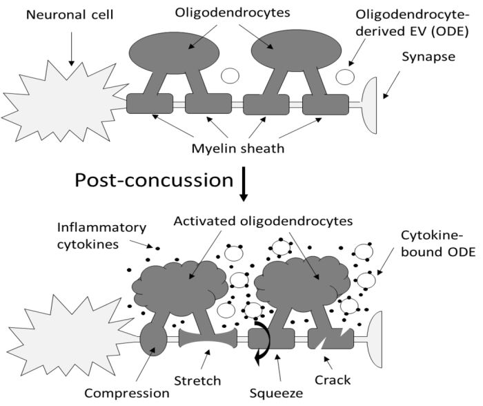
“Axons are long fibers conducting nerve impulses from nerve cells to synaptic ends. Like electric wires, axons are insulated by the myelin sheath produced by oligodendrocytes (ODC) in the brain or Schwann cells in the periphery. The myelin sheath is vulnerable to mechanical stresses after head injuries, as well as targets for autoimmune attack in multiple sclerosis and degeneration in various white matter diseases.
145850ce6289d06e5318d35f

It is challenging to definitively validate axonal neuroinflammation, because axonal neuroinflammation is only diagnosed at post-mortem autopsy, or wait for more than a decade to potentially witness progression to chronic traumatic encephalopathy, or white matter dementia. Advanced imaging analysis of computed tomography and magnetic resonance imaging are not sensitive enough to identify such microscopic abnormalities.

We developed a sandwich immunoassay detecting dual signals of myelin oligodendrocyte glycoprotein (MOG) and interleukin 1B (IL1B) in human plasma, [IL1B on MOG]. MOG is a transmembrane protein specifically expressed in ODC and Schwann cells membranes, and doesn’t freely exist in plasma. We found that serum from capillary blood is acceptable, and we tested control and athlete samples using only 5 mL samples. When we tested 63 control plasma samples, values were widely distributed over 2 logs, so we focused on longitudinal studies.

Damaged neurons are not easily detectable using conventional physical examinations, because the brain’s inherent adaptability allows it to compensate for localized damage by finding alternate routes. While this adaptability is advantageous, it also means that these concealed lesions can go unnoticed, potentially leading to future complications.

Elevation of [IL1B on MOG] was seen in some athletes who did not show concussion or traumatic brain injury (TBI). While the occurrence of concussion is relatively limited, potential prevalence of subconcussion or subconcussive condition is expected to be substantially higher.

If [IL1B on MOG] levels remain unchanged during this early post-concussion period (2-4 weeks), it may suggest that neuroinflammation has not been induced, potentially providing reassurance for the athletes to return to play. Conversely, if [IL1B on MOG] levels increase within this timeframe, it may indicate the need for intervention or closer monitoring. Thus, there is compelling potential for incorporating this test into concussion guidelines.”

https://www.researchsquare.com/article/rs-3997676/v1 “An approach for the analysis of axonal neuroinflammation by measuring dual biomarkers of oligodendrocytes and inflammatory cytokine in human plasma”


A rodent study investigated the immune system’s influence on oligodendrocyte lineage cells after TBI:

“White matter injury is thought to be a major contributor to long-term cognitive dysfunctions after TBI. This damage occurs partly due to apoptotic death of oligodendrocyte lineage cells (OLCs) after injury, triggered directly by the trauma or in response to degenerating axons.

Our data indicates that depletion of the gut microbiota after TBI impaired remyelination, reduced OLCs proliferation, and required the presence of T cells. This suggests that T cells are an important mechanistic link by which the gut microbiota modulate oligodendrocyte response and white matter recovery after TBI.

Our findings suggest that oligodendrocytes are not passive in the neuroinflammatory and degenerative environment caused by brain trauma, but instead could exert an active role in modulation of immune response.”

https://www.researchsquare.com/article/rs-4289147/v1 “Gut Microbiota Shape Oligodendrocyte Response after Traumatic Brain Injury”


A rodent study investigated whether oligodendrocyte precursor cells had myelination-independent roles in brain aging:

“OPCs, the source cells of myelin-forming cells in the central nervous system, have been linked to brain aging by their compromised differentiation and regeneration capability. Our results demonstrate that macroautophagy influx declines in aged OPCs, which results in the accumulation of senescent OPCs in aged brains. Senescent OPCs impair neuronal plasticity and exacerbate neurodegeneration, eventually leading to cognitive decline.

Inactivation of autophagy in OPCs exhibits a limited effect on myelin thickness but a loss of myelin in middle-aged mice. The loss of myelin observed is an adaptational change to suppressed neuronal plasticity. However, neither the number of OLs nor oligodendrogenesis is altered by inactivation of autophagy in adult OPCs.

The present study indicates that the intervention of senescent OPCs is an additional promising therapeutic strategy for aging and aging-related cognitive deficits. Autophagy regulates senescence by impairing protein turnover, mitochondrial homeostasis, oxidative stress, and maintaining senescence-associated secretory phenotype. Further investigation remains on whether autophagy in OPCs shares the exact mechanism to promote senescence as that in other types of cells.

Considering autophagy declines with aging, our study brings a novel mechanism in brain aging. Declined autophagy causes senescence of OPCs, which impairs neuronal plasticity and exacerbates neurodegeneration via CCL3/5-CCR5 signaling.”

https://www.researchsquare.com/article/rs-3926942/v1 “Impaired Macroautophagy in Oligodendrocyte Precursor Cells Exacerbates Aging-related Cognitive Deficits via a Senescence Associated Signaling”

PXL_20240418_104114528.MP

Maintaining your myelin, Part 1

Three papers on myelin and oligodendrocytes, starting with a 2023 review:

“Myelin is the spiral ensheathment of axons by a lipid and cholesterol-rich glial cell membrane that reduces capacitance and increases resistance of the axonal membrane. Axonal myelination speeds up nerve conduction velocity as a function of axon diameter.

While myelination proceeds rapidly after birth in the peripheral nervous system, central myelination is a spatially and temporally more regulated process. Ongoing myelination of the human brain has been documented at up to 40 years of age. This late myelination in the adult cortex is followed by exhaustion of oligodendrocyte precursor cells (OPC) with senescence and a gradual loss of myelin integrity in the aging brain.

The brain is well known for its high energy demands, specifically in gray matter areas. In white matter tracts, energy consumption is lower. Myelination poses a unique challenge for axonal energy generation where myelin sheaths cover more than 95% of the axonal surface areas.

Oligodendrocytes help support axonal integrity. Oligodendrocytes survive well in the absence of mitochondrial oxidative phosphorylation, and without signs of myelin loss, cell death, neurodegeneration or secondary inflammation.

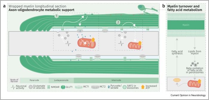
Glycolysis products of oligodendroglial origin are readily metabolized in axonal mitochondria. Oligodendroglial metabolic support is critical for larger and faster-spiking myelinated axons that also have a higher density of mitochondria. An essential requirement for the direct transfer of energy-rich metabolites from oligodendrocytes to the myelinated axonal compartment is ‘myelinic channels’ within the myelin sheath.

Interactions of oligodendrocytes and myelin with the underlying axon are complex and exceed the transfer of energy-rich metabolites. Continuous turnover of myelin membranes by lipid degradation and fatty acid beta-oxidation in mitochondria and peroxisomes leads to recycling of acetate residues by fatty acid synthesis and membrane biogenesis.

1-s2.0-S0959438823001071-gr2_lrg

In human multiple sclerosis (MS) and its animal model myelin oligodendrocyte glycoprotein-experimental autoimmune encephalomyelitis (MOG-EAE), acute inflammatory demyelination is followed by axonal degeneration in lesion sites that is mechanistically not fully understood. It is widely thought that demyelination and the lack of an axon-protective myelin sheath in the presence of numerous inflammatory mediators are the main causes of axon loss.

But unprotected axons improve rather than worsen the overall clinical phenotype of EAE mice which exhibited the same degree of autoimmunity. Thus, ‘bad myelin is worse than no myelin’ because MS-relevant myelin injuries perturb the integrity of myelinic channels and metabolic support.

Dysfunctional or injured oligodendrocytes that do not allow for compensation by any other cell types turn the affected myelin ensheathment into a burden of the underlying axonal energy metabolism, which causes irreversible axon loss. Any loss of myelin integrity, as seen acutely in demyelinating disorders or more gradually in the aging brain, becomes a risk factor for irreversible neurodegeneration.”

https://www.sciencedirect.com/science/article/pii/S0959438823001071 “Expanding the function of oligodendrocytes to brain energy metabolism”


A 2024 review focused on myelin and oligodendrocyte plasticity:

“This review summarizes our current understanding of how myelin is generated, how its function is dynamically regulated, and how oligodendrocytes support the long-term integrity of myelinated axons.

Apart from its unique ultrastructure, there are several other exceptional features of myelin. One is certainly its molecular composition. Another is its extraordinary stability. This was compellingly illustrated when 5000-year-old myelin with almost intact ultrastructure was dissected from a Tyrolean Ice Man.

Myelin is a stable system in contrast to most membranes. However, myelin is compartmentalized into structurally and biochemically distinct domains. Noncompacted regions are much more dynamic and metabolically active than tightly compacted regions that lack direct access to the membrane trafficking machinery of oligodendrocytes.

The underlying molecular basis for stability of myelin is likely its lipid composition with high levels of saturated, long chain fatty acids, together with an enrichment of glycosphingolipids (∼20% molar percentage of total lipids) and cholesterol (∼40% of molar percentage of total lipids). In addition, myelin comprises a high proportion of plasmalogens (ether lipids) with saturated long-chain fatty acids. In fact, ∼20% of the fatty acids in myelin have hydrocarbon chains longer than 18 carbon atoms (∼1% in the gray matter) and only ∼6% of the fatty acids are polyunsaturated (∼20% in gray matter).

With maturation of oligodendrocytes, the plasma membrane undergoes major transformations of its structure. Whereas OPCs are covered by a dense layer of large and negatively charged self-repulsive oligosaccharides, compacted myelin of fully matured oligodendrocytes lacks most of these glycoprotein and complex glycolipids.

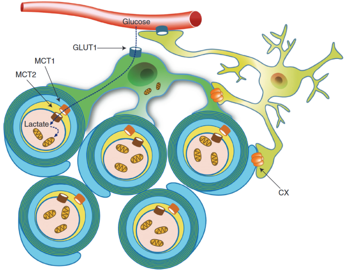
Schematic depiction of an oligodendrocyte that takes up blood-derived glucose and delivers glycolysis products (pyruvate/lactate) via monocarboxylate transporters (MCT1 and MCT2) to myelinated axons. Oligodendrocytes and myelin membranes are also coupled by gap junctions to astrocytes, and thus indirectly to the blood–brain barrier.

oligodendrocyte

Adaptive myelination refers to dynamic events in oligodendroglia driven by extrinsic factors such as experience or neuronal activity, which subsequently induces changes in circuit structure and function. Understanding how these adaptive changes in neuron-oligodendroglia interactions impact brain function remains a pressing question for the field.

Transient social isolation during adulthood results in chromatin and myelin changes, but does not induce consequent behavioral alterations. When mice undergo a social isolation paradigm during early life development, they similarly exhibit deficits in prefrontal cortex function and myelination, but these deficiencies do not recover with social reintroduction. This implicates a critical period for social deprivation effects on myelin dynamics. Experience-dependent changes in myelin dynamics may depend on not only the age, brain region, and cell type studied, but also the specific myelin structural change assessed.

Local synaptic neurotransmitter release along an axon not only affects the number of OPCs and oligodendrocytes associated with that axon and local synthesis of myelin proteins, but also drives preferential selection of active axons for myelination over the ensheathment of electrically silenced neighboring axons. Neuronal activity–induced plasticity may preferentially impact brain regions that remain incompletely myelinated compared to more fully myelinated tracts.

Whereas the myelin sheath has been regarded for a long time as an inert insulating structure, it has now become clear that myelin is metabolically active with cytoplasmic-rich pathways, myelinic channels, for movement of macromolecules into the periaxonal space. The myelin sheath and its subjacent axon need to be regarded as one functional unit, which are not only morphological but also metabolically coupled.”

https://cshperspectives.cshlp.org/content/early/2024/04/15/cshperspect.a041359 “Oligodendrocytes: Myelination, Plasticity, and Axonal Support” (not freely available) Thanks to Dr. Klaus-Armin Nave for providing a copy.


A 2024 rodent study investigated oligodendrocyte precursor cell transcriptional and epigenetic changes:

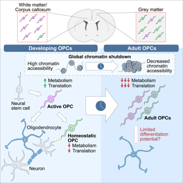
“We used single-cell RNA sequencing (scRNA-seq), single-cell ATAC sequencing (scATAC-seq), and single-cell spatial transcriptomics to characterize murine cortical OPCs throughout postnatal life. One group (active, or actOPCs) is metabolically active and enriched in white matter. The second (homeostatic, or hOPCs) is less active, enriched in gray matter, and predicted to derive from actOPCs. Relative to developing OPCs, both actOPCs and hOPCs are less active metabolically and have less open chromatin.

In adulthood, these two groups are transcriptionally but not epigenetically distinct, indicating that they may represent different states of the same OPC population. If that is the case, then one model is that the parenchymal environment maintains adult OPCs within an hOPC state, whereas those OPCs recruited into white matter or exposed to demyelinated axons may transition toward an actOPC state in preparation for making new oligodendrocytes. We do not yet know the functional ramifications of these differences, but this finding has clear implications for the development of therapeutic strategies for adult remyelination.

opcs

Another finding is that developing but not adult actOPC chromatin is preferentially open for binding motifs associated with neural stem cells, transit-amplifying precursors, and neurogenesis. Although this may simply reflect their origin as the immediate progeny of neonatal neural precursor cells, it may also explain why developing but not adult OPCs have the capacity to make neurons in culture.

If we could, at least in part, reverse the global chromatin shutdown that occurs between development and adulthood, then perhaps adult OPCs may reacquire the ability to make neurons or become better able to generate new oligodendrocytes for remyelination.”

https://www.cell.com/stem-cell-reports/fulltext/S2213-6711(24)00077-8 “Single-cell approaches define two groups of mammalian oligodendrocyte precursor cells and their evolution over developmental time”

Continued in Part 2.


PXL_20240414_103442372

What can be done today to fulfill early unmet needs?

Got agitated earlier this week watching Tucker Carlson’s freely-available interview with a maniac who thinks he’s graduated into a higher state by worshiping the Great AI (Artificial Intelligence, aka Automated Internet, inhabited solely by robots) which will dictate every aspect of what to do with his life. Nevermind that behind the Great AI curtain are the same people who have lied to billions of us, especially during every day of this decade.

Are his current set of beliefs better than previous ones he had of putting a chip into everybody’s brain? What’s wrong with getting to live your own life?

5000

What I saw expressed in the interview was an exhausting pursuit of substitutes for feeling loved. I doubt that many others saw the same, because feeling unloved is so devastating we’ll do anything to avoid it.

But re-experiencing early memories and feelings of unmet needs in a therapeutic setting is the way to keep them from subsequently running our lives. Otherwise, we’ll develop unfulfilling substitutes for what we missed, with misdirected ideas and beliefs accompanied by their unconscious act-outs.

While speaking with a mother who is doing a terrific job of meeting her six-month-old’s needs, I attempted to contrast this interview with the experiences she and her husband are giving their child. Maybe if they read this post, my poor explanation will become clearer.


Wild persimmon trees’ eclipse shadows

PXL_20240408_192336638

Changing a cancerous phenotype

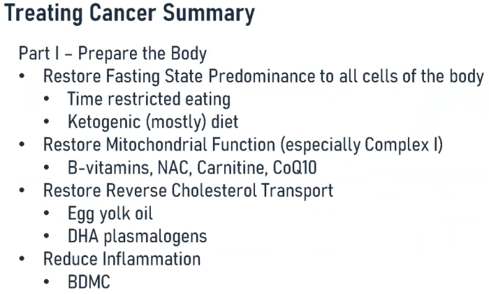
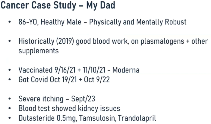
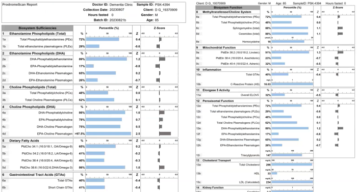
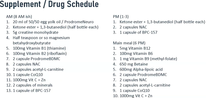
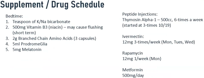
A 2024 Dr. Goodenowe presentation to a professional audience. He ended the presentation by using his 86-year-old father as a case study of treatment to create an inhospitable environment for cancer.

1. Get the body ready

slide 189

2. Starve the cancer and boost the immune system

slide 190

3. Characteristics

slide 191

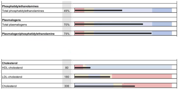
4. 2019 sample biochemistry

slide 192

5. 2023 biochemistry (compare HDL (33 vs. 80), see off-the-chart hsCRP, Hcy 16)

slide 193

6. Treatment details #1

slide 197

7. Treatment details #2

slide 198

https://drgoodenowe.com/tfim-2024-recording-now-available/ “Breaking Cancer: The Biochemistry of Cancer Risk Assessment, Prevention, and Treatment—Real Knowledge That You Can Use In Your Practice”


PXL_20240408_185424838

Eat broccoli sprouts to maintain your cells

Two more papers cited Precondition your defenses with broccoli sprouts, starting with a 2024 review of broccoli compounds’ influences on autophagy and cellular function:

“Promotion of autophagy has been related to lifespan expansion, tumor suppression, and maintenance of metabolic health. Alterations in this pathway have been related to human diseases or pathological states including neurodegenerative diseases, stroke, metabolic alterations, or cancer.

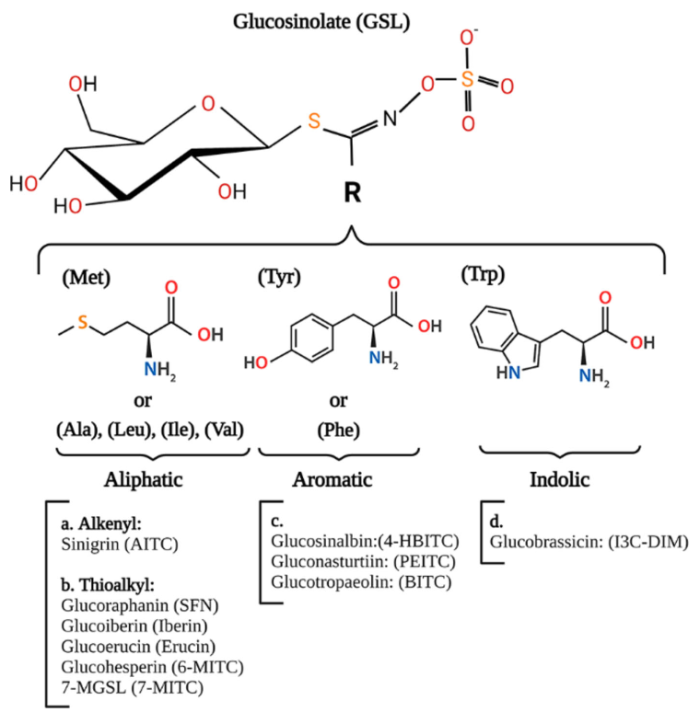
We describe the different types of glucosinolates (GSL), grouped depending on the structure of their side chain, with special attention to those GSL and their derived isothiocyanate (ITC) which have been suggested to be of relevance to treat or prevent human diseases, their structure, and plant source.

gsl-itc

It has been shown that SFN activates TFEB, boosting expression of genes required for autophagosome and lysosome biogenesis. SFN induced a short burst of ROS necessary for TFEB activation, and TFEB activity was required for SFN-induced NRF2 activation and protection against acute and chronic oxidative stress.

TFEB was also required for SFN-induced removal of excessive mitochondrial ROS, indicating an important role for mitophagy in SFN-induced antioxidant response. Thus, direct activation of NRF2 by SFN or other ITC can promote autophagy.

Research on autophagy has been characterized by controversies regarding autophagy mediating survival or cell death, or its role in health and disease, not only because autophagy is a complicated process with context dependent roles depending on the cell type or the step of the autophagic pathway being modulated, but also, because in occasions, autophagy is not measured correctly.

An interesting area of research would be to decipher effects of NRF2-regulated or NRF2-independent autophagy induction by ITC, and whether these effects would determine the role of the autophagic process in cellular survival or death. Also, it is needed to clarify which of the effects regulated by ITC are mediated by autophagy, and which ones are not, and the importance of autophagy induction in the therapeutic effects mediated by ITC.”

https://link.springer.com/article/10.1007/s11101-024-09944-w “Glucosinolates, isothiocyanates, and their role in the regulation of autophagy and cellular function” (not freely available)

This paper’s contact coauthor (who provided access to the full paper) is also the contact for Our model clinical trial for Changing to a youthful phenotype with broccoli sprouts.


The coauthors of Exercise substitutes? published a 2024 human cell study:

“While physical activity is an excellent inducer of mitochondrial turnover, its ability to ubiquitously activate and enhance mitochondrial turnover prevents definitive differentiation of the contribution made by each pathway. We employed three agents which are activators of important biological markers involved in antioxidant signaling, mitochondrial autophagy, and mitochondrial biogenesis.

Results suggest that early time points of treatment increase upstream pathway activity, whereas later time points represent increased phenotypic expression of related downstream markers. Findings suggest that spatiotemporal progression of these mechanisms following drug treatment is another important factor to consider when examining subcellular changes towards mitochondrial turnover in muscle.”

https://www.sciencedirect.com/science/article/pii/S2666337624000398 “Sulforaphane, Urolithin A, and ZLN005 induce time-dependent alterations in antioxidant capacity, mitophagy, and mitochondrial biogenesis in muscle cells”


PXL_20240330_175846440