Clearing out the 2019 queue of interesting papers

I’m clearing out the below queue of 27 studies and reviews I’ve partially read this year but haven’t taken the time to curate. I have a pesky full-time job that demands my presence elsewhere during the day. :-\

Should I add any of these back in? Let’s be ready for the next decade!


Early life

https://link.springer.com/article/10.1007/s12035-018-1328-x “Early Behavioral Alterations and Increased Expression of Endogenous Retroviruses Are Inherited Across Generations in Mice Prenatally Exposed to Valproic Acid” (not freely available)

https://www.sciencedirect.com/science/article/pii/S0166432818309392 “Consolidation of an aversive taste memory requires two rounds of transcriptional and epigenetic regulation in the insular cortex” (not freely available)

https://www.nature.com/articles/s41380-018-0265-4 “Intergenerational transmission of depression: clinical observations and molecular mechanisms” (not freely available)

mother

https://www.ncbi.nlm.nih.gov/pmc/articles/PMC6454089/ “Epigenomics and Transcriptomics in the Prediction and Diagnosis of Childhood Asthma: Are We There Yet?”

https://www.ncbi.nlm.nih.gov/pmc/articles/PMC6628997/Placental epigenetic clocks: estimating gestational age using placental DNA methylation levels”

https://www.ncbi.nlm.nih.gov/pmc/articles/PMC6770436/ “Mismatched Prenatal and Postnatal Maternal Depressive Symptoms and Child Behaviours: A Sex-Dependent Role for NR3C1 DNA Methylation in the Wirral Child Health and Development Study”

https://www.sciencedirect.com/science/article/pii/S0889159119306440 “Environmental influences on placental programming and offspring outcomes following maternal immune activation”

https://academic.oup.com/mutage/article-abstract/34/4/315/5581970 “5-Hydroxymethylcytosine in cord blood and associations of DNA methylation with sex in newborns” (not freely available)

https://physoc.onlinelibrary.wiley.com/doi/full/10.1113/JP278270 “Paternal diet impairs F1 and F2 offspring vascular function through sperm and seminal plasma specific mechanisms in mice”

https://onlinelibrary.wiley.com/doi/full/10.1111/nmo.13751 “Sex differences in the epigenetic regulation of chronic visceral pain following unpredictable early life stress” (not freely available)

https://www.ncbi.nlm.nih.gov/pmc/articles/PMC6811979/ “Genome-wide DNA methylation data from adult brain following prenatal immune activation and dietary intervention”

https://link.springer.com/article/10.1007/s00702-019-02048-2miRNAs in depression vulnerability and resilience: novel targets for preventive strategies”


Later life

https://www.ncbi.nlm.nih.gov/pmc/articles/PMC6543991/ “Effect of Flywheel Resistance Training on Balance Performance in Older Adults. A Randomized Controlled Trial”

https://www.mdpi.com/2411-5142/4/3/61/htm “Eccentric Overload Flywheel Training in Older Adults”

https://www.nature.com/articles/s41577-019-0151-6 “Epigenetic regulation of the innate immune response to infection” (not freely available)

https://link.springer.com/chapter/10.1007/978-981-13-6123-4_1 “Hair Cell Regeneration” (not freely available)

https://www.ncbi.nlm.nih.gov/pmc/articles/PMC6422915/Histone Modifications as an Intersection Between Diet and Longevity”

https://www.sciencedirect.com/science/article/abs/pii/S0306453019300733 “Serotonin transporter gene methylation predicts long-term cortisol concentrations in hair” (not freely available)

https://www.sciencedirect.com/science/article/abs/pii/S0047637419300338 “Frailty biomarkers in humans and rodents: Current approaches and future advances” (not freely available)

https://onlinelibrary.wiley.com/doi/full/10.1111/pcn.12901 “Neural mechanisms underlying adaptive and maladaptive consequences of stress: Roles of dopaminergic and inflammatory responses

https://www.ncbi.nlm.nih.gov/pmc/articles/PMC6627480/ “In Search of Panacea—Review of Recent Studies Concerning Nature-Derived Anticancer Agents”

https://www.sciencedirect.com/science/article/abs/pii/S0028390819303363 “Reversal of oxycodone conditioned place preference by oxytocin: Promoting global DNA methylation in the hippocampus” (not freely available)

https://www.futuremedicine.com/doi/10.2217/epi-2019-0102 “Different epigenetic clocks reflect distinct pathophysiological features of multiple sclerosis”

https://www.ncbi.nlm.nih.gov/pmc/articles/PMC6834159/ “The Beige Adipocyte as a Therapy for Metabolic Diseases”

https://www.sciencedirect.com/science/article/abs/pii/S8756328219304077 “Bone adaptation: safety factors and load predictability in shaping skeletal form” (not freely available)

https://www.nature.com/articles/s41380-019-0549-3 “Successful treatment of post-traumatic stress disorder reverses DNA methylation marks” (not freely available)

https://www.sciencedirect.com/science/article/abs/pii/S0166223619301821 “Editing the Epigenome to Tackle Brain Disorders” (not freely available)
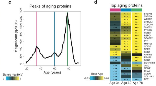
A blood plasma aging clock

This 2019 Stanford human study developed an aging clock using blood plasma proteins:

“We measured 2,925 plasma proteins from 4,331 young adults to nonagenarians [18 – 95] and developed a novel bioinformatics approach which uncovered profound non-linear alterations in the human plasma proteome with age. Waves of changes in the proteome in the fourth, seventh, and eighth decades of life reflected distinct biological pathways, and revealed differential associations with the genome and proteome of age-related diseases and phenotypic traits.

To determine whether the plasma proteome can predict chronological age and serve as a “proteomic clock,” we used 2,858 randomly selected subjects to fine-tune a predictive model that was tested on the remaining 1,473 subjects. We identified a sex-independent plasma proteomic clock consisting of 373 proteins. Subjects that were predicted younger than their chronologic age based on their plasma proteome performed better on cognitive and physical tests.

The 3 age-related crests were comprised of different proteins. Few proteins, such as GDF15, were among the top 10 differentially expressed proteins in each crest, consistent with its strong increase across lifespan. Other proteins, like chordin-like protein 1 (CHRDL1) or pleiotrophin (PTN), were significantly changed only at the last two crests, reflecting their exponential increase with age.

We observed a prominent shift in multiple biological pathways with aging:

  • At young age (34 years), we observed a downregulation of proteins involved in structural pathways such as the extracellular matrix. These changes were reversed in middle and old ages (60 and 78 years, respectively).
  • At age 60, we found a predominant role of hormonal activity, binding functions and blood pathways.
  • At age 78, key processes still included blood pathways but also bone morphogenetic protein signaling, which is involved in numerous cellular functions, including inflammation.

These results suggest that aging is a dynamic, non-linear process characterized by waves of changes in plasma proteins that are reflective of a complex shift in the activity of biological processes.”

https://www.biorxiv.org/content/10.1101/751115v1.full “Undulating changes in human plasma proteome across lifespan are linked to disease”


A non-critical review of the study was published by the Life Extension Advocacy Foundation. Frequent qualifiers like “could,” “may,” and “possible” were consistent with the confirmation biases of their advocacy.

There were several misstatements of what the study did, including the innumerate:

  1. “used around half of the participant data to build a “proteomic clock”
  2. tested it on the other half of the participants
  3. a total of 3000 proteins”

Per the above study quotation, the numbers were actually:

  1. Closer to two thirds (2,858 ÷ 4,331), not “around half”;
  2. The other third (1,473 ÷ 4,331), not “the other half”; and
  3. 2,925 not 3000.

The final paragraph and other parts of the review bordered on woo. Did a review of the findings have to fit LEAF’s perspective?


In contrast, Josh Mitteldorf did his usual excellent job of providing contexts for the study with New Aging Clock based on Proteins in the Blood, emphasizing comparisons with epigenetic clock methodologies:

“For some of the proteins that feature prominently in the clock, we have a good understanding of their metabolic function, and for the most part they vindicate my belief that epigenetic changes are predominantly drivers of senescence rather than protective responses to damage.

Wyss-Coray compared the proteins in the new (human) proteome clock with the proteins that were altered in the (mouse) parabiosis experiments, and found a large overlap [46 proteins change in the same direction and define a conserved aging signature]. This may be the best evidence we have that the proteome changes are predominantly causal factors of senescence.

46 plasma proteins

Almost all the proteins identified as changing rapidly at age 78 are increasing. In contrast, a few of the fastest-changing proteins at age 60 are decreasing (though most are increasing). GDF15 deserves a story of its own.

The implication is that a more accurate clock can be constructed if it incorporates different information at different life stages. None of the Horvath clocks have been derived based on different CpG sites at different ages, and this suggests an opportunity for a potential improvement in accuracy.”

A commentator linked the below study:

https://www.sciencedirect.com/science/article/pii/S0092867419308323 “GDF15 Is an Inflammation-Induced Central Mediator of Tissue Tolerance” (not freely available)

which prompted his response:

“Thanks, Lee! This is just the kind of specific information that I was asking for. It would seem we should construct our clocks without GDF15, which otherwise might loom large.”

Epigenetic inheritance and microRNAs

This 2019 Canadian rodent study found:

“Folic acid (FA) supplementation mitigates sperm miRNA profiles transgenerationally following in utero paternal exposure to POPs [persistent organic pollutants]. Across the F1 – F4 generations, sperm miRNA profiles were less perturbed with POPs + FA compared to sperm from descendants of dams treated with POPs alone..and only in F1 sperm.

The POPs mixture represents the pollutant composition found in Ringed seal blubber of Northern Quebec which is a traditional food of Inuit people in that region.

F0 founder dams were gavaged with the POPs mixture corresponding to 500 µg PCBs/kg body weight or corn oil (CTRL) thrice weekly and were fed the AIN-93G diet containing either 2 mg/kg (1X) or 6 mg/kg (3X) of FA ad libitum. Treatments were only administered to F0 founder dams for 9 weeks in total; 5 weeks before mating to untreated males at postnatal day 90 and until parturition. Subsequent lineages, F1 through F4, were neither exposed to POPs nor 3X FA – instead they received 1X FA diet ad libitum.”


Folic acid’s mechanisms weren’t clear:

“The protective role of FA supplementation in the F1 sperm may be partly explained by its antioxidant activity if the miRNA changes are caused by oxidative stress induced by POPs exposure. If, however, the miRNA changes in POPs exposed sperm are due to an altered methylation capacity or dysregulated nucleotide synthesis or mutations, then the increased availability of methyl groups provided by FA supplementation may mitigate the POPs effect by supporting DNA repair through nucleotide synthesis. Additional studies of the interaction between POPs and FA are required.”

Epigenetic inheritance mechanisms were also unclear:

“It remains puzzling how environmentally perturbed paternal miRNAs can persist across multiple generations. To become heritable, parts of the sperm chromatin must escape reprogramming, leading to the possibility that sperm miRNA profiles are subsequently modified by environmental factors. There are clear examples of sperm DNA methylation that escape reprogramming and histones can be involved.”

The study may have produced more clarity had its design investigated DNA methylation as Epigenetic transgenerational inheritance extends to the great-great-grand offspring did. That study also had an intercross breeding scheme with the populations for the F1 – F3 generations before an outcross for the F4 generation because:

“An intercross within the exposure lineage population (with no sibling or cousin breeding to avoid inbreeding artifacts) provides the optimal phenotypes (i.e. pathology) and germline epigenetic alterations.”

Which breeding scheme do you think would more fairly represent the humans of this study? I’d guess that intercross would – if all Inuits eat Ringed seal blubber and have children with other Inuits.

https://academic.oup.com/eep/article/5/4/dvz024/5677505 “Folic acid supplementation reduces multigenerational sperm miRNA perturbation induced by in utero environmental contaminant exposure”

Using oxytocin receptor gene methylation to pursue an agenda

A pair of 2019 Virginia studies involved human mother/infant subjects:

“We show that OXTRm [oxytocin receptor gene DNA methylation] in infancy and its change is predicted by maternal engagement and reflective of behavioral temperament.”

https://www.ncbi.nlm.nih.gov/pmc/articles/PMC6795517 “Epigenetic dynamics in infancy and the impact of maternal engagement”

“Infants with higher OXTRm show enhanced responses to anger and fear and attenuated responses to happiness in right inferior frontal cortex, a region implicated in emotion processing through action-perception coupling.

Infant fNIRS [functional near-infrared spectroscopy] is limited to measuring responses from cerebral cortex. It is unknown whether OXTR is expressed in the cerebral cortex during prenatal and early postnatal human brain development.”

https://www.sciencedirect.com/science/article/pii/S187892931830207X “Epigenetic modification of the oxytocin receptor gene is associated with emotion processing in the infant brain”


Both studies had weak disclosures of limitations on their findings’ relevance and significance. The largest non-disclosed contrary finding was from the 2015 Early-life epigenetic regulation of the oxytocin receptor gene:

These results suggest that:

  • Blood Oxtr DNA methylation may reflect early experience of maternal care, and
  • Oxtr methylation across tissues is highly concordant for specific CpGs, but
  • Inferences across tissues are not supported for individual variation in Oxtr methylation.

That rat study found that blood OXTR methylation of 25 CpG sites couldn’t accurately predict the same 25 CpG sites’ OXTR methylation in each subject’s hippocampus, hypothalamus, and striatum (which includes the nucleus accumbens) brain areas. Without significant effects in these limbic system structures, there couldn’t be any associated behavioral effects.

But CpG site associations and correlations were deemed good in the two current studies because they cited:

“Recent work in prairie voles has found that both brain- and blood-derived OXTRm levels at these sites are negatively associated with gene expression in the brain and highly correlated with each other.”

https://www.sciencedirect.com/science/article/pii/S0306453018306103 “Early nurture epigenetically tunes the oxytocin receptor”

The 2018 prairie vole study – which included several of the same researchers as the two current studies – found four nucleus accumbens CpG sites that had high correlations to humans. Discarding one of these CpG sites allowed their statistics package to make a four-decimal place finding:

“The methylation state of the blood was also associated with the level of transcription in the brain at three of the four CpG sites..whole blood was capable of explaining 94.92% of the variance in Oxtr DNA methylation and 18.20% of the variance in Oxtr expression.”

Few limitations on the prairie vole study findings were disclosed. Like the two current studies, there wasn’t a limitation section that placed research findings into suitable contexts. So readers didn’t know researcher viewpoints on items such as:

  • What additional information showed that 3 of the 30+ million human CpGs accurately predicted specific brain OXTR methylation and expression from saliva OXTR methylation?
  • What additional information demonstrated how “measuring responses from cerebral cortex” although “it is unknown whether OXTR is expressed in the cerebral cortex” provided detailed and dependable estimates of limbic system CpG site OXTR methylation and expression?
  • Was the above 25-CpG study evidence considered?

Further contrast these three studies with a typical, four-point, 285-word limitation section of a study like Prenatal stress heightened adult chronic pain. The word “limit” appeared 6 times in that pain study, 3 times in the current fNIRS study, and 0 times in the current maternal engagement and cited prairie vole studies.

Frank interpretations of one’s own study findings to acknowledge limitations is one way researchers can address items upfront that will be questioned anyway. Such analyses also indicate a goal to advance science.

An out-of-date review of epigenetic transgenerational inheritance

This December 3, 2019, French review title was “Transgenerational Inheritance of Environmentally Induced Epigenetic Alterations during Mammalian Development”:

“We attempt to summarize our current knowledge about transgenerational inheritance of environmentally induced epigenetic changes. While the idea that information can be inherited between generations independently of DNA’s nucleotide sequence is not new, the outcome of recent studies provides a mechanistic foundation for the concept.

Systematic resetting of epigenetic marks between generations represents the largest hurdle to conceptualizing epigenetic inheritance. Our understanding of rates and causes of epimutations remains rudimentary.

Environmental exposure to toxicants could promote changes in germline cells at any developmental stage, with more dramatic effects being observed during embryonic germ cell reprogramming. Epigenetic factors and their heritability should be considered during disease risk assessment.”


This review showed an inexplicable lack of thorough research. 2017 was its latest citation of epigenetic transgenerational inheritance studies from Washington State University labs of Dr. Michael Skinner. I’ve curated six of the labs’ 2019 studies!

  1. Transgenerational diseases caused by great-grandmother DDT exposure;
  2. Another important transgenerational epigenetic inheritance study;
  3. The transgenerational impact of Roundup exposure;
  4. Epigenetic transgenerational inheritance mechanisms that lead to prostate disease;
  5. A transgenerational view of the rise in obesity; and
  6. Epigenetic transgenerational inheritance extends to the great-great-grand offspring.

This lack led to – among other items – equivocal statements where current definitive evidence could have been cited. This review was submitted on October 31, 2019, and all above studies were available.


The publisher provided insight into the peer review process via https://www.mdpi.com/2073-4409/8/12/1559/review_report:

  • Peer reviewer 1: “Taking into account that this is not my main area of expertise..Do the authors really believe in that?”
  • Peer reviewer 2 provided a one-paragraph non-review.
  • Peer reviewer 3: “The authors are missing a large sector of what types of environmental factors can influence methylation and do not acknowledge that other sources exist.”

Authors responded with changes or otherwise addressed peer reviewer comments.

https://www.mdpi.com/2073-4409/8/12/1559/htm “Transgenerational Inheritance of Environmentally Induced Epigenetic Alterations during Mammalian Development”

Prenatal stress heightened adult chronic pain

This 2019 McGill rodent study found:

Prenatal stress exacerbates pain after injury. Analysis of mRNA expression of genes related to epigenetic regulation and stress responses in the frontal cortex and hippocampus, brain structures implicated in chronic pain, showed distinct sex and region-specific patterns of dysregulation.

In general, mRNA expression was most frequently altered in the male hippocampus and effects of prenatal stress were more prevalent than effects of nerve injury. Recent studies investigating chronic pain-related pathology in the hippocampus in humans and in rodent models demonstrate functional abnormalities in the hippocampus, changes in associated behavior, and decreases in adult hippocampal neurogenesis.

The change in expression of epigenetic- and stress-related genes is not a consequence of nerve injury but rather precedes nerve injury, consistent with the hypothesis that it might play a causal role in modulating the phenotypic response to nerve injury. These findings demonstrate the impact of prenatal stress on behavioral sensitivity to a painful injury.

Decreased frontal mRNA expression of BDNF and BDNF IV in male offspring following neuropathic pain or prenatal stress respectively. Relative mRNA expression of other stress-related genes (GR17, FKBP5) and epigenetic-related genes (DNMTs, TETs, HDACs, MBDs, MeCP2) in male offspring.

A drastic decrease in expression of HDAC1 was observed in all groups compared to sham-control animals. CCI: chronic constriction injury.”


The study’s design was similar to the PRS (prenatal restraint stress) model, except that the PRS procedure covered gestational days 11 to 21 (birth):

“Prenatal stress was induced on Embryonic days 13 to 17 by restraining the pregnant dams in transparent cylinder with 5 mm water, under bright light exposure, 3 times per day for 45 min.”

None of the French, Italian, and Swiss PRS studies were cited.

The limitation section included:

  1. “Although our study shows significant changes in expression of epigenetic enzymes, it didn’t examine the impact of these changes on genes that are epigenetically regulated by this machinery or their involvement in intensifying pain responses.
  2. The current study is limited by the focus on changes in gene expression which do not necessarily correlate with changes in protein expression.
  3. Another limitation of this study is the inability to distinguish the direct effects of stress in utero vs. changes in the dam’s maternal behavior due to stress during pregnancy; cross-fostering studies are needed to address this issue.
  4. Functional experiments that involve up and down regulation of epigenetic enzymes in specific brain regions are required to establish a causal role for these processes in chronic pain.”

What do you think about possible human applicability of this study’s “effects of prenatal stress were more prevalent than effects of nerve injury” finding?

Are there any professional therapeutic frameworks that instruct trainees to recognize that if a person’s mother was stressed while pregnant, their prenatal experiences could cause more prevalent biological and behavioral effects than a recent injury?

https://www.sciencedirect.com/science/article/pii/S0166432819315219 “Prenatal maternal stress is associated with increased sensitivity to neuropathic pain and sex-specific changes in supraspinal mRNA expression of epigenetic- and stress-related genes in adulthood” (not freely available)

An epigenetic clock review by committee

This 2019 worldwide review of epigenetic clocks was a semi-anonymous mishmash of opinions, facts, hypotheses, unwarranted extrapolations, and beliefs. Diversity of viewpoints among the 21 coauthors wasn’t evident.

1. Citations of coauthors’ works seemed excessive, and they apologized for omissions. However:

  • Challenge 5 was titled “Single-cell analysis of aging changes and disease” and
  • Table 1 “Major biological and analytic issues with epigenetic DNA methylation clocks” had single-cell analysis as the Proposed solution to five Significant issues.

Yet studies such as High-Resolution Single-Cell DNA Methylation Measurements Reveal Epigenetically Distinct Hematopoietic Stem Cell Subpopulations were unmentioned.

2. Some coauthors semi-anonymously expressed faith that using current flawed methodologies in the future – only more thoroughly, with newer equipment, etc. – would yield better results. If all 21 coauthors were asked their viewpoints of Proposed solutions to the top three Significant issues of epigenetic clocks, what would they emphasize when quoted?

3. Techniques were praised:

“Given the precision with which DNA methylation clock age can be estimated and evolving measures of biological, phenotype-, and disease-related age (e.g., PhenoAge, GrimAge)..”

Exactly why these techniques have at times produced inexplicable results wasn’t examined, though. Two examples:

  • In Reversal of aging and immunosenescent trends, Levine PhenoAge methodology estimated that the 51-65 year old subjects’ biological ages at the beginning of the study averaged 17.5 years less than their chronological age. Comparing that to Horvath average biological age of 3.95 years less raised the question: exactly why did PhenoAge show such a large difference?
  • The paper mentioned GrimAge methodology findings about “smoking-related changes.” But it didn’t explain why GrimAge methylation findings most closely associated with smoking history also accurately predicted future disease risk with non-smokers.

Eluding explanations for these types of findings didn’t help build confidence in methodologies.

4. A more readable approach to review by committee could have coauthors – in at least one section – answer discussion questions, as Reversing epigenetic T cell exhaustion did with 18 experts.

https://genomebiology.biomedcentral.com/articles/10.1186/s13059-019-1824-y “DNA methylation aging clocks: challenges and recommendations”