Year Three of Changing to a youthful phenotype with sprouts

1. I’ve continued daily practices from Year Two for 13 times longer now than any sulforaphane clinical trial. The main difference over the past year is that I eat AGE-less chicken vegetable soup 3-4 times a week rather than twice a day. That was just too boring, plus I stopped eating lunch. My other dinners are often steamed vegetables and seafood.

I frame these efforts as tactics in a strategy of delaying my body from doing more to kill itself every year:

  • Eat Avena nuda oats for breakfast;
  • Eat 3-day-old hulled Avena sativa oat sprouts twice a day;
  • Eat AGE-less chicken vegetable soup 3-4 times a week
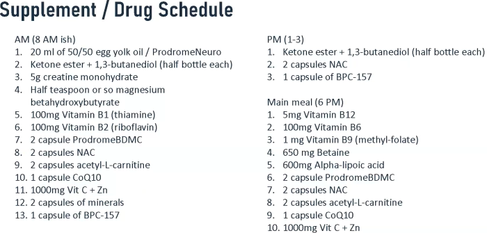
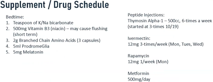
  • Take supplements that promote healthspan twice a day;
  • Exercise at least 30 minutes daily;
  • Take yeast cell wall β-glucan daily, with nothing else an hour before or after; and
  • Avoid undue stress by working from home 40 hours a week in my 26th year as a professional software developer.

Tactics’ main components activate AMPK, Nrf2, and associated signaling pathways, and inhibit pro-inflammatory pathways such as NF-κB. But fixing inflammation doesn’t repair all existing damage.

I haven’t had another three-year period in my life where I wasn’t sick even once. I’ll take that as a win, and wonder whether previous damage could have been resolved if I had started earlier.

2. One place I take clues from are successful anti-aging animal research efforts such as a study reviewed here earlier this month. Last curated in Improve your internal environment, improve its constituents’ functions, it used plasma fraction treatment. Plasma fraction eclipsed a caloric restriction treatment’s previous record for maximum species lifespan by 5%.

This type of research clearly isn’t a priority for official sponsors to fund, though. Take responsibility for your own one precious life.

Eat broccoli sprouts to protect your lungs

This 2023 human cell study investigated sulforaphane’s effects on tuberculosis infections:

“Basic research efforts on tuberculosis (TB) immunotherapy are currently only the tip of the iceberg. This study highlights the association between autophagy-related genes and immune infiltration in TB, an infectious pathogen that has been around for tens of thousands of years.

Sulforaphane (SFN) is readily absorbed into the bloodstream by the intestine due to its lipophilic nature. Experiments in this study TB patient cells showed that SFN could promote autophagy in macrophages infected with Mycobacterium abscessus (Mab). Intracellular bacterial load of macrophages was associated with SFN-enhanced cellular autophagic processes.

1-s2.0-S156757692300276X-gr4

The relationship between autophagy and immune cells is complex, and recurrence of tuberculosis is significantly influenced by intracellular mycobacteria of macrophages. Macrophages have longer lifespans than neutrophils, and provide shelter for mycobacteria as they are better suited than neutrophils to establish strategies for targeting autophagy.

This is one of the reasons why autophagy in macrophages was the focus of this study. Appropriate autophagy is beneficial for the body and controls Mtb replication, but autophagic programmed cell death can activate tissues to produce an excessive inflammatory response, resulting in severe damage to lung tissues.

Autophagy-related genes regulated by SFN have good diagnostic potential, with FOXO1 potentially serving as a target for TB immunotherapy. Downstream targets of FOXO1 include important pro-inflammatory signaling molecules such as IL-1β and TNF-α, which are important for control of mycobacterium.”

https://www.sciencedirect.com/science/article/pii/S156757692300276X “Identifying autophagy-related genes as potential targets for immunotherapy in tuberculosis”

Take yeast cell wall β-glucan, too, and train your immune system.

Eat broccoli sprouts for depression, Part 3

Here are two papers published after Part 2 that cited the Part 1 rodent study, starting with a 2023 rodent study performed by several Part 1 coauthors:

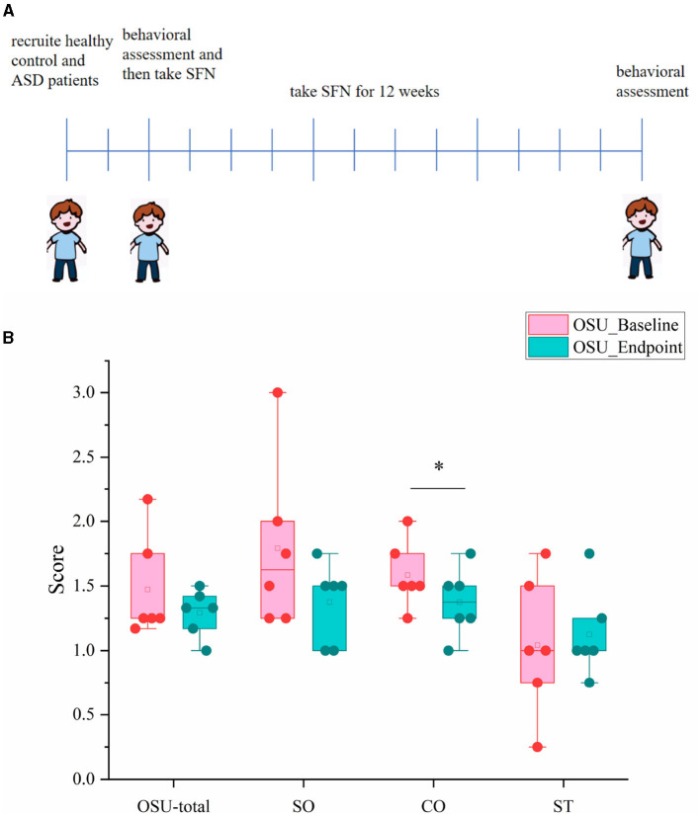
“We used a low-dose LPS-induced endotoxaemia model to mimic clinical characteristics of sepsis. We found that adolescent LPS treatment was sufficient to increase levels of inflammatory factor TNF-α in both the medial prefrontal cortex (mPFC) and hippocampus at post-natal day P22.

P21 LPS-treated mice were injected with sulforaphane (SFN) or saline intraperitoneally at P49 and then subjected to subthreshold social defeat stress (SSDS). We found that SFN preventative treatment significantly:

  • Decreased the social avoidance, anhedonia, and behavioural despair detected by the social interaction test, sucrose preference test, tail suspension test, and forced swim test, respectively.
  • Decreased anxiety-like behaviours without affecting locomotor activities.
  • Increased Nrf2 and brain-derived neurotrophic factor (BDNF) levels in the mPFC of P21 LPS-treated mice after SSDS compared with saline control mice.

The above results suggest that activation of the Nrf2-BDNF signalling pathway prevents the effect of adolescent LPS-induced endotoxaemia on stress vulnerability during adulthood.

sulforaphane and stress vulnerability

These results suggest that early adolescence is a critical period during which inflammation can promote stress vulnerability during adulthood. This might be due to increased inflammatory response in the mPFC, and mediated by decreased levels of Nrf2 and BDNF. These findings may shed light on the potential use of SFN as an alternative preventative intervention for inflammation-induced stress vulnerability.”

https://link.springer.com/article/10.1007/s00213-022-06285-4 “Lipopolysaccharide-induced endotoxaemia during adolescence promotes stress vulnerability in adult mice via deregulation of nuclear factor erythroid 2-related factor 2 in the medial prefrontal cortex” (not freely available)

This study demonstrated that adolescent diseases and stresses don’t necessarily develop into adult social problems. A timely intervention may even prevent future adult problems.

The one-time 10 mg/kg sulforaphane dose was the same as Part 1’s dose, a human equivalent of which is (10 mg x .081) x 70 kg = 57 mg.

I’d like to know more about how subjects’ memories of adverse events were retained, and subsequently affected their biology and behavior. Pretty sure limbic structures like the hypothalamus as well as lower brain structures played a part.


A 2022 review summarized what was known up to that time regarding Nrf2 and depression:

“Sulforaphane, an organosulfur compound isolated from Brassicaceae plants, is a potent natural NRF2 activator. Sulforaphane:

  • Exerts antidepressant- and anxiolytic-like activities and inhibits HPA axis and inflammatory response.
  • Has both therapeutic and prophylactic effects on inflammation-related depression.
  • Confers stress resilience.
  • Protects neurons via autophagy and promotes mitochondrial biogenesis by activating Nrf2.”

https://www.sciencedirect.com/science/article/pii/S2213231722002944 “Nrf2: An all-rounder in depression”


PXL_20230306_202857493

Oat β-glucan effects

Three papers on oat β-glucan’s effects in humans, starting with a 2023 study that compared different doses:

“Two randomized, double-blind, controlled studies were conducted with asymptomatic subjects between 20 and 40 years of age, male or female, normal weight or overweight.

In the first study – a crossover trial comprising two days of testing (β-glucan and control) separated by at least one week – 14 subjects ingested a breakfast with or without β-glucan from oats (5.2 g). Results indicate that acute intake of 5 g β-glucan slows transit time and decreases hunger sensation and postprandial glycaemia without affecting bile-acid synthesis. These changes were associated with decreased plasma insulin, C-peptide, and ghrelin, and increased plasma gastric inhibitory polypeptide  and pancreatic polypeptide.

In the second study, 32 subjects were distributed into 2 groups to ingest daily foods with (3 g/day) or without β-glucan for 3 weeks. Results indicate a regular daily intake of 3 g β-glucan is not sufficient to have an effect on fecal microbiota composition, suggesting that health-promoting effects at 3 g/d are probably due more to their physiological effect in the proximal part of the gastrointestinal tract than to their prebiotic effect in the colon.”

https://www.mdpi.com/2304-8158/12/4/700 “Modulation of Postprandial Plasma Concentrations of Digestive Hormones and Gut Microbiota by Foods Containing Oat ß-Glucans in Healthy Volunteers”


I’ll use a 2021 study Rapid Determination of β-Glucan Content of Hulled and Naked Oats Using near Infrared Spectroscopy Combined with Chemometrics to estimate my daily β-glucan intake. Those researchers tested 100 varieties of Avena nuda that varied between 3.12% and 5.22% β-glucan. My intake from 82 g (dry weight) of hulless oats (cinnamon sprinkled for taste) is probably between (82 g x .0312) = 3 g and (82 g x .0522) = 4 g.

They also tested 79 varieties of hulled Avena sativa that varied between 3.1% and 5.5% β-glucan. Oat sprouts analysis tested a Avena sativa variety where the β-glucan content decreased from 3.48% to 2.10% over four days of sprouting, a 40% reduction.

My daily β-glucan intake from 40 g (dry weight) of three-day-old hulled oat sprouts is probably 1 g [(40 g x .031) x .6 = 1 g and (40 g x .055) x .6 = 1 g]. That’s okay, because oat sprouts have other benefits per Oat sprouts analysis and Advantages of 3-day-old oat sprouts over oat grains.

My daily oat β-glucan intake is 4 – 5 grams. I’ve maintained that for two years, and don’t see any reason to stop.


A second 2023 paper from a clinical trial investigated effects of combining oat bran along with orange juice:

“Orange juice (OJ) is a rich dietary source of bioactive flavanones, and consuming OJ has been associated with beneficial effects including decreased inflammation and improved lipid profiles. However, dietary recommendations are to limit OJ consumption to one serving per day due to high sugar and low fiber content. Metabolic concerns are increased postprandial insulin response to a high sugar load which in individuals at risk may promote insulin resistance.

Consumption of 22 g oat bran containing 6 g of β-glucan together with 500 mL of OJ by healthy subjects impacts on OJ flavanone bioavailability with the 0-24 post-intake excretion of phase II metabolites, such as hesperetin-7-glucuronide, being reduced ~3-fold. This was not a consequence of bran affecting the rate of gastric transport, and underlying mechanisms behind reduced excretion of OJ flavanone metabolites remain a matter of conjecture.

The pool of bound phenolics in bran linked to polysaccharides appears not to be converted to free phenolics. It was rather principally a consequence of a bran-mediated increase in quantities of flavanones passing from the upper to the lower bowel where they were subjected to microbiota-mediated catabolism.”

https://www.sciencedirect.com/science/article/abs/pii/S0891584923000515 “Bioavailability of orange juice (poly)phenols: β-glucan-rich oat bran decreases urinary excretion of flavanone phase II metabolites and enhances excretion of microbiota-derived phenolic catabolites” (not freely available) Thanks to Dr. José Manuel Moreno-Rojas for providing a copy.

This paper referenced a preliminary study by many of the same coauthors that found oat bran with 3 g of β-glucan didn’t have similar effects.


A 2022 meta-analysis investigated differences between whole oats and purified β-glucan:

“This systematic review and meta-analysis evaluated the impact of oats or β-glucan supplements on the lipid profile. Our findings show that both oat and isolated β-glucan interventions can improve lipid profiles, specifically total cholesterol and low density lipoprotein cholesterol (LDL) concentrations, and should be incorporated into one’s regular eating habits.

Interventions ranged from 14 to 84 days in length. Quantity of β-glucan ingested (oats and isolated β-glucan) ranged from 1.2 g/day to 11.2 g/day.

Limitations and additional considerations include:

  • We did not have enough studies that matched total fiber intake between intervention and control groups, and so could not evaluate if results were exclusively influenced by oat/isolated β-glucan supplementation, or if other types of dietary fiber would have a similar impact on lipidemia.
  • Mechanisms of changes in concentrations of triglycerides (TG) are linked to carbohydrates. An increase in availability of glucose in serum, resulting from absorption of carbohydrates, stimulates secretion of insulin and, as a result, synthesis of fatty acids in the liver is increased. Mixed results found in this and other meta-analyses regarding TG may be related to the fact that oats and isolated β-glucan were frequently administered through day-to-day processed foods which have sugar and other types of refined flour in their recipes.
  • Different oat cooking procedures, processing methods, and molecular weights modify viscosity and impact in cholesterol concentrations differently. Less processed oats appear to be more effective than processed oat products in improving lipidemia. Higher molecular weight is associated with increased viscosity, and greater reduction in LDL. Also, the process used to treat oats affects its molecular weight, and the highest viscosities were observed as a consequence of dry processes in comparison to ones that exhibit enzymatic activity.
  • Reducing saturated fat intake may be, in combination with increased viscous fiber intake from oats or isolated β-glucan, the most effective way to improve dyslipidemia. In future studies, amount and type of fat in diet should be evaluated and considered accordingly.”

http://dx.doi.org/10.1016/j.clnesp.2022.12.019 “The separate effects of whole oats and isolated beta-glucan on lipid profile: A systematic review and meta-analysis of randomized controlled trials” (not freely available)


PXL_20230309_202539345

Take inulin for your brain

This 2023 rodent study investigated effects of inulin on gut microbiota and brain inflammation:

“Microglia are the first immune responders in the brain. Their activation leading to neuroinflammation can promote homeostasis, but if unchecked can be pathological.

We evaluated anti-inflammatory effects of short-chain fatty acids (SCFAs) on lipopolysaccharide (LPS)-stimulated microglia from mice fed inulin, a soluble fiber fermented by intestinal microbiota to produce SCFAs, and SCFAs applied to primary microglia in vitro:

  • Feeding mice inulin increased SCFAs in the cecum and in plasma collected from the hepatic portal vein.
  • Microglia isolated from mice fed inulin and stimulated with LPS in vitro secreted less tumor necrosis factor α (TNF-α) compared to microglia from mice not given inulin.
  • Mice fed inulin and injected i.p with LPS ex vivo secretion of TNF-α by isolated microglia was lower than that secreted by microglia from mice not fed inulin and injected with LPS.

41598_2022_27086_Fig8_HTML

in vitro treatment of primary microglia with acetate and butyrate either alone or in combination downregulated microglia cytokine production, with effects being additive. SCFAs reduced histone deacetylase activity and nuclear factor-κB nuclear translocation after LPS treatment in vitro.

If SCFAs produced in the gut regulate microglia directly, it is likely through an epigenetic mechanism following diffusion.”

https://www.nature.com/articles/s41598-022-27086-x “Inhibition of inflammatory microglia by dietary fiber and short-chain fatty acids”


Mice typically eat 4-5 grams of chow daily. A human equivalent of this study’s 2.5% inulin treatment would be:

  • (5,000 mg x .025) = 125 mg;
  • (125 mg x .081) = 10.125 mg;
  • (10.125 mg x 70 kg) = 709 mg.

A daily intake of < 1 gram of inulin isn’t very much. I take < 10 grams.

PXL_20221230_151820047

Ancient parasite DNA within us

Two 2023 papers on endogenous retroviruses (ERVs) and aging relationships, starting with the Introduction section of a comprehensive study:

“Several causal determinants of aging-related molecular changes have been identified, such as epigenetic alterations and stimulation of senescence-associated secretory phenotype (SASP) factors. Although the majority of these studies describe aging determinants originating primarily from protein-coding genes, the non-coding part of the genome has started to garner attention as well.

ERVs belonging to long terminal repeat (LTR) retrotransposons are a relic of ancient retroviral infection, fixed in the genome during evolution, comprising about 8% of the human genome. As a result of evolutionary pressure, most human ERVs (HERVs) accumulate mutations and deletions that prevent their replication and transposition function. However, some evolutionarily young subfamilies of HERV proviruses, such as the recently integrated HERVK, maintain open reading frames encoding proteins required for viral particle formation.

In this study, using cross-species models and multiple techniques, we revealed an uncharacterized role of endogenous retrovirus resurrection as a biomarker and driver for aging. Specifically, we identified endogenous retrovirus expression associated with cellular and tissue aging and that the accumulation of HERVK retrovirus-like particles (RVLPs) mediates the aging-promoting effects in recipient cells. More importantly, we can inhibit endogenous retrovirus-mediated pro-senescence effects to alleviate cellular senescence and tissue degeneration in vivo, suggesting possibilities for developing therapeutic strategies to treat aging-related disorders.”

https://www.cell.com/cell/fulltext/S0092-8674(22)01530-6 “Resurrection of endogenous retroviruses during aging reinforces senescence”


This first paper’s foreword summarized their many experiments and findings:

“The study found that HERVK transcripts, viral proteins, and RVLPs were highly activated in prematurely aged human mesenchymal progenitor cells (hPMCs). This was similarly observed in aged human primary fibroblasts and hPMCs. They also discovered that decreasing silencing epigenetic marks DNA methylation and H3K9me3 while increasing H3K36me3 enabled HERVK expression.

erv aging mechanism

These observations also raise several intriguing questions:

  • HERVK is occasionally activated and eventually suppressed under physiological conditions, for example, in human embryonic cells. It would be fascinating to probe the possibility of mimicking physiological conditions in order to turn off the positive feedback between HERVK and senescence.
  • ERVs are hallmarks of aging in different species, including human, primate, and mouse. Future quantification of the absolute physiological level of ERVs across a broad population of various ages might provide further insights into the relationship between ERVs and organismal age.”

https://academic.oup.com/lifemedi/advance-article/doi/10.1093/lifemedi/lnad001/6982772 “Endogenous retroviruses make aging go viral”


Previously curated papers on these subjects include:

A study of our evolutionary remnants

“Repressive epigenetic marks associated with ERVs, particularly LTRs, show a remarkable switch in silencing mechanisms, depending on evolutionary age:

  • Young LTRs tend to be CpG-rich and are mainly suppressed by DNA methylation, whereas
  • Intermediate age LTRs are associated predominantly with histone modifications, particularly histone H3 lysine 9 (H3K9) methylation.
  • Evolutionarily old LTRs are more likely inactivated by accumulation of loss-of-function genetic mutations.”

Starving awakens ancient parasite DNA within us

Reality is sometimes stranger than what fiction writers dream up. 🙂

PXL_20230209_210243470

Improve your internal environment, improve its constituents’ functions

A third update to Signaling pathways and aging:

“Sima, who was born on 28 February 2019, has lived for 47 months, surpassing the 45.5 months believed to be the oldest age recorded in scientific literature for a female Sprague-Dawley rat, the researchers say. So far, Sima has outlived her closest rival in the study by nearly six months.

‘The real point of our experiments is not so much to extend lifespan, but to extend youthspan, to rejuvenate people, to make their golden years really potentially golden years, instead of years of pain and decrepitude,’ Katcher said. ‘But the fact is, if you manage to do that, you also manage to lengthen life, and that’s not a bad side-effect.'”

https://www.theguardian.com/science/2023/feb/08/anti-ageing-scientists-extend-lifespan-of-oldest-living-lab-rat “Anti-ageing scientists extend lifespan of oldest living lab rat”


Whale funeral

PXL_20230208_211431462

Eat broccoli sprouts to protect your brain from stroke

Starting this blog’s ninth year with a 2022 rodent study of sulforaphane neuroprotection:

“An example of endogenous neuroprotection is ischemia-resistance of the hippocampal regions comprising the CA2, CA3, CA4 and dentate gyrus subfields (here abbreviated to CA2-4,DG) which can be contrasted with the ischemia-vulnerable CA1 region, which is noted in rodents as well as humans.

As with CA2-4,DG, nuclear Nrf2 levels are also higher in the olfactory bulb, while in the cortex, striatum, and cerebellum, they are similar to ones observed in the CA1 region.

brain area comparative Nrf2 activity

We found an in vitro dose-dependent response to administration of sulforaphane on neuronal viability, with an optimal effect noted where the dose was 10 µM. A protective effect was also evident in vivo when a single 5 mg/kg dose of sulforaphane was administered intraperitoneally with delay to ischemia.

Morphology of the CA1 region stratum pyramidale was significantly improved in comparison to ischemia-operated group, with mean numbers of proper cells being 35 ± 19 and 20 ± 7, respectively, for subjects injected during ischemia or 30 min into reperfusion. Morphology of the CA2-4,DG region did not reveal change between the ischemia-operated, SFN-injected, and control groups.

We suggest that high levels of nuclear Nrf2 activity in CA2-4,DG may guarantee resistance of this region to I/R episode, while at the same time offering a potential explanation for the phenomenon of differential sensitivities of hippocampal regions. Our results are in line with the existing view that Nrf2 activation may represent a promising therapeutic strategy against cerebral ischemia.

The uniqueness of Nrf2 lies in its pleiotropic action and subsequent regulation of multiple cytoprotective pathways. This may support more efficient neuroprotection compared to single-target strategies.”

https://link.springer.com/article/10.1007/s12035-022-03166-x “Is Nrf2 Behind Endogenous Neuroprotection of the Hippocampal CA2-4,DG Region?”


Winter beach shock therapy

PXL_20230129_174306200

Broccoli sprout beer?

This 2023 study investigated supplementing broccoli into beer:

“The objective of this work was to evaluate alternative broccoli materials (sprouts and by-products powder), which could be a sustainable dietary source of bioactive sulforaphane (SFN). Results showed high concentrations of SFN in beers supplemented with broccoli sprouts and powder (5.00 and 2.54 mg/L, respectively, previous to bottling), indicating that formation rate for SFN when adding sprouts and by-products powder were almost 38.5% and 19.5%, correspondingly. These concentrations remained stable until bottling, where they were reduced by >50%.

1-s2.0-S2212429222007180-gr1_lrg

Beers supplemented with broccoli presented a higher alcohol content (6.6%, on average) than control beer, regardless of the broccoli material used. Sensory analysis revealed positive attributes (colour, floral, fruity, and aftertaste as a result of the profile of volatile compounds) of beers developed using sprouts. Future research is still needed to stabilize SFN during bottling.”

https://www.sciencedirect.com/science/article/pii/S2212429222007180 “Broccoli products supplemented beers provide a sustainable source of dietary sulforaphane”


PXL_20221003_163955758

Week 144 of Changing to a youthful phenotype with sprouts

Two papers, starting with a 2023 study that investigated the same red radish cultivar as Sulforaphene, a natural analog of sulforaphane:

“Availability of microgreen products is constantly rising, i.e., they are offered for sale in local farmers markets, specialty stores, and in chain grocery stores. Due to the low demands required for their cultivation and easily available LED settings, microgreens are increasingly grown on a small scale in homes and after harvesting, they are stored in kitchen refrigerators at 4 °C.

The aim of this study was to simulate such cultivation and storage conditions to examine antioxidant capacity of home-grown radish microgreens. Seven-day-old radish microgreens, grown under purple and white LED light, were harvested and stored at 4 °C for two weeks.

Measurements of total antioxidant capacity and bioactive substances were conducted on the harvesting day and on the 3rd, 7th, and 14th day of storage. All three radish cultivars (Raphanus sativus L.) with different leaf colorations:

  • Purple radish (R. sativus cult. China Rose, cvP);
  • Red radish (R. sativus cult. Sango, cvR); and
  • Green radish (Raphanus sativus var. longipinnatus, Japanese white or daikon radish, cvG)

were purchased commercially from a local supplier.

The highest contents of total soluble phenolics, proteins, and sugars, dry matter, and monomeric anthocyanin content, as well as higher overall antioxidant capacity determined in the red radish cultivar (cvR), distinguished this cultivar as the most desirable for human consumption regardless of the cultivation light spectrum.”

https://www.mdpi.com/2311-7524/9/1/76 “Antioxidant Capacity and Shelf Life of Radish Microgreens Affected by Growth Light and Cultivars”


A 2021 review summarized what was known about radishes up to then. Here’s part of its Discussion section:

“It is worth considering radish’s organoleptic characteristics since its particular flavor can influence its acceptability among consumers. The main compound associated with its characteristic pungent flavor is raphasatin, which we have found to be the most reported isothiocyanate produced from the breakdown of glucoraphasatin.

Glucoraphasatin ranked as one of the most concentrated glucosinolates in radish, particularly in its sprouts, but also present in other parts like roots and seeds. Pungency differs among radish cultivars, environmental growth factors, agronomic, and cooking practices.”

1-s2.0-S0924224421003058-gr3_lrg

https://www.sciencedirect.com/science/article/pii/S0924224421003058 “Nutritional and phytochemical characterization of radish (Raphanus sativus): A systematic review”


Seeds I’ve sprouted this year so far, left to right – red radish (Sango), broccoli, red cabbage (Red Acre), yellow mustard, oat (Avena sativa):

PXL_20230106_121548495

Red radish had similar growth characteristics as broccoli. Started with 3.6 grams of seeds, which increased to 22.2 g after three days using the same soaking and rinsing protocol I use for other sprouts.

PXL_20230109_121815772

The taste of red radish was too sharp for me to eat by themselves, so I combined them with my broccoli / red cabbage / mustard sprout mix. Bumped up microwaving time to 48 seconds in a 1000 W microwave while staying short of the 60°C (140°F) myrosinase cliff.

The whole mix still had a strong radish taste, though. It was as if two whole red radishes were sliced into a small salad.

Can’t add anything more to dampen that taste and expect beneficial compounds to be unaffected. From Week 19:

A 2018 Netherlands study Bioavailability of Isothiocyanates From Broccoli Sprouts in Protein, Lipid, and Fiber Gels found:

Compared to the control broccoli sprout, incorporation of sprouts in gels led to lower bioavailability for preformed sulforaphane and iberin.”

IAW, eating protein, fats, and fiber along with microwaved broccoli sprouts wouldn’t help. A 2018 review with some of the same researchers Isothiocyanates from Brassica Vegetables-Effects of Processing, Cooking, Mastication, and Digestion offered one possible explanation for protein acting to lower broccoli sprout compounds’ bioavailability:

“In vitro studies show that ITCs can potentially react with amino acids, peptides, and proteins, and this reactivity may reduce the ITC bioavailability in protein‐rich foods. More in vivo studies should be performed to confirm the outcome obtained in vitro.”

Mixing in red radish sprouts also gave me an upset stomach five of the six mornings. So I won’t continue to sprout red radish.

That said, I’d definitely consider sprouting red radish again to accelerate isothiocyanate treatment of problems where symptoms are much worse than an upset stomach, such as:

  • Neurogenerative diseases with their cognitive decline;
  • Immune system disorders;
  • Bacterial and viral infections; and
  • Other damage caused by oxidative stress conditions in eyes, vascular system, kidney function, etc.

Piping in the New Year

PXL_20230101_193048797

Sulforaphene, a natural analog of sulforaphane

Three papers on sulforaphene, starting with a 2022 in vitro digestion study by Our model clinical trial for Changing to a youthful phenotype with broccoli sprouts group:

“This work aims to assess anti-inflammatory potential of bioactive compounds of cruciferous sprouts red radish (RRS) and red cabbage (RCS) in their bioaccessible form (obtained by the digestion of aqueous extracts). We used a well-established in vitro inflammation cellular model consisting of human macrophage-like HL60 cells stimulated with LPS, which mimics systemic chronic inflammatory conditions present in certain non-communicable diseases such as cardiovascular disease, cancer, and diabetes.

d2fo02914f-f1

Composition of RRS and RCS digestates extracts presented differences with a 20% lower content of total isothiocyanates (ITCs) in RRS than in RCS. However, there was more variability in the compounds present in RRS than in RCS extract digestates, including sulforaphene (SFE) and 3,3′-diindolylmethane (DIM), which were exclusively present in RRS.

RCS extract showed a trend of decreasing both TNF-α and IL-6 production under LPS-stimulated conditions, and this inhibitory effect was mainly observed at final protein expression. This activity at higher rates might be related to the inhibitory ability of iberin upon TLRs dimerization, impairing the NF-κB signaling pathway.

On the other hand, RRS exhibited a significant dose–response inhibition of IL-6 production levels. This difference in better performance of RRS compared to RCS could be exerted by the higher concentration of sulforaphane, and the exclusive presence of SFE, DIM, and anthocyanins in RRS.”

https://pubs.rsc.org/en/content/articlelanding/2023/FO/D2FO02914F “Anti-inflammatory potential of digested Brassica sprout extracts in human macrophage-like HL-60 cells”

I was surprised that this study didn’t detect anthocyanins in 8-day-old red cabbage sprout digestates, as they are visibly present in red cabbage sprouts. For example, from Week 56:

PXL_20210504_212505224


Reference 32 of this study was a 2021 review:

“Sulforaphene (SRP), as a product derived from glucoraphenin in the presence of myrosinase, mainly exists in cruciferous plants, especially in dried and mature seeds of radish. The most abundant ITC in juice of R. sativus L. coming from Sango freeze-dried sprouts is SRP. There is no safe and efficient SRP chemical synthesis which could be industrialized.

Structural variation in ITCs, such as the presence of particular functional group, molecular size, and length of a hydrocarbon chain, often results in very diverse antimicrobial activities. SRP, which is similar to sulforaphane in chemical structure but has an extra double bond, shows a much higher antimicrobial activity. However, the exact explanation for this enhanced microbial activity remains unknown.”

https://www.tandfonline.com/doi/full/10.1080/15422119.2021.1944209 “Sulforaphene: Formation, stability, separation, purification, determination and biological activities” (not freely available) Thanks to Dr. Jie Zhang for providing a copy.

Eat broccoli sprouts instead of antibiotics had two papers on ITCs’ antimicrobial actions.


A third paper was a 2022 cell study:

“Acne is a chronic inflammatory disease of the sebaceous gland attached to hair follicles. Cutibacterium acnes is a major cause of inflammation caused by acne.

It is well known that C. acnes secretes a lipolytic enzyme to break down lipids in sebum, and free fatty acids produced at this time accelerate the inflammatory reaction. There are several drugs used to treat acne; however, each one has various side effects.

We examined effects of sulforaphene (SFEN) on bacterial growth and inflammatory cytokine production induced by C. acnes. SFEN showed antibacterial activity against C. acnes and controlled the inflammatory response on keratinocytes and monocytes. This finding means that SFEN has potential as both a cosmetic material for acne prevention and a pharmaceutical material for acne treatment.”

https://www.jmb.or.kr/journal/download_pdf.php?doi=10.4014/jmb.2209.09051 “Sulforaphene Attenuates Cutibacterium acnes-Induced Inflammation”


I ordered the Sango variety of red radish seeds used in this first study, to arrive in two weeks. I expect that their flavor and sulforaphene combination will be a good substitute for the mainly-flavor mustard third of my 3-day-old sprouts brocolli / red cabbage / mustard sprouts morning mix.

Home sprouting cupboard setup, with Avena sativa twice-daily hulled oats sprouts on top:

PXL_20221208_123517082

Do broccoli sprouts treat asthma?

This 2022 rodent study investigated sulforaphane’s effects on airway disease:

“Sulforaphane has been studied in numerous preclinical and clinical models of lung damage and airway diseases. The lack of definitive findings from clinical studies to date most likely reflects issues with extract preparations and dosage regimes.

We investigated effects of administration of L-sulforaphane (LSF), which is also known as (R)-sulforaphane, in a murine model of ovalbumin (OVA)-induced chronic allergic airways disease (AAD). This model of chronic AAD recapitulates several features of human asthma including airway inflammation, airway remodeling, and airway hyper-responsiveness.

Our findings confirmed the efficacy of LSF in attenuating pathologies associated with AAD, involving activation of antioxidant and anti-inflammatory pathways. Inhibition of HDAC enzymes by LSF and accumulation of acetylated core histones and α-tubulin in vivo following LSF administration represent an important epigenetic regulatory mechanism. LSF and its metabolites may modulate HDAC6 and HDAC8 enzymes by binding to the catalytic site.

sulforaphane asthma

Our findings along with accumulated evidence, highlight the clinical potential of sulforaphane as either a prophylactic or a therapeutic in the context of AAD.”

https://link.springer.com/article/10.1007/s00018-022-04609-3 “Sulforaphane prevents and reverses allergic airways disease in mice via anti-inflammatory, antioxidant, and epigenetic mechanisms” (not freely available)


PXL_20221214_200748143

Eat mushrooms every day?

Three 2022 papers on amino acid ergothioneine, starting with a human study:

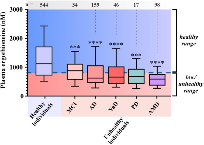
“We examined temporal relationships between plasma ergothioneine (ET) status and cognition in a cohort of 470 elderly subjects attending memory clinics in Singapore. All participants underwent baseline plasma ET measurements as well as neuroimaging for cerebrovascular disease (CeVD) and brain atrophy. Neuropsychological tests of cognition and function were assessed at baseline and follow-up visits for up to five years.

Lower plasma ET levels were associated with poorer baseline cognitive performance and faster rates of decline in function as well as in multiple cognitive domains including memory, executive function, attention, visuomotor speed, and language. In subgroup analyses, longitudinal associations were found only in non-demented individuals.

Mediation analyses showed that effects of ET on cognition seemed to be largely explainable by severity of concomitant CeVD, specifically white matter hyperintensities, and brain atrophy. Our findings support further assessment of plasma ET as a prognostic biomarker for accelerated cognitive and functional decline in pre-dementia and suggest possible therapeutic and preventative measures.”

https://www.mdpi.com/2076-3921/11/9/1717 “Low Plasma Ergothioneine Predicts Cognitive and Functional Decline in an Elderly Cohort Attending Memory Clinics”


Earlier this year, two of the study’s coauthors put together a collection of 11 ergothioneine papers:

“One catalyst for this upsurge of interest was the discovery in 2005 of a transporter for ET (OCTN1, often now called the ergothioneine transporter, ETT), which accounts for the fact that animals (including humans) take up and avidly retain ET from the diet. The presence of a specific transporter together with the avid retention of ET in the body implies that this compound is important to us.

To quote an old phrase ‘correlation does not imply causation.’ Low ET levels may predispose to disease, but disease could also lead to low ET levels. Possible reasons could include:

  • Alterations in diet due to illness so that less ET is consumed;
  • Decreases in ETT activity in the gut (leading to less ET uptake) or kidney (impairing ET reabsorption) with age and disease.
  • Changes in gut microbiota might influence uptake and accumulation in the body.
  • ET is being consumed as it scavenges oxygen radicals and other reactive oxygen species, the production of which is known to increase in these diseases and during ageing in general.

Only the gold standard of placebo-controlled double-blinded clinical studies can definitively establish the value (if any) of ET in preventing or treating human disease. Several such trials are being planned or in progress; we await the results with interest, and a streak of optimism.”

https://febs.onlinelibrary.wiley.com/doi/10.1002/1873-3468.14350 “Ergothioneine, where are we now?”


One of the collection’s papers focused on what ETT research findings could or could not be replicated:

“ETT is not expressed ubiquitously and only cells with high ETT cell-surface levels can accumulate ET to high concentration. Without ETT, there is no uptake because the plasma membrane is essentially impermeable. We review substrate specificity and localization of ETT, which is prominently expressed in neutrophils, monocytes/macrophages, and developing erythrocytes.

Comparison of transport efficiency (TE) for acknowledged substrates of the ETT. Bar length represents approximate TE of wild-type human ETT.

feb214269-fig-0001-m

We have not found in the literature any other ET transporters. However, it is highly probable that additional ET transporters work in the human body:

  • Uptake of ET from the small intestine into epithelial cells occurs through apically localized ETT. The very hydrophilic ET cannot then exit these cells toward the blood without help – a basolateral efflux transporter is required.
  • After oral administration of 3H-ET, a considerable amount of ET was still absorbed into the body in the ETT KO [knockout] mice. There must be another transporter for apical uptake at least in the small intestine of the mouse.
  • When ET was administered intravenously, ETT KO mice showed no change in ET concentration in the brain compared to wild type. The little ET that enters the brain must therefore pass through the BBB via a different transporter.”

https://febs.onlinelibrary.wiley.com/doi/10.1002/1873-3468.14269 “The ergothioneine transporter (ETT): substrates and locations, an inventory”


It’s persuasive that there’s an evolutionarily conserved transmitter specific to ergothioneine. It isn’t persuasive that this compound once consumed is almost always in stand-by mode to do: what?

Ergothioneine isn’t a substitute for the related glutathione, especially since its supply isn’t similarly available from an endogenous source. It isn’t an active participant in day-to-day human life.

Still, I hedge my bets. I eat ergothioneine every day via white button mushrooms in AGE-less chicken vegetable soup at a cost of about $1.30.

PXL_20221210_191511270

What do we know about human aging from mouse models?

Here is a 2021 rodent study and relevant parts from 3 of its 26 citing papers:

“A long line of evidence has established the laboratory mouse as the prime model of human aging. However, relatively little is known about detailed behavioral and functional changes that occur across their lifespan, and how this maps onto the phenotype of human aging.

To better understand age-related changes across the lifespan, we characterized functional aging in male C57BL/6J mice of five different ages (3, 6, 12, 18, and 22 months of age) using a multi-domain behavioral test battery. Assessment of functional aging in humans and mice: age-related patterns were determined based on representative data (Table 2), and then superimposed onto survival rate. (A) Body weight, (B) locomotor activity, (C) gait velocity, (D) grip strength, (E) trait anxiety, (F) memory requiring low attention level, and (G) memory requiring high attention level.

fnagi-13-697621-g012

These functional alterations across ages are non-linear, and patterns are unique for each behavioral trait. Physical function progressively declines, starting as early as 6 months of age in mice, while cognitive function begins to decline later, with considerable impairment present at 22 months of age.

Functional aging of male C57BL/6J mice starts at younger relative ages compared to when it starts in humans. Our study suggests that human-equivalent ages of mice might be better determined on the basis of its functional capabilities.”

https://www.frontiersin.org/articles/10.3389/fnagi.2021.697621/full “Functional Aging in Male C57BL/6J Mice Across the Life-Span: A Systematic Behavioral Analysis of Motor, Emotional, and Memory Function to Define an Aging Phenotype”


“Studies in mice show that physical function (i.e., locomotor activity, gait velocity, grip strength) begins to deteriorate around post-natal day (PND) 180, but cognitive functions (i.e., memory) do not exhibit impairment until roughly PND 660. Our results should be considered within the context of behavior changing throughout vole adulthood. Caution should be taken to avoid categorizing the oldest age group in our study as ‘elderly’ or ‘geriatric.'”

https://journals.plos.org/plosone/article?id=10.1371/journal.pone.0276897 “Behavioral trajectories of aging prairie voles (Microtus ochrogaster): Adapting behavior to social context wanes with advanced age”


“We used adult mice ranging in age from 5-6 months, not enough to modify experimental autoimmune encephalomyelitis progression. Mice are considered adult after 8 weeks; however, rapid growth for most biological processes is observed until 3 months of age, while past 6 months, mice might be affected by senescence.”

https://www.frontiersin.org/articles/10.3389/fimmu.2022.1036680/full “Age related immune modulation of experimental autoimmune encephalomyelitis in PINK1 knockout mice”


“Locomotor activity and gait velocity of 12 months old male C57BL/6 correlates with an elderly human being aged 60 or older, supporting that the ~15 months old mice we used in our study were aged mice at the time of tissue collection.”

https://www.mdpi.com/1422-0067/23/20/12461 “Genomic Basis for Individual Differences in Susceptibility to the Neurotoxic Effects of Diesel Exhaust”


PXL_20221122_200643133

Broccoli sprouts activate the AMPK pathway, Part 4

Today someone viewed the 2020 Part 3 of Broccoli sprouts activate the AMPK pathway which lacked citations at the time. Checking again, here are three citing 2022 papers, starting with a review:

“Nrf2 is an important transcription factor that regulates expression of a large number of genes in healthy and disease states. Nrf2 regulates expression of several key components of oxidative stress, mitochondrial biogenesis, mitophagy, autophagy, and mitochondrial function in all organs of the human body, and in the peripheral and central nervous systems.

Overall, therapeutic drugs including sulforaphane that target Nrf2 expression and Nrf2/ARE pathway are promising. This article proposes additional research in Nrf2’s role within Parkinson’s disease, Huntington’s disease, and ischemic stroke in preclinical mouse models and humans with age-related neurodegenerative diseases.”

https://www.sciencedirect.com/science/article/pii/S1568163722001982 “Role of Nrf2 in aging, Alzheimer’s and other neurodegenerative diseases” (not freely available) Thanks to Dr. P. Hemachandra Reddy for providing a copy.


One of the Part 3 study’s coauthors contributed to this very detailed review:

“Due to observed overlapping cellular responses upon AMPK or NRF2 activation and common stressors impinging on both AMPK and NRF2 signaling, it is plausible to assume that AMPK and NRF2 signaling may interdepend and cooperate to readjust cellular homeostasis.

1-s2.0-S089158492200497X-gr3_lrg

The outcome and underlying signaling events of AMPK-NRF2 crosstalk may diverge between:

  1. in vitro and in vivo studies (one cell type in isolation vs inter-organ crosstalk in living organisms);
  2. Different cell types/organs/organisms of different cultivation conditions, genetic background, age or sex;
  3. Different stress-regimens (chronic vs acute, nature of stress (lipotoxicity, redox stress, xenobiotic, starvation, etc));
  4. Different modes of Nrf2 or AMPK activation and inhibition (genetic vs pharmacological, constitutive vs transient/intermittent, systemic vs organ-specific, electrophilic vs PPI, allosteric vs covalent, or pan vs subtype-specific);
  5. Different target genes with distinct promoter and enhancer structure; or
  6. Different timing of activation.

The latter should deserve increased attention as Nrf2 is one of the most cycling genes under control of the circadian clock. Feeding behavior, metabolism and hence AMPK activity follow and substantiate the biological clock, indicating an entangled circadian regulation of metabolic and redox homeostasis.”

https://www.sciencedirect.com/science/article/pii/S089158492200497X “AMPK and NRF2: Interactive players in the same team for cellular homeostasis?”


A third citing paper was a study of lens cells that provided an example of similar metformin effects noted in Part 2 of Broccoli sprouts activate the AMPK pathway:

“Loss of Nrf2 and Nrf2 antioxidant genes expression and activity in aging cells leads to an array of oxidative-induced deleterious responses, impaired function, and aging pathologies. This deterioration is proposed to be the primary risk factor for age-related diseases such as cataracts.

AMPK regulates energy at physiological levels during metabolic imbalance and stress. AMPK is a redox sensing molecule, and can be activated under cellular accumulation of reactive oxygen species, which are endogenously produced due to loss of antioxidant enzymes.

The therapeutic potential of AMPK activation has context-dependent beneficial effects, from cell survival to cell death. AMPK activation was a requisite for Bmal1/Nrf2-antioxidants-mediated defense, as pharmacologically inactivating AMPK impeded metformin’s effect.

Using lens epithelial cell lines (LECs) of human or mouse aging primary LECs along with lenses as model systems, we demonstrated that metformin could correct deteriorated Bmal1/Nrf2/ARE pathway by reviving AMPK-activation and transcriptional activities of Bmal1/Nrf2, resulting in increased antioxidants enzymatic activity and expression of Phase II enzymes. Results uncovered crosstalk between AMPK and Bmal1/Nrf2/antioxidants mediated by metformin for blunting oxidative/aging-linked pathobiology.”

https://www.mdpi.com/2073-4409/11/19/3021/htm “Obligatory Role of AMPK Activation and Antioxidant Defense Pathway in the Regulatory Effects of Metformin on Cellular Protection and Prevention of Lens Opacity”


PXL_20221027_185754842