A free educational series recorded in 2021-2022 available at https://drgoodenowe.com/dr-goodenowes-educational-seminars/ takes the viewer through underlying research and principles of Dr. Goodenowe’s approach to health. It’s advertised as lasting four hours, but took me two days to view.
The series’ discussions and references are background material to better understand later presentations and interviews. Points of interest included:
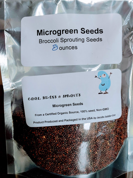
- Seminar B100 shows that the metabolomic profile of people who regularly eat broccoli is different than others.
- B109 clarifies how peroxisomal function is improved through resistance exercise and intermittent fasting.
- C103 and C104 show how plasmalogens act against neurodegeneration (Parkinson’s disease and multiple sclerosis).
Texts below videos are additional information, not transcripts. C101 text is historically informative.
The B200 ProdromeScan tutorial will take more study. But unlike Labcorp tests, ordering a ProdromeScan requires using a practitioner in Dr. Goodenowe’s network.
I sent the following to Prodrome customer service earlier this month:
Please add me to your approved list for ProdromeScan.
Customer service replied:
“We only add health professionals to an approved list, not individuals.”
I responded:
Good morning. I looked at the websites of doctors who are associated with Dr. Goodenowe who are near me. All of them are too compromised for me to establish a doctor / patient relationship. But I’m glad they left up their blog posts from earlier this decade so I could see who they really were before I reached out to them.
I request an exception to the policy.
Customer service replied:
“There is no exception that can be made to this policy. You need to be a patient of a certified practitioner.”
I’ll escalate my request before my 90-day trial of Prodrome Glia and Neuro products ends so I can get an appropriate metabolomic status. Right now, I won’t involve someone I can’t trust just to know my ProdromeScan information that’s additional to next week’s Labcorp tests.
My treatment-result metabolomic data is probably not mature today on Day 29 of ProdromeGlia and ProdromeNeuro supplementation, resistance exercise, and intermittent fasting. I otherwise wouldn’t have experienced these two events:
I have a quibble with the series’ recommendations for taking N-acetyl cysteine. Relevant views and research:
Switch on your Nrf2 signaling pathway pointed out:
“We use NAC in the lab all the time because it stops an Nrf2 activation. So that weak pro-oxidant signal that activates Nrf2, you switch it off by giving a dose of NAC. It’s a potent antioxidant in that right, but it’s blocking signalling. And that’s what I don’t like about its broad use.”
If someone bombs themself everyday with antioxidants, they’re doing nothing to improve training of their endogenous systems’ defensive functions. What happens when they stop bombing? One example was a 2022 human study that found GlyNAC-induced improvements dissolved back to baseline after supplements stopped.
Also, Precondition your defenses with broccoli sprouts highlighted NAC’s deleterious effects on autophagy and lysosome functions:
“TFEB activity is required for sulforaphane (SFN)-induced protection against both acute oxidant bursts and chronic oxidative stress. SFN-induced TFEB nuclear accumulation was completely blocked by pretreatment of cells by N-acetyl-cysteine (NAC), or by other commonly used antioxidants. NAC also blocked SFN-induced mRNA expression of TFEB target genes, as well as SFN-induced autophagosome formation.”
If a secondary goal of taking NAC per is also necessary for the formation of glutathione, taurine can do that without an antioxidant bomb. Taurine supplementation will free up cysteine to do things other than synthesize taurine, like synthesize glutathione.