Trained immunity mechanisms

This 2021 cell study investigated how inflammatory memory is established, maintained, and recalled:

“Cells retain a memory of inflammation that equips them to react quickly and broadly to diverse secondary stimuli. Temporal, dynamic changes to chromatin accessibility, histone modifications, and transcription factor (TF) binding occur during inflammation, post-resolution, and in memory recall following injury.

Epigenetic records of inflammation have been found in innate immune cells, including macrophages, monocytes, and natural killer cells, as well as CD8+ and regulatory T cells, granulocyte-monocyte progenitors, and long-term hematopoietic stem cells. Inflammatory memory was recently extended to epithelial barrier tissues, which are the first line of defense against infectious pathogens and noxious agents.

Epigenetic memory of an inflammatory experience is rooted in chromatin of a cell via retention of chromatin accessibility, histone marks, and key TFs that endow it with heightened responsiveness to diverse secondary stimuli. AP-1 (activating protein-1) is a collective term referring to transcription factors composed of JUN, FOS, or ATF (activating transcription factor) subunits that bind to a common DNA site.

1-s2.0-S1934590921002861-fx1_lrg

We unearth an essential, unifying role for the general stress-responsive transcription factor FOS, which partners with JUN and cooperates with stimulus-specific STAT3 to establish memory. JUN then remains with other homeostatic factors on memory domains, facilitating rapid FOS re-recruitment and gene re-activation upon diverse secondary challenges.

We offer a comprehensive, potentially universal mechanism behind inflammatory memory and less discriminate recall phenomena with implications for tissue fitness in health and disease:

  1. Stimulus-specific STAT3 and broad stress factor AP1 co-establish memory domains;
  2. Stem cell factors access open memory domains and remain bound after inflammation;
  3. FOS activates open memory domains, enabling secondary responses to diverse stimuli; and
  4. AP1 mediates epigenetic inflammatory memory across cell types, stimuli, and species.”

https://www.sciencedirect.com/science/article/abs/pii/S1934590921002861 “Establishment, maintenance, and recall of inflammatory memory” (not freely available)


Take responsibility for your own one precious life. Train your immune system every day with yeast cell wall β-glucan.

PXL_20211003_110857218

Dementia blood factors

This 2021 human study performed blood metabolite analyses:

“Dementia is a collective term to describe various symptoms of cognitive impairment in a condition in which intelligence is irreversibly diminished due to acquired organic disorders of the brain, characterized by deterioration of memory, thinking, behavior, and the ability to perform daily activities.

In this study, we conducted nontargeted, comprehensive analysis of blood metabolites in dementia patients. Effort expended in this ‘no assumptions’ approach is often recompensed by identification of diagnostic compounds overlooked by targeted analysis.

The great variability of data in Figure 1 reflects genuine individual variation in metabolites, which were accurately detected by our metabolomic analysis. These data demonstrate that compounds having small to large individual variability are implicated in dementia.

dementia blood factors

7 group A compounds – plasma-enriched dementia factors – increased in dementia patients and might have a negative toxic impact on central nervous system (CNS) functions by themselves or their degradation products.

26 group B to E metabolites may be beneficial for the CNS, as their quantity all declined in dementia patients:

  • Red blood cell (RBC)-enriched group B metabolites all containing the trimethyl-ammonium ion may protect the CNS through their antioxidative and other activity.
  • Group C compounds, also RBC-enriched, have cellular functions implicated in energy, redox, and so forth, and may be important for maintaining CNS brain functions.
  • Group D’s 12 plasma compounds (amino acids, nucleosides, choline, and carnitine) – half of which had been reported as Alzheimer’s disease (AD)-related markers – may underpin actions of other metabolites for supply and degradation. Consistency of group D plasma metabolites as dementia markers but not group B and C RBC metabolites validated the method of searching dementia markers that we employed in the present study.
  • Group E compounds, caffeine and and its derivative dimethyl-xanthine, declined greatly in dementia subjects. Caffeine is an antagonist of adenosine, consistent with the present finding that adenosine belongs to group A compounds.

Twelve [groups B + C] of these 33 compounds are RBC-enriched, which has been scarcely reported. The majority of metabolites enriched in RBCs were not identified in previous studies.

Nine compounds possessing trimethylated ammonium ions are amphipathic compounds (with both hydrophilic and lipophilic properties) and form the basis of lipid polymorphism. All of them showed a sharp decline in abundance in dementia subjects.

amphipathic compounds

These amphipathic compounds may have similar roles, forming a higher-ordered, assembled structure. They might act as major neuroprotectants or antioxidants in the brain, and their levels are sensitive to both antioxidants and ROS.

We speculate the 7 group A compounds pathologically enhance or lead to severe dementia such as AD. This presumed dementia deterioration by group A factors is opposed if group B to E metabolites are sufficiently supplied.

However, group A markers were not found in frail subjects. If the change in group A is causal for dementia, then a cognitive cause in frailty may be distinct from that of dementia.”

https://www.pnas.org/content/118/37/e2022857118 “Whole-blood metabolomics of dementia patients reveal classes of disease-linked metabolites”


Dementia subjects (ages 75-88) lived in an Okinawa hospital. Healthy elderly (ages 67-80) and young (ages 28-34) subjects lived in a neighboring village. Of the 24 subjects, 3 dementia and 1 healthy elderly were below a 18.5 to <25 BMI range, and none were above.

Get neuroprotectants working for you. Previous relevant curations included:

The Illusion of Knowledge: The paradigm shift in aging research that shows the way to human rejuvenation

Dr. Harold Katcher increased interviews to coincide with release of his book this month. Here’s one in four parts that provides highlights of his rejuvenation research progress:


Previously curated papers of his work include:

Epigenetic clocks so far in 2021

2021’s busiest researcher took time out this month to update progress on epigenetic clocks:

Hallmarks of aging aren’t all associated with epigenetic aging.

epigenetic aging vs. hallmarks of aging

Interventions that increase cellular lifespan aren’t all associated with epigenetic aging.

epigenetic aging vs. cellular lifespan

Many of his authored or coauthored 2021 papers developed human / mammalian species relative-age epigenetic clocks.

epigenetic clock mammalian maximum lifespan

Relative-age epigenetic clocks better predict human results from animal testing.

pan-mammalian epigenetic clock


Previously curated papers that were mentioned or relevant included:

Gut microbiota and critical development periods

This 2021 rodent study focused on global histone acetylation as a model to understand roles of microbially produced short-chain fatty acids in liver function:

“Despite the utility of germ-free mice in probing complex interactions between gut microbiota and host physiology, germ-free mice are developmentally, physiologically, and metabolically unique when compared with their conventionally housed counterparts. We sought to determine whether antibiotic-mediated microbiota depletion would affect global hepatic histone acetylation states through SCFA-dependent mechanisms, as previously observed in germ-free mice.

The inability of antibiotic-mediated microbiota depletion to recapitulate findings observed in germ-free mice suggests that the transition from a germ-free to a colonized mouse leads to resilient alterations in hepatic histone acetylation states that cannot be altered by further modulating the microbial environment. This finding is distinct from other germ-free phenotypes that are considered to be partially reversible, with clear alterations in their function observed after antibiotic treatment.

histone acetylation

Comparing antibiotic-treated and untreated mice that both received CCl4 at 24 and 48 hours after injury, there were almost no histone acetylation differences. This demonstrates that hepatic injury leads to a global shift in histone acetylation that is primarily independent of gut microbiota.

Major chromatin reorganization driven by histone acetylation leads to markers of differentiation, and addition of targeted differentiation signals induces events to stabilize these histone acetylation patterns – a key feature of embryonic development and terminal cellular differentiation. Differences in histone acetylation patterns seen between germ-free and conventionally raised mice may be a developmental-like effect of hepatocytes not yet exposed to microbial by-products.

Results suggest that microbial and dietary modifications to the gut microbiome in conventionally raised mice are not a means to modulate global hepatic histone acetylation. Microbiota-dependent landscaping of the hepatic epigenome appears static in nature, while the hepatic transcriptome is responsive to alterations in the gut microbiota, yet independent of global histone acetylation.

Findings underscore significant differences between these model systems that should be taken into account when considering their relevance to human biology.”

https://aasldpubs.onlinelibrary.wiley.com/doi/10.1002/hep.32043 “Global Microbiota-Dependent Histone Acetylation Patterns Are Irreversible and Independent of Short Chain Fatty Acids” (not freely available) Thanks to Dr. Elliot S. Friedman for providing a copy.


1. By describing “a key feature of embryonic development,” this study provided a gut microbiota-liver analogy of critical periods. If developmental events don’t happen when they are required, it’s probable that their window is missed, and won’t reopen later for a second chance at normalizing.

2. Many studies used a germ-free animal model, such as:

This study provided evidence for a limitation of this model, especially when extrapolating germ-free animal results to humans without similarly testing humans.


PXL_20210914_165000840_exported_1399

Eat oat avenanthramides for your gut microbiota

This 2021 paper covered a 2016 human clinical trial, and several in vitro and rodent follow-up studies:

“Oat has been widely accepted as a key food for human health. It is becoming increasingly evident that individual differences in metabolism determine how different individuals benefit from diet. Both host genetics and gut microbiota play important roles on metabolism and function of dietary compounds.

Results:

  • Avenanthramides (AVAs), the signature bioactive polyphenols of whole-grain (WG) oat, were not metabolized into their dihydro forms, dihydro-AVAs (DH-AVAs), by both human and mouse S9 fractions.
  • DH-AVAs were detected in colon and distal regions, but not in proximal and middle regions of the perfused mouse intestine, and were in specific pathogen–free (SPF) mice but not in germ-free (GF) mice.
  • A kinetic study of humans fed oat bran showed that DH-AVAs reached their maximal concentrations at much later time points than their corresponding AVAs (10.0–15.0 hours vs. 4.0–4.5 hours, respectively).
  • We observed interindividual variations in metabolism of AVAs to DH-AVAs in humans.
  • Faecalibacterium prausnitzii was identified as the individual bacterium to metabolize AVAs to DH-AVAs by 16S rRNA sequencing analysis.
  • Moreover, as opposed to GF mice, F. prausnitzii–monocolonized mice were able to metabolize AVAs to DH-AVAs.

AVA metabolizers

These findings demonstrate that intestinal F. prausnitzii is indispensable for proper metabolism of AVAs in both humans and mice. We propose that abundance of F. prausnitzii can be used to subcategorize individuals into AVA metabolizers and nonmetabolizers after WG oat intake.

Our findings pave the way to use AVAs and DH-AVAs as exposure biomarkers to reflect WG oat intake, which could more accurately record WG oat intake. Whether production of DH-AVAs is part of the beneficial effect of oats on human health will require further investigation.”

https://academic.oup.com/jn/article/151/6/1426/6165027 “Avenanthramide Metabotype from Whole-Grain Oat Intake is Influenced by Faecalibacterium prausnitzii in Healthy Adults”

Commentary at Faecalibacterium prausnitzii Abundance in Mouse and Human Gut Can Predict Metabolism of Oat Avenanthramides.


This study advanced an understanding of inter-individual variability, rather than usual practices that try to sweep individual differences under a statistical rug. Study designs such as four mentioned in Part 2 of Switch on your Nrf2 signaling pathway could have benefited from a similar approach to their research areas.

Not sure why it took over five years to get this paper published after its clinical trial’s January 21, 2016 completion. Meanwhile, science marched on to study effects of specific F. prausnitzii strains, providing results such as three human studies curated in Gut microbiota strains:

  • The third 2018 study found:

    “Only a small number of bacteria with genetic capacity for producing SCFAs were able to take advantage of this new resource and become dominant positive responders. The response, however, was strain specific: only one of the six strains of Faecalibacterium prausnitzii was promoted.”

  • The second 2021 study investigated 135 known strains of F. prausnitzii; and
  • The first 2021 study found beneficial F. prausnitzii strains not yet covered in genomic databases.

Resistant starch therapy recommended de-emphasizing relative gut microbiota abundance measurements, because:

“Relative abundances of smaller keystone communities (e.g. primary degraders) may increase, but appear to decrease simply because cross-feeders [like F. prausnitzii] increase in relative abundance to a greater extent. These limitations illustrate the necessity of sufficiently powering resistant starch interventions where microbiome composition is the primary endpoint, collecting critical baseline data and employing appropriate statistical techniques.”


Four humpback whales successively diving for lunch

PXL_20210914_164307307_exported_16255

PXL_20210914_164307307_exported_26282

PXL_20210914_164307307_exported_41871

PXL_20210914_164307307_exported_50365

Natural products vs. neurodegenerative diseases

I was recently asked about taking rapamycin for its effects on mTOR. I replied that diet could do the same thing. Here’s a 2021 review outlining such effects:

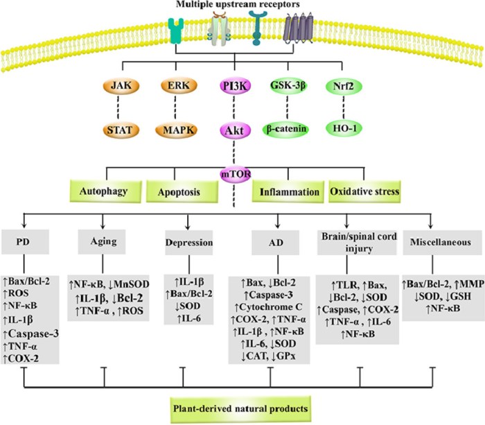
“As common, progressive, and chronic causes of disability and death, neurodegenerative diseases (NDDs) significantly threaten human health, while no effective treatment is available. Recent studies have revealed the role of phosphoinositide 3-kinase (PI3K)/Akt (Protein kinase B)/mammalian target of rapamycin (mTOR) in some diseases and natural products with therapeutic potentials.

Growing evidence highlights the dysregulated PI3K/Akt/mTOR pathway and interconnected mediators in pathogenesis of NDDs. Side effects and drug-resistance of conventional neuroprotective agents urge the need for providing alternative therapies.

1-s2.0-S0944711321002075-ga1_lrg

Polyphenols, alkaloids, carotenoids, and terpenoids have shown to be capable of a great modulation of PI3K/Akt/mTOR in NDDs. Natural products potentially target various important oxidative/inflammatory/apoptotic/autophagic molecules/mediators, such as Bax, Bcl-2, p53, caspase-3, caspase-9, NF-κB, TNF-α, GSH, SOD, MAPK, GSK-3β, Nrf2/HO-1, JAK/STAT, CREB/BDNF, ERK1/2, and LC3 towards neuroprotection.

This is the first systematic and comprehensive review with a simultaneous focus on the critical role of PI3K/Akt/mTOR in NDDs and associated targeting by natural products.”

https://www.sciencedirect.com/science/article/abs/pii/S0944711321002075 “Natural products attenuate PI3K/Akt/mTOR signaling pathway: A promising strategy in regulating neurodegeneration” (not freely available) Thanks to Dr. Sajad Fakhri for providing a copy.


Natural products mentioned in this review that I eat in everyday foods are listed below. The most effective ones are broccoli and red cabbage sprouts, and oats and oat sprouts:

  • Artichokes – luteolin;
  • Blackberries – anthocyanins;
  • Blueberries – anthocyanins, gallic acid, pterostilbene;
  • Broccoli and red cabbage sprouts – anthocyanins, kaempferol, luteolin, quercetin, sulforaphane;
  • Carrots – carotenoids;
  • Celery – apigenin, luteolin;
  • Green tea – epigallocatechin gallate;
  • Oats and oat sprouts – avenanthramides;
  • Strawberries – anthocyanins, fisetin;
  • Tomatoes – fisetin.

Four humpback whales

PXL_20210914_170732350_exported_43137

Screenshot_20210914-121800

All about vasopressin

This 2021 review subject was vasopressin:

“Vasopressin is a ubiquitous molecule playing an important role in a wide range of physiological processes, thereby implicated in pathomechanisms of many disorders. The most striking is its central effect in stress-axis regulation, as well as regulating many aspects of our behavior.

Arginine-vasopressin (AVP) is a nonapeptide that is synthesized mainly in the supraoptic, paraventricular (PVN), and suprachiasmatic nucleus of the hypothalamus. AVP cell groups of hypothalamus and midbrain were found to be glutamatergic, whereas those in regions derived from cerebral nuclei were mainly GABAergic.

In the PVN, AVP can be found together with corticotropin-releasing hormone (CRH), the main hypothalamic regulator of the HPA axis. The AVPergic system participates in regulation of several physiological processes, from stress hormone release through memory formation, thermo- and pain regulation, to social behavior.

vasopressin stress axis

AVP determines behavioral responses to environmental stimuli, and participates in development of social interactions, aggression, reproduction, parental behavior, and belonging. Alterations in AVPergic tone may be implicated in pathology of stress-related disorders (anxiety and depression), Alzheimer’s, posttraumatic stress disorder, as well as schizophrenia.

An increasing body of evidence confirms epigenetic contribution to changes in AVP or AVP receptor mRNA level, not only during the early perinatal period, but also in adulthood:

  • DNA methylation is more targeted on a single gene; and it is better characterized in relation to AVP;
  • Some hint for bidirectional interaction with histone acetylation was also described; and
  • miRNAs are implicated in the hormonal, peripheral role of AVP, and less is known about their interaction regarding behavioral alteration.”

https://www.mdpi.com/1422-0067/22/17/9415/htm “Epigenetic Modulation of Vasopressin Expression in Health and Disease”


Find your way, regardless of what the herd does.

PXL_20210911_103344386

Choosing your gut immune response

This 2021 paper reviewed evidence for immune system effects associated with specific gut areas:

“The intestinal immune system must not only contend with continuous exposure to food, commensal microbiota, and pathogens, but respond appropriately according to intestinal tissue differences. The entire intestine, inclusive of its lymph nodes, is considered a immunosuppressive organ overall compared to most other tissues, indicating that a state of tolerance to food and commensals – yet vigilance toward pathogens – was an evolutionarily stable strategy.

By operating in compartments, the immune system may generate multiple immune outcomes, even with simultaneous opposite goals e.g., tolerance or inflammation. Generation of unique immunologic niches within the intestine is influenced by a combination of tissue intrinsic properties, extrinsic environmental factors, and regionalized immune populations.

intestinal immune compartmentalization

Complexity of intrinsic and extrinsic driving forces shaping an intestinal niche makes it very challenging to determine causality in disease development and predicting effective therapeutic approaches. We really only stand at the beginning of understanding this interplay.”

https://www.nature.com/articles/s41385-021-00420-8 “Intestinal immune compartmentalization: implications of tissue specific determinants in health and disease”


I patterned this post after Choosing your future with β-glucan:

“So where do you choose to be? In an 80% survival group who were administered β-glucan before they encountered a serious infection? Or in a < 20% survival group who didn’t take β-glucan?”

and Long-lasting benefits of a common vaccine:

“As inferred by “induction of trained immunity by both Bacillus Calmette-Guerin tuberculosis vaccine and β-glucan” many of these findings also apply to yeast cell wall β-glucan treatments.”

This paper’s food allergy references were interesting. It’s an area that personally requires further work, although avoidance has historically been effective.

This paper briefly mentioned broccoli’s effects in the proximal small intestine. It wasn’t informative per gut compartment with this year’s focus on making my gut microbiota happy, such as what our colonic microbiota can do to reciprocate their host giving them what they want.

This review’s human studies referenced what could be done post-disease like surgery etc. in different gut compartments. Very little concerned an individual taking responsibility for their own one precious life to prevent such diseases in the first place. Its Conclusions section claim was a fallacy:

“..very challenging to determine causality in disease development and predicting effective therapeutic approaches.”

PXL_20210911_104042916

Take taurine for your mitochondria

This 2021 review summarized taurine’s beneficial effects on mitochondrial function:

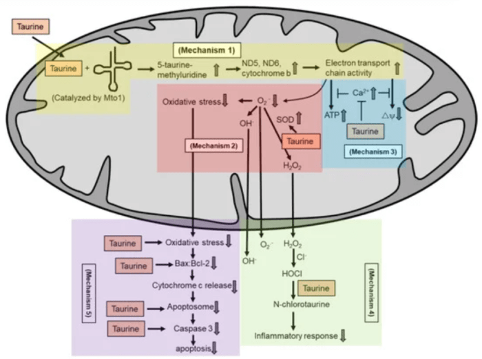
“Taurine supplementation protects against pathologies associated with mitochondrial defects, such as aging, mitochondrial diseases, metabolic syndrome, cancer, cardiovascular diseases and neurological disorders. Potential mechanisms by which taurine exerts its antioxidant activity in maintaining mitochondria health include:

  1. Conjugates with uridine on mitochondrial tRNA to form a 5-taurinomethyluridine for proper synthesis of mitochondrial proteins (mechanism 1), which regulates the stability and functionality of respiratory chain complexes;
  2. Reduces superoxide generation by enhancing the activity of intracellular antioxidants (mechanism 2);
  3. Prevents calcium overload and prevents reduction in energy production and collapse of mitochondrial membrane potential (mechanism 3);
  4. Directly scavenges HOCl to form N-chlorotaurine in inhibiting a pro-inflammatory response (mechanism 4); and
  5. Inhibits mitochondria-mediated apoptosis by preventing caspase activation or by restoring the Bax/Bcl-2 ratio and preventing Bax translocation to the mitochondria to promote apoptosis.

taurine mechanisms

An analysis on pharmacokinetics of oral supplementation (4 g) in 8 healthy adults showed a baseline taurine content in a range of 30 μmol to 60 μmol. Plasma content increased to approximately 500 μmol 1.5 h after taurine intake. Plasma content subsequently decreased to baseline level 6.5 h after intake.

We discuss antioxidant action of taurine, particularly in relation to maintenance of mitochondria function. We describe human studies on taurine supplementation in several mitochondria-associated pathologies.”

https://www.mdpi.com/1420-3049/26/16/4913/html “The Role of Taurine in Mitochondria Health: More Than Just an Antioxidant”


I take a gram of taurine at breakfast and at dinner along with other supplements and 3-day-old Avena sativa oat sprouts. Don’t think my other foods’ combined taurine contents are more than one gram, because none are found in various top ten taurine-containing food lists.

As a reminder, your mitochondria came from your mother, except in rare cases.

Choosing appropriate dietary fibers

This 2021 rodent study investigated effects of dietary fibers on Type 2 diabetes:

“Nine types of dietary fibers were used to investigate and evaluate their effects on type-2 diabetic rats via physiology, genomics, and metabolomics.

In human clinical trials, supplementation with dietary fibers was found inversely associated with risks of diabetes, along with improvement on glycemic control, lipid profiles, and host homeostasis. However, mixed fibers with diverse types from dietary sources are generally used for treatment intervention in clinical trials, and effects of individual dietary fibers on T2D are seldom discussed.

We found that supplementation with β-glucan, arabinogalactan, guar gum, and apple pectin had favorable effects on alleviating T2D:

upset plot

Non-bioactive dietary fibers (NBDF) were glucomannan, arabinoxylan, carrageenan, xylan, and xanthan gum.

dietary fibers effects

Relatively high viscosity was an important driving factor of dietary fibers for hypoglycemic effects. Supplementation with β-glucan, arabinogalactan, guar gum, and apple pectin tended to restore gut microbiota composition.

Our study uncovered effects of different dietary fibers on T2D, along with their potential mechanisms. Different dietary fibers influenced host metabolism via different metabolic pathways.”

https://pubs.acs.org/doi/10.1021/acs.jafc.1c01465 “Bioactive Dietary Fibers Selectively Promote Gut Microbiota to Exert Antidiabetic Effects” (not freely available). Thanks to Dr. Yonggan Sun for providing a copy.


I eat oat β-glucan three times a day – Avena nuda whole oats for breakfast, and twice daily 3-day-old Avena sativa hulled 3-day-old oat sprouts. Not to be confused with training my immune system with daily yeast cell wall β-glucan.

I recommend “Section 6. Biological functions” of the 2021 Plants arabinogalactans: From structures to physico-chemical and biological properties (not freely available), which reviewed:

  • ACE inhibitory;
  • Anti-cancer;
  • Anti-complementary;
  • Anti-diabetic;
  • Anti-ulcer;
  • Antiaging;
  • Antinociceptive;
  • Antioxidant;
  • Antitumor;
  • Antitussive;
  • Antiviral;
  • Complementary system;
  • Complement fixation;
  • Gastrointestinal-protective;
  • Hepatoprotective;
  • Hypoglycemic;
  • Immunomodulating;
  • Immunostimulatant;
  • Immune enhancing;
  • Intestinal immune system;
  • Phagocytosis stimulating; and
  • Prebiotic activities

properties of different arabinogalactans. Thanks to Professor Michaud for providing a copy.

Arabinogalactans were favored in both papers, yet few are commercially available. In January 2021 I used an arabinogalactan supplement, but it was too expensive to continue. Maybe multiple processing steps were a cost factor?

arabinogalactan processing

Changing your immune system / gut microbiota interactions with diet

This 2021 human clinical trial investigated associations between gut microbiota and host adaptive immune system components:

“Diet modulates gut microbiome, and gut microbes impact the immune system. We used two gut microbiota-targeted dietary interventions – plant-based fiber or fermented foods – to determine how each influences microbiome and immune system in healthy adults. Using a 17-week randomized, prospective study design combined with -omics measurements of microbiome and host and extensive immune profiling, we found distinct effects of each diet:

  • Those in the high-fiber diet arm increased their fiber consumption from an average of 21.5±8.0 g per day at baseline to 45.1±10.7 g per day at the end of the maintenance phase.
  • Participants in the high-fermented food diet arm consumed an average of 0.4±0.6 servings per day of fermented food at baseline, which increased to an average of 6.3±2.9 servings per day at the end of the maintenance phase.
  • Participants in the high-fiber diet arm did not increase their consumption of fermented foods (Figure 1.C dashed line), nor did participants consuming the high-fermented food diet increase their fiber intake.

fiber vs fermented

Fiber-induced microbiota diversity increases may be a slower process requiring longer than the six weeks of sustained high consumption achieved in this study. High-fiber consumption increased stool microbial protein density, carbohydrate-degrading capacity, and altered SCFA production, indicating that microbiome remodeling was occurring within the study time frame, just not through an increase in total species.

Comparison of immune features from baseline to the end of the maintenance phase in high-fiber diet participants revealed three clusters of participants representing distinct immune response profiles. No differences in total fiber intake were observed between inflammation clusters. A previous study demonstrated that a dietary intervention, which included increasing soluble fiber, was less effective in improving inflammation markers in individuals with lower microbiome richness.

In both diets, an individual’s microbiota composition became more similar to that of other participants within the same arm over the intervention, despite retaining the strong signal of individuality.

Coupling dietary interventions to longitudinal immune and microbiome profiling can provide individualized and population-wide insight. Our results indicate that fermented foods may be valuable in countering decreased microbiome diversity and increased inflammation.”

https://www.cell.com/cell/fulltext/S0092-8674(21)00754-6 “Gut-microbiota-targeted diets modulate human immune status” (not freely available). See https://www.biorxiv.org/content/10.1101/2020.09.30.321448v2.full for the freely available preprint version.


Didn’t care for this study’s design that ignored our innate immune system components yet claimed “extensive immune profiling.” Not.

There was sufficient relevant evidence on innate immunity cells – neutrophils, monocytes, macrophages, natural killer cells, and dendrites – when the trial started five years ago. But maybe this didn’t satisfy study sponsors?

This study found significant individual differences in the high-fiber group. These individual differences failed to stratify into subgroup p-value significance.

I won’t start eating fermented dairy or fermented vegetable brines to “counter decreased microbiome diversity and increased inflammation.” I’m rolling the die with high-fiber intake (2+ times more grams than this clinical trial, over a 3+ times longer period so far).

Changing to a high-fiber diet this year to increase varieties and numbers of gut microbiota is working out alright. No worries about “increased inflammation” because twice-daily 3-day-old microwaved broccoli sprouts since Day 70 results from Changing to a youthful phenotype with broccoli sprouts have taken care of inflammation for 15 months now.

What effects have this year’s diet changes had on my adaptive and innate immune systems? 2021’s spring allergy season wasn’t pleasant. But late summer’s ragweed onslaught hasn’t kept me indoors – unlike other years – despite day after day of readings like today’s:

ragweed

Regarding an individual’s starting point and experiences, those weren’t the same as family, friends, significant other, identified group members, or strangers. Each of us has to find our own way to getting well.

Agenda-free evidence may provide good guidelines. So does how you feel.

Seeds vs. sprouts: red cabbage and broccoli

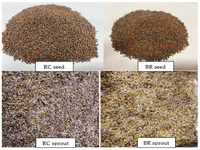
This 2021 study compared properties of red cabbage and broccoli seeds and sprouts:

“Antioxidant and antidiabetic properties and metabolite profiling of ethanol extracts of red cabbage (RC) and broccoli (BR) seeds and sprouts were investigated:

  • BR seeds had the highest total phenolic and flavonoid contents;
  • BR sprouts had the highest saponin content;
  • RC sprouts demonstrated the highest antioxidant capacity;
  • BR and RC sprouts showed the most potent inhibition against α-glucosidase and pancreatic lipase; and
  • BR seeds demonstrated the lowest AGE inhibition.

RC and BR seeds vs sprouts

In vitro assessment of antidiabetic potential of extracts revealed that sprouts demonstrated better potential as antioxidant, α-glucosidase, and pancreatic lipase inhibitors compared to raw seeds. Amino acids and phenolic compounds were the most improved metabolites in the germination process.

Germination not only enhanced levels of metabolites, but also synthesized new compounds in seeds. Germination effectively enhanced functional properties and metabolite profiles of broccoli and red cabbage seeds, making their sprouts more applicable as functional ingredients.”

https://www.mdpi.com/2076-3921/10/6/852/htm “UHPLC-ESI-QTOF-MS/MS Metabolite Profiling of the Antioxidant and Antidiabetic Activities of Red Cabbage and Broccoli Seeds and Sprouts”


I asked coauthors for sprout ages and pertinent growing conditions for the above-pictured sprouts. I’ll guess > 3-days-old, temperature 25° C, and relative humidity 90%. What would you guess?

Update: Two coauthors replied:

“Red Cabbage and Broccoli were germinated for 6 and 7 days respectively. Temperature ranged between 20-23 °C in the dark.”

PXL_20210825_102832166

Preventing human infections with dietary fibers

This 2020 review covered interactions of gut microbiota, intestinal mucus, and dietary fibers. I’ve outlined its headings and subheadings, and ended with its overview:

“I. Dietary fibers and human mucus-associated polysaccharides: can we make an analogy?

I.1 Brief overview of dietary fibers and mucus polysaccharides structures and properties

I.I.1 Dietary fibers

  • Dietary fiber intake and health effects

I.I.2 Intestinal mucus polysaccharides

  • Structure
  • Main functions

I.2 Similarities and differences between dietary fibers and mucus carbohydrates

  • Origin and metabolism
  • Structure

II. Interactions of dietary fibers and mucus-associated polysaccharides with human gut microbiota

II-1 Substrate accessibility and microbial niches

  • Dietary fibers
  • Mucus polysaccharides

II-2 Recognition and binding strategies

  • Dietary fibers
  • Mucus polysaccharides

II-3 Carbohydrate metabolism by human gut microbiota

II-3.1 Specialized carbohydrate-active enzymes

II-3.2 Vertical ecological relationships in carbohydrate degradation

  • Dietary fibers
  • Mucus polysaccharides

II-3.3 Horizontal ecological relationships in carbohydrate degradation

II.4 Effect of carbohydrates on gut microbiota composition and sources of variability

II.4.1 Well-known effect of dietary fibers on the gut microbiota

II.4.2 First evidences of a link between mucus polysaccharides and gut microbiota composition

III. Gut microbiota, dietary fibers and intestinal mucus: from health to diseases?

[no III.1]

III.2 Current evidences for the relationship between dietary fibers, mucus and intestinal-inflammatory related disorder

III.2.1 Obesity and metabolic-related disorders

  • Dietary fibers
  • Mucus polysaccharides

III.2.2 Inflammatory bowel diseases

  • Dietary fibers
  • Mucus polysaccharides

III.2.3 Colorectal cancer

  • Dietary fibers
  • Mucus polysaccharides

IV. How enteric pathogens can interact with mucus and dietary fibers in a complex microbial background?

IV.1 Mucus-associated polysaccharides: from interactions with enteric pathogens to a cue for their virulence?

IV.1.1 Pathogens binding to mucus

  • Binding structures
  • Sources of variations

IV.1.2 Mucus degradation by pathogens

  • Bacterial mucinases
  • Glycosyl hydrolases

IV.1.3 Mucus-based feeding of pathogens

  • Primary degraders or cross-feeding strategies
  • Importance of microbial background

IV.1.4 Pathogens and inflammation in a mucus-altered context

IV.1.5 Modulation of virulence genes by mucus degradation products

IV.2 How can dietary fiber modulate enteric pathogen virulence?

IV.2.1 Direct antagonistic effect of dietary fibers on pathogens

  • Bacteriostatic effect
  • Inhibition of cell adhesion
  • Inhibition of toxin binding and activity

IV.2.2 Indirect effect of dietary fibers through gut microbiota modulation

  • Modulation of microbiota composition
  • Modulation of gut microbiota activity

IV.2.3 Inhibition of pathogen interactions with mucus: a new mode of dietary fibers action?

  • Binding to mucus: dietary fibers acting as a decoy
  • Inhibition of mucus degradation by dietary fibers

V. Human in vitro gut models to decipher the role of dietary fibers and mucus in enteric infections: interest and limitations?

V.1 Main scientific challenges to be addressed

V.2 In vitro human gut models as a relevant alternative to in vivo studies

V.3 In vitro gut models to decipher key roles of digestive secretions, mucus and gut microbiota

V.4 Toward an integration of host responses

V.5 From health to disease conditions

dietary fibers prevent infections

Overview of the potential role of dietary fibers in preventing enteric infections. Reliable and converging data from scientific literature are represented with numbers in circles, while data more hypothetical needing further investigations are represented with numbers in squares.

  1. Some dietary fibers exhibit direct bacteriostatic effects against pathogens.
  2. Dietary fiber degradation leads to short-chain fatty acids (SCFAs) production that can modulate pathogens’ virulence.
  3. By presenting structure similarities with receptors, some dietary fibers can prevent pathogen adhesin binding to their receptors.
  4. By the same competition mechanism, dietary fibers can also prevent toxins binding to their receptors.
  5. Dietary fibers are able to promote gut microbiota diversity.
  6. Dietary fibers may promote growth of specific strains with probiotic properties and therefore exhibit anti-infectious properties.
  7. Suitable dietary fiber intake prevents microbiota’s switch to mucus consumption, limiting subsequent commensal microbiota encroachment and associated intestinal inflammation.
  8. Dietary fibers may prevent pathogen cross-feeding on mucus by limiting mucus degradation and/or by preserving diversity of competing bacterial species.
  9. By preventing mucus over-degradation by switcher microbes, dietary fibers can hamper pathogen progression close to the epithelial brush border, and further restrict subsequent inflammation.”

https://doi.org/10.1093/femsre/fuaa052 “Tripartite relationship between gut microbiota, intestinal mucus and dietary fibers: towards preventive strategies against enteric infections” (not freely available)


There were many links among gut microbiota studies previously curated. For example, Go with the Alzheimer’s Disease evidence found:

“Akkermansia cannot always be considered a potentially beneficial bacterium. It might be harmful for the gut–brain axis in the context of AD development in the elderly.”

The current review provided possible explanations:

“Akkermansia muciniphila could be considered as a species that fulfills a keystone function in mucin degradation. It is a good example of a mucus specialist.”

Points #7-9 of the above overview inferred that insufficient dietary fiber may disproportionately increase abundance of this species. But Gut microbiota strains also found that effects may be found only below species at species’ strain levels.

These reviewers provided copies in places other than what’s linked above. Feel free to contact them for a copy.


Moon bandit

PXL_20210822_100718644.NIGHT

No magic bullet, only magical thinking

Consider this a repost of Dr. Paul Clayton’s blog post The Drugs Don’t Work:

“The drug industry has enough funds to:

  • Rent politicians;
  • Subvert regulatory agencies;
  • Publish fake data in the most august peer-reviewed literature; and
  • Warp the output of medical schools everywhere.

Their products are a common cause of death. Every year, America’s aggressively modern approach to disease kills over 100,000 in-hospital patients, and twice that number of out-patients.

In 1900, a third of all deaths occurred in children under the age of 5. By 2000 this had fallen to 1.4%. The resulting 30-year increase in average life expectancy fed into the seductive and prevailing myth that we are all living longer; which is manifestly untrue. Improvements in sanitation were far more significant in pushing infections back than any medical developments.

There is currently no pharmaceutical cure for Alzheimer’s or Parkinsonism, nor can there be when these syndromes are in most cases driven by multiple metabolic distortions caused by today’s diet. The brain is so very complex, and it can go wrong in so many ways. The idea that we can find a magic bullet for either of these syndromes is ill-informed and philosophically mired in the past.

It is also dangerous. There is a significant sub-group of dementia sufferers whose conditions are driven and exacerbated by pharmaceuticals. Chronic use of a number of commonly prescribed drugs – and ironically, anti-Parkinson drugs – increases the risk of dementia by roughly 50%.

Big Pharma’s ability to subvert regulatory authorities is even more dangerous. The recent FDA approval of Biogen’s drug aducanumab is a scandal; not one member of the FDA Advisory Committee voted to approve this ineffective product, and three of them resigned in the aftermath of the FDA’s edict. This ‘anti-Alzheimer’s’ drug, which will earn Biogen $56,000 / patient / year, was licensed for financial reasons; it reduced amyloid plaque but was clinically ineffective.

So did the eagerly awaited gantenerumab and solanezumab. But they, too, failed to produce any significant clinical benefit.”


A knee-replacement patient enduring her daily workout

PXL_20210822_102713662