Add vitamin C to broccoli sprouts?

A 2026 paper presented results of a clinical trial that selectively added myrosinase enzyme via mustard powder and vitamin C to measure in vivo effects on glucoraphanin conversion to sulforaphane:

“Effects of exogenous myrosinase (Myr) on conversion efficiency of glucoraphanin (GR) to sulforaphane (SF) was compared to gut microbial Myr-like activity. In a randomized, double-blind, crossover study, sixteen subjects (9 F: 7 M) received a single oral dose of GR in 385 mg broccoli seed extract (BSE) with 72.5 mg Myr-containing mustard seed powder, or broccoli seed extract alone, both with 100 mg ascorbic acid.

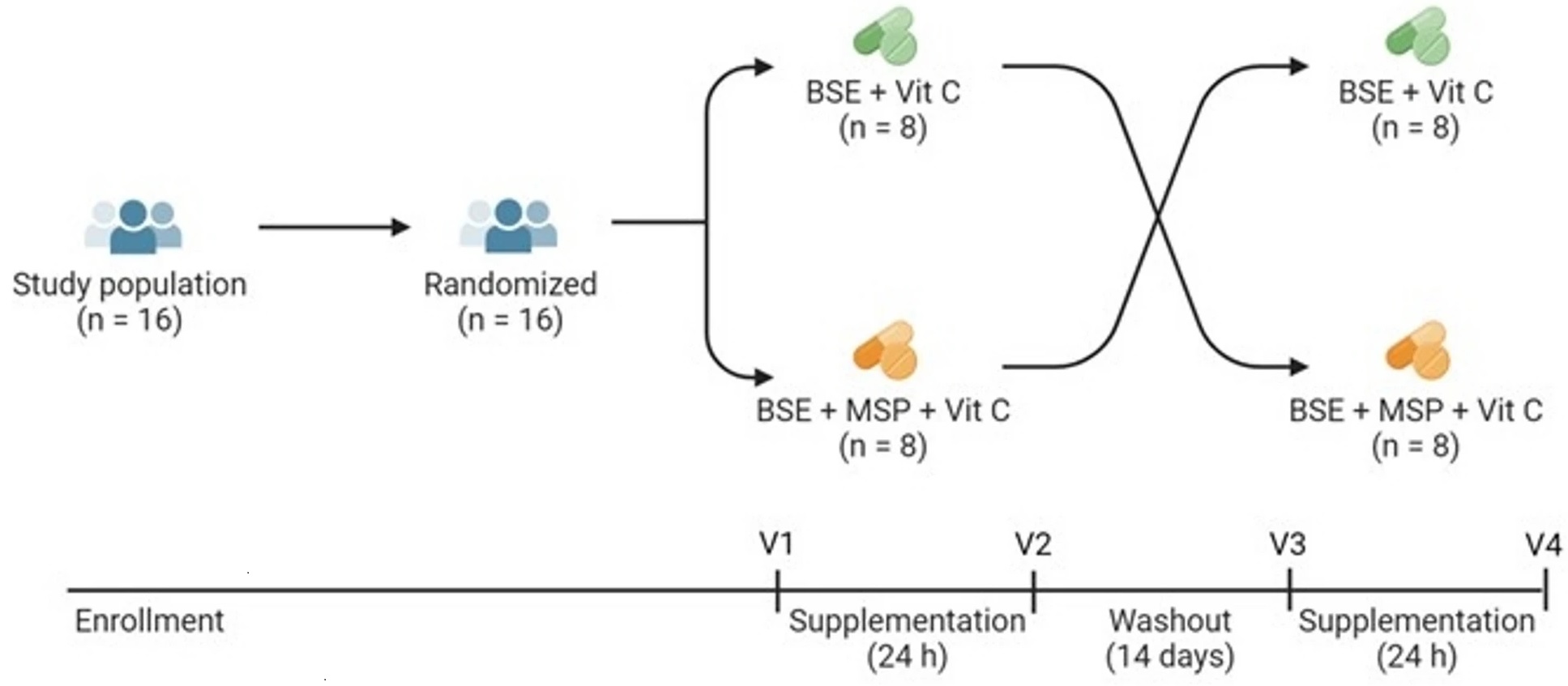
GR + Myr, on average, doubled the bioavailability of SF (39.8 ± 3.1%) compared to GR alone (18.6 ± 3.1%), and increased the conversion rate in the first 8 h (25.4% ± 2.7%) compared to GR alone (8.0% ± 2.7) based on measurement of urinary metabolites. The majority of subjects given GR as BSE with exogenous Myr and Vit C, converted GR to SF notably faster (e.g., within the first 8 h), than those given GR and Vit C alone.

One of the most likely explanations for the pronounced differences is that when SF was produced as a result of added exogenous Myr, it was produced and absorbed in the small intestine and metabolized primarily to its glutathione (GSH) derivatives and excreted in urine. This is a more rapid process than when GR passes into the large intestine and then is acted upon by the greater bacterial population within that terminal segment of the gastrointestinal system.

No differences were observed in the 8 to 24 h urine collection (Time 24 h) between the two treatments: 11.7 ± 1.3% for GR + Myr vs. 8.9 ± 1.3% for GR alone. Bacterial communities did not differ between low/high GR converters following supplementation.

Many bacteria which persist in the small intestine and upper large intestine, as well as in mucosal-associated fractions, are not well represented in feces. The lack of anatomically specific microbial communities in the human gut limits our knowledge of GR conversion in people.”

https://www.nature.com/articles/s41598-026-39389-4 “Exogenous myrosinase from mustard seed increases bioavailability of sulforaphane from a glucoraphanin-rich broccoli seed extract in a randomized clinical study”


1. Lost in this study’s shuffle was the reason why sulforaphane’s effects are beneficial in the first place. As Switch on your Nrf2 signaling pathway pointed out:

“We use N-acetylcysteine (NAC) in the lab all the time because it stops an Nrf2 activation. So that weak pro-oxidant signal that activates Nrf2, you switch it off by giving a dose of NAC. It’s a potent antioxidant in that right, but it’s blocking signalling. And that’s what I don’t like about its broad use.”

It’s relevant to the Nrf2 activating effects of sulforaphane when antioxidant vitamin C taken to increase myrosinase hydrolyzation of glucoraphanin to sulforaphane if this vitamin C dose may also block Nrf2 activation. An increase of sulforaphane and its metabolites wouldn’t physiologically matter if their beneficial effects were simultaneously blocked.

2. Another indicator that these researchers lost the plot was shown when they asserted: “These data suggest that the present study may not have included sufficient AA to optimize Myr enzyme activity in vivo, another consideration for future studies” based on comparing the in vitro Reference 21 ascorbic acid doses (0, 11, 44, 88, or 154 mg AA/capsule). I don’t have access to Reference 21 to see whether it also didn’t assess Nrf2 activation.

3. For comparison of this study’s 50 mg glucoraphanin dose on two non-consecutive days, the cited Our model clinical trial for Changing to a youthful phenotype with broccoli sprouts provided daily 30 grams of fresh broccoli sprouts that contained an estimated 51 mg of glucoraphanin for ten weeks. Although no broccoli sprout preparation or intake guidelines were enforced, unassisted glucoraphanin conversion to sulforaphane had many beneficial effects in that trial.

4. These researchers stated “No differences were observed in the 8 to 24 h urine collection (Time 24 h) between the two treatments.” Whether the faster small intestine absorption of sulforaphane had benefits over the slower large intestine absorption wasn’t investigated.

5. I’ve previously corresponded with one of this study’s coauthors, and think their research group could do better work without the retired broccoli sprout expert. Maybe they would take more confident ownership of their work if they let him ride off into the sunset, and decide for themselves what predictable findings would be in their research effort’s scope?

Maybe they wouldn’t let fester the same old issues in his papers? It doesn’t say anything good about current research if its main findings just repeat last decade’s findings.

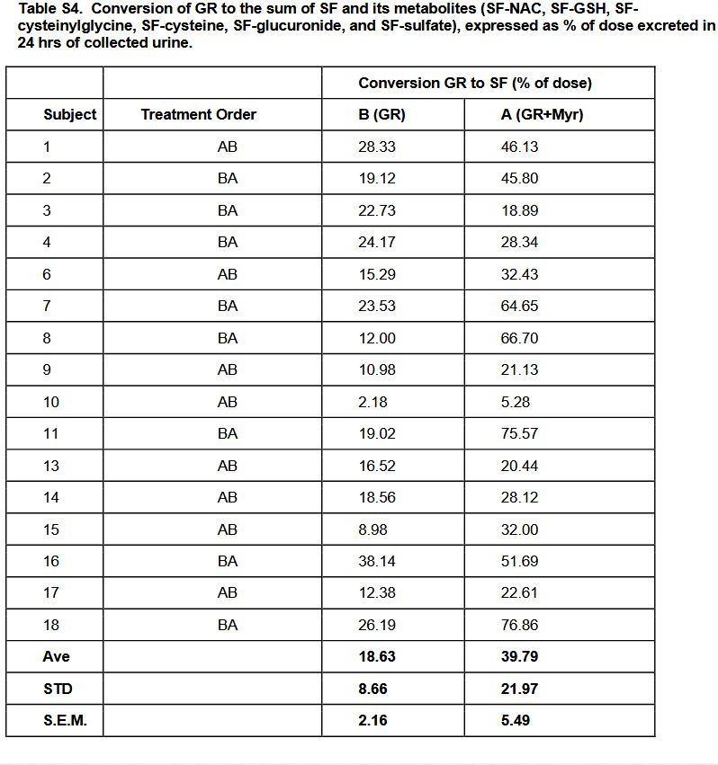
For example, Table S4 has stories that weren’t told. It conforms to the expert’s cited 2015 study findings, but presenting averages doesn’t reveal causes for individual differences. So questions on the individual level continue to be unanswered, such as:

  • Subject 8 had more than a fivefold increase of glucoraphanin-only conversion to glucoraphanin + myrosinase conversion. What blocked the other subjects from achieving similar results?
  • Why was Subject 10 so far behind everyone else’s capabilities? They would have had to increase their glucoraphanin-only conversion by fourfold just to get to the next lowest person’s level, Subject 15, but sixfold to get to Subject 15’s glucoraphanin + myrosinase conversion level.

6. Every researcher wants to have an impact on their field. The time to think over how to newly research possible impactful outcomes is before the study starts.

Since I’m in my seventh year of eating broccoli sprouts every day, I would have paid close attention to more rigorous bioavailability, more exact microbiota collection techniques, or detailed exploration of differences in people’s responses to the same treatments, all of which were predictable issues beforehand. I didn’t really care for listing them in the Discussion section, then dismissing investigations of these findings for some future research to explore.


Self-reinforcing feedback loops of aging

A 2026 rodent study investigated age-associated queuine decline. Queuine is characterized as a “longevity vitamin” in the cited 2018 review Prolonging healthy aging: Longevity vitamins and proteins, along with ergothioneine, astaxanthin, and taurine.

“The contribution of transfer RNA (tRNA)-specific modifications to aging remains largely unexplored. We systematically profile tRNA modifications across multiple organs, species, and senescence models, and identify mannosyl-queuosine (manQ) as the first tRNA-specific modification that consistently declines with age.

Across species, queuine supplementation extends lifespan and enhances healthspan. In naturally aging mice, long-term oral administration beginning at 16-months-old (human equivalent 50 years) extends mean lifespan by 15.3%, reduces DNA methylation age, improves cognitive and motor performance, strengthens antioxidant defenses, remodels the gut microbiota, and alleviates inflammation and metabolic dysfunction without detectable toxicity.

These findings establish tRNA epitranscriptomic remodeling as a previously unrecognized layer of aging regulation, and identify restoration of manQ through queuine supplementation as a multi-system strategy to delay aging.

manQ hypomodification is selective rather than reflecting global tRNA depletion. Aging preferentially reduces the manQ-containing tRNAAsp fragment while leaving the corresponding unmodified tRNAAsp fragment, and other queuosine-modified tRNAs, relatively unchanged.

This pattern supports a regulated defect in modification homeostasis rather than a generalized change in transcript abundance. Such specificity argues that manQ loss is not merely a passive consequence of tissue degeneration, but instead represents a conserved, biologically meaningful aging-associated event with mechanistic impact.

Because proteostasis intersects with multiple canonical hallmarks (e.g. mitochondrial dysfunction, impaired stress resilience, and altered intercellular communication), translation-coupled proteome destabilization offers a unifying explanation for how a single tRNA modification defect can elicit multi-system consequences. In this view, manQ decline is not merely one of many molecular changes observed in aging, but rather a proximate determinant capable of amplifying downstream hallmarks through a common axis of proteome quality control.

Our findings further suggest that manQ depletion may engage self-reinforcing feedback loops that accelerate aging trajectories. This architecture offers a conceptual framework in which aging progressively erodes ‘epitranscriptomic integrity’ at the tRNA level, pushing translation toward an error-prone regime that accelerates proteostatic collapse and functional decline.

A distinctive implication of this work is that queuine introduces a microbiota-host epitranscriptomic axis into aging biology. Queuine is produced by gut microbiota and cannot be synthesized de novo by mammals. These findings expand the conceptual scope of geroscience by placing a microbiota-derived nutrient upstream of translational quality control.

Queuine supplementation offers a distinct therapeutic logic: rather than modulating a single signaling cascade, it restores a tRNA modification state that governs translational fidelity – an upstream determinant of proteome quality that can, in principle, influence multiple downstream hallmarks concurrently. These findings highlight an intervention paradigm centered on restoring molecular fidelity, rather than suppressing a single downstream phenotype, as a strategy to delay systemic aging.”

https://www.biorxiv.org/content/10.64898/2026.03.22.713446v1.full “Evolutionarily Conserved Decline of tRNA Mannosyl-Queuosine Links Translational Regulation to Aging and Is Reversed by Queuine”

Treat your gut microbiota well. Give them what they want, and expect reciprocity.


The hops compound xanthohumol

Two 2026 papers, with the first an in vitro study of over 2000 compounds to select those that best inhibit BACH1:

“BACH1 regulates the cellular oxidative stress responses by suppressing expression of cytoprotective genes. Dysregulated BACH1 activity has been implicated in a range of pathologies, including chronic inflammatory diseases, fibrosis, and cancer, making it a promising therapeutic target.

We identified four structurally distinct compounds that robustly inhibit BACH1 function. Notably, these compounds simultaneously activate transcription factor NRF2, suggesting the potential for a broader modulation of oxidative stress pathways.

However, while NRF2 induces expression of genes that protect against oxidative stress and inflammation and suppress ferroptosis, BACH1 represses them. While NRF2 broadly activates cytoprotective genes, BACH1 inhibition triggers a more restricted response but with a strong upregulation of HMOX1 (significantly stronger than the one obtained upon NRF2 activation).

As such, combined NRF2 activation and BACH1 inactivation is expected to produce a more potent antioxidant and anti-inflammatory effect than targeting either factor alone. Moreover, BACH1 regulates unique targets not shared with NRF2 and can dominantly repress genes even in the presence of active NRF2. This confers BACH1 inhibition distinct therapeutic value, particularly in contexts such as cancer cell invasion, where its suppression yields anti-metastatic effects.

  1. Auranofin is an FDA-approved gold salt used in rheumatoid arthritis whose primary mechanism of action is inhibition of thioredoxin reductases (TrxRs).
  2. Xanthohumol is a natural compound, prenylated chalcone, that belongs to the flavonoid family, with reported antimicrobial, anti-inflammatory, and antioxidant activities, and demonstrated safety in phase I and phase II trials.
  3. Alantolactone is a natural compound, member of the sesquiterpene lactone class with anti-inflammatory and antioxidant effects, and has been tested in several animal models without reported toxicity.
  4. CH55 is a synthetic retinoid with high affinity for RAR-α and RAR-β and antifibrotic activity and has not been yet tested in vivo.

In summary, this work establishes a robust screening platform for identification of functional BACH1 inhibitors, and provides new chemical scaffolds with potential for future therapeutic development.”

https://papers.ssrn.com/sol3/papers.cfm?abstract_id=6439073 “Development of a High-Throughput Screening Platform for Identification of Functional BACH1 Inhibitors Reveals Compounds with Anti-Invasive Potential”

Xanthohumol is ninth on the Nrf2 activator list. BACH1 interactions were also covered in Part 3 of Broccoli sprouts activate the AMPK pathway.


A 2026 paper that was too recent to be referenced in the above study described two xanthohumol clinical trials. The first trial was designed to assess bioavailability in healthy people (6 men and 6 women), and the second was designed to determine bioactivity in 16 healthy women:

“The aim of the present project was to systematically investigate bioavailability of native xanthohumol compared to micellar xanthohumol at two doses (86 mg vs. 172 mg) in a randomized crossover trial. We furthermore examined short-term effects of xanthohumol on resting energy expenditure (REE), blood pressure (BP), and heart rate (HR) in a randomized placebo-controlled crossover study.

Micellar solubilization significantly increased area under the curve (AUC), maximum plasma concentration of xanthohumol (Cmax), timepoint of maximum plasma concentration of xanthohumol (tmax), and apparent bioavailability compared to native xanthohumol. The dose also significantly influenced plasma kinetics, but apparent bioavailability and tmax were dose-independent in contrast to AUC and Cmax. In our subsequent study, xanthohumol did not affect REE, substrate oxidation, BP, or HR.

Two properties of xanthohumol impair its bioavailability. First, xanthohumol is relatively unstable in an acidic environment, and second, xanthohumol is highly lipophilic and hydrophobic, insoluble in the aqueous environment of the intestinal lumen, and poorly absorbed into enterocytes.

In addition to determining typical plasma kinetic parameters (e.g., Cmax, tmax, and AUC), we also calculated the amount of xanthohumol absorbed using maximum plasma xanthohumol concentration. These estimated amounts are minimum quantities of xanthohumol that had to be absorbed to achieve observed plasma xanthohumol concentrations.

Results of these calculations emphasized the relatively poor bioavailability of xanthohumol in humans. Only ∼0.1% and ∼1.2% of native and micellar xanthohumol were absorbed, respectively.

However, a limitation of this plasma estimation is that fractional absorption of xanthohumol was underestimated because distribution of xanthohumol in cells and tissues was not taken into account. It is likely that more xanthohumol was actually absorbed. Whether the bioavailability of xanthohumol is sufficient for physiological efficacy must be investigated further in humans.

In conclusion, oral bioavailability of micellar xanthohumol was higher than that of native xanthohumol. Systemic availability of xanthohumol did not differ between men and women. Our study provides no evidence that xanthohumol acutely affects REE, BP, and HR.”

https://onlinelibrary.wiley.com/doi/10.1002/mnfr.70413 “The Bioavailability of Xanthohumol in Humans and the Influence of Formulation and Dose: Randomized Controlled Trial Data”