Part 3 of Rejuvenation therapy and sulforaphane

Part 1 focused on the study’s clinical biomarkers. Part 2 highlighted its epigenetic clocks. Now we’ll look at rejuvenation of cognitive function.

Charts for this study’s most relevant human aging applications – measured by the new human-rat relative biological age clock – were in supplementary data due to combining study untreated tissue samples into clock training data. Reanalyses showed:

“Using the final version of the epigenetic clocks, we find that treatment effects become even more significant especially for the hypothalamus.”

Human-rat relative clock percentages of rejuvenation were:

  • “Blood 70.6%
  • Liver 79.4%
  • Heart 61.6%
  • Hypothalamus 20.9%”

The Discussion section addressed hypothalamus rejuvenation:

“Why does plasma fraction treatment not reduce brain epigenetic age by the same magnitude as it does other organs? We can only begin to address this question after having first understood what epigenetic aging entails.

As it stands, our knowledge in this area remains limited, but it is nevertheless clear that:

  1. Epigenetic aging is distinct from the process of cellular senescence and telomere attrition,
  2. Several types of tissue stem cells are epigenetically younger than non-stem cells of the same tissue,
  3. A considerable number of age-related methylation sites, including some clock CpGs, are proximal to genes whose proteins are involved in the process of development,
  4. Epigenetic clocks are associated with developmental timing, and
  5. Relate to an epigenomic maintenance system.

Collectively, these features indicate that epigenetic aging is intimately associated with the process of development and homeostatic maintenance of the body post-maturity.

  • While most organs of the body turnover during the lifetime of the host, albeit at different rates, the brain appears at best to do this at a very much slower rate.
  • While most tissues harbor stem cells that are necessary for replenishment and turnover, stem cells in adult brain have only been detected in a defined and very limited area of the subventricular zone, olfactory bulb (in rats), hippocampus and hypothalamic proliferative region.

As such, if plasma fraction treatment’s rejuvenating effect is:

  • Mediated through the process of development and
  • Involves tissue stem cells

then its effect on epigenetic age of the brain would appear to be modest, which indeed it does.

It is to be noted however, that improving brain function does not depend on neurogenesis as much as it does on synapse formation and factors such as NMDA receptors, which decline in density with age.

Assessment of plasma fraction treatment on cognitive function (learning and memory). Rats were subjected to Barnes maze test – nine consecutive days of test where the time (in seconds) required by rats to find the escape hole (latency) was recorded and plotted. Error bars depict 2 standard errors.

Within a month of plasma fraction treatment, rats exhibited significantly reduced latency to escape, i.e., they learned and remembered better. After the second month, treated rats began with a slightly reduced latency period compared to untreated old rats, and once again, they learned much faster than the latter.

By the third month, it was clear that treated rats remembered the maze much better than untreated ones even from the first day of test as their latency period was significantly reduced. By the end of the test period, their latency was similar to that of young rats. This feature was sustained and repeated in the fourth month.”

Not sure why there’s a 62-day gap between “Second month” and “Third month.” Maybe it had something to do with “First month” starting 10 days after the first treatment and “Third month” similarly starting 13 days after the second treatment?


Many of us know older people who lived well past their time of good cognitive function:

  • We see how they’re helpless and dependent; and
  • We see how others take advantage of them in their morbidity phase, where healthspan stops increasing but lifespan continues.

We can make personal plans for that day, sure. But let’s also put some urgency into applying this study’s new human-rat relative biological age clock, and make:

“A step change in aging research. Although conservation of aging mechanism could be equally deduced from the existence of multiple individual clocks for other mammals (mouse, dog), the single formula of the human-rat clock that is equally applicable to both species effectively demonstrates this fact.”

Part 2 of Rejuvenation therapy and sulforaphane

A rejuvenation therapy and sulforaphane focused on the study’s clinical biomarkers and not its biological age measurements. This Part 2 curation of the study highlights its epigenetic clocks because:

“While clinical biomarkers have obvious advantages (being indicative of organ dysfunction or disease), they are neither sufficiently mechanistic nor proximal to fundamental mechanisms of aging to serve as indicators of them. It has long been recognized that epigenetic changes are one of several primary hallmarks of aging.

DNA methylation (DNAm) epigenetic clocks capture aspects of biological age. The discrepancy between DNAm age and chronological age (term as ‘epigenetic age acceleration’) is predictive of all-cause mortality. Pathologies and conditions that are associated with epigenetic age acceleration includes, but are not limited to, cognitive and physical functioning, centenarian status, Down syndrome, HIV infection, obesity, and early menopause.

The [new] human-rat clocks apply to both species. The two human-rat pan-tissue clocks are distinct, by way of measurement parameters. One estimates absolute age (in units of years), while the other estimates relative age, which is the ratio of chronological age to maximum lifespan; with values between 0 and 1. This ratio allows alignment and biologically meaningful comparison between species with very different lifespan (rat and human), which is not afforded by mere measurement of absolute age.

Relative age estimation was made using the formula: Relative age = Age / maxLifespan where the maximum lifespan for rats and humans were set to 3.8 years and 122.5 years, respectively.”

From Supplementary Table 3, old control and old treatment subjects were males 109 weeks old, 55% of their maximum lifespan (109 / 197.6). Young control subjects were males 30 weeks old, 15% of their maximum lifespan.

The money charts for this study’s human aging applications – measured by the new human-rat relative biological age clock – were buried in Supplementary Figure 12, bar plots M through P:

“Human-rat clock measure of relative age defined as age/maximum species lifespan. Each bar-plot reports the mean value and one standard error.”

From Supplementary Table 8, the percentages of rejuvenation for the above bar plots, calculated as “(100 * (1 – Old Treated / Old Control)” were:

  • “Blood 70.6%
  • Liver 79.4%
  • Heart 61.6%
  • Hypothalamus 20.9%”

Let’s return to clinical biomarkers for comparison purposes. The current study measured pro-inflammatory cytokine IL-6 blood plasma levels at every time point, but didn’t publish numbers. Bar plots and narrative were:

“Inflammation is an important response that helps protect the body, but excess inflammation especially in terms of duration of this response can have very detrimental effects instead. This occurs when inflammation fails to subside and persists indefinitely; a condition referred to as chronic inflammation, which for reasons not well-understood, increases with age and is associated with a multitude of conditions and pathologies.

The levels of two of the most reliable and common biomarkers of chronic inflammation, interleukin 6 (IL-6) and tumor necrosis factor α (TNF-α), are found to be considerably higher in old rats, and these were very rapidly diminished, within days by plasma fraction treatment, to comparable levels with those of young rats. This was especially stark with IL-6.

In time, the levels of these inflammatory factors began to rise gradually, but they were once again very effectively reduced following the second administration of the plasma fraction on the 95th day.”

Let’s compare the above IL-6 graphic with IL-6 concentration improvements of our 2018 model clinical trial, Effects of long-term consumption of broccoli sprouts on inflammatory markers in overweight subjects, calculated as (100 * (1 – Day _ mean / Day 0 mean):

Mean pg/ml | % improvement | Period | Broccoli sprout consumption

  • 4.594 | 0% | Day 0 | “One week before the beginning of the intervention period, subjects were asked to avoid the consumption of Brassica vegetables (broccoli, radish, cauliflower, Brussel sprouts, mustards, among others) and their derived products.”
  • 1.748 | 62.0% | Day 0 to 70 | Subjects ate 30 g raw broccoli sprouts every day, and stopped eating them after Day 70.
  • 0.896 | 80.5% | Day 0 to 90 | “After the intervention period, a follow-up recovery period for all subjects continued for another 90 days with no ingestion of broccoli sprouts.”
  • 2.170 | 52.8% | Day 0 to 160 | Subjects had not eaten broccoli sprouts after Day 70.

Results between the studies were similar in that:

  1. IL-6 levels improved during early treatments through rat Day 8 and human Day 70, respectively.
  2. IL-6 levels continued decreasing shortly after treatments for 7 days (through rat Day 15) and 20 days (through human Day 90), respectively.
  3. IL-6 levels rose after rat Day 15 and human Day 90, respectively, but were still significantly below Day 0 values at rat Day 95 and human Day 160.

The current study measured Nrf2 but didn’t publish numbers. Bar plots and narrative were:

“The reduction of these inflammation markers is consistent with the profile of the nuclear factor erythroid 2-like 2 protein (Nrf2), which plays a major role in resolving inflammation, in part by inhibiting the expression of IL-6 and TNF-α. Nrf2 also induces the expression of antioxidants that neutralizes ROS [reactive oxygen species], which is also a significant feature in inflammation.”

A PubMed search on “nrf2 sulforaphane human” didn’t turn up relevant 2020 human in vivo studies. I disregarded reviews, cancer studies, disproven hypotheses, and other compounds listed in the below graphic.

I won’t repeat the entire Nrf2 section from the Part 1 curation, just one graphic and paragraph:

It [sulforaphane] is not only a potent Nrf2 inducer but also highly bioavailable [around 80%], so that modest practical doses can produce significant clinical responses. Other Nrf2 activators [shown in the above image] not only lack potency but also lack the bioavailability to be considered as significant intracellular Nrf2 activators.”


As noted in Reviewing clinical trials of broccoli sprouts and their compounds, there are no sulforaphane clinical trials that also use epigenetic clocks. Broccoli sprouts and their compounds’ effects on human aging is an area that hasn’t drawn attention and funding.

What effects may broccoli sprout compounds have on human aging? With this new human-rat relative biological age clock, researchers can get reliable answers from rat studies, with human clinical trials needed only to confirm those findings!

As rejuvenation research continues, what could people do easily, cheaply, and today for our long-term selves? Don’t know about the hypothalamus, but our blood, liver, and heart biological ages may decrease as we reduce inflammation and oxidative stress by eating broccoli sprouts.

I’m at a similar percentage of species maximum lifespan as were the study’s treated subjects. It’s my choice as to what my healthspan will be.

There isn’t evidence today to definitively say that changing my inflammatory phenotype with broccoli sprouts has had / will have rejuvenation effects on biological ages of my cells, organs, and body. But if eating broccoli sprouts every day not only reduces chronic inflammation and oxidative stress as expected, but also makes me younger, I could probably learn to live with that. 🙂

Continued with Part 3 of Rejuvenation therapy and sulforaphane.

A rejuvenation therapy and sulforaphane

The founder of the epigenetic clock methodology with the coauthor of Aging as an unintended consequence released a 2020 rodent study “Reversing age: dual species measurement of epigenetic age with a single clock” at https://www.biorxiv.org/content/10.1101/2020.05.07.082917v1.full.pdf:

“We employed six clocks to investigate the rejuvenation effects of a plasma fraction treatment in different rat tissues. Two of these epigenetic clocks apply to both humans and rats.

The treatment more than halved the epigenetic ages of blood, heart, and liver tissue. A less pronounced, but statistically significant, rejuvenation effect could be observed in the hypothalamus.

The treatment was accompanied by progressive improvement in the function of these organs as ascertained through numerous biochemical/physiological biomarkers and behavioral responses to assess cognitive functions. Cellular senescence, which is not associated with epigenetic aging, was also considerably reduced in vital organs.

Plasma fraction treatment consists of two series of intravenous injections of plasma fraction. Rats were injected four times on alternate days for 8 days. A second identical series of injections were administered 95 days later. In its entirety, the experiment lasted 155 days.

Overall, this study demonstrates that a plasma-derived treatment markedly reverses aging according to epigenetic clocks and benchmark biomarkers of aging.”

The study hasn’t been peer reviewed, so can’t be viewed yet as conclusive. Given that researchers’ single-most valuable asset is their reputations, though, will the findings have major revisions?


I was alerted to the study by Josh Mitteldorf’s blog post Age Reduction Breakthrough, who did his usual excellent curation:

“Most of the explosion in aging research (and virtually all the venture capital startups) are looking to treat aging at the cellular level. Their paradigm is that aging is an accumulation of molecular damage, and they see their job as engineering of appropriate repair mechanisms.

The truth, as Katcher [the lead lab researcher] understands it, is that, to a large extent, aging is coordinated system-wide via signal molecules in the blood. The problem is that there are thousands of constituents represented in tiny concentrations in blood plasma, but conveying messages that cells read. Which of these are responsible for aging?

The two-species clock[s] was [were] a significant innovation, a first bridge for translating results from an animal model into their probable equivalent in humans. Besides the methylation clock[s], the paper presents evidence of rejuvenation by many other measures. For example:

  • IL-6, a marker of inflammation, was restored to low youthful levels;
  • Glutathione (GSH), superoxide dismutase (SOD), and other antioxidants were restored to higher youthful levels;
  • In tests of cognitive function (Barnes maze), treated rats scored better than old rats, but not as well as young rats.;
  • Blood triglycerides were brought down to youthful levels;
  • HDL cholesterol rose to youthful levels; and
  • Blood glucose fell toward youthful levels.

These results bring together three threads that have been gaining credibility over the last decade. Mutually reinforcing, the three have a strength that none of them could offer separately.

  1. The root cause of aging is epigenetic progression = changes in gene expression over a lifetime.
  2. Methylation patterns in nuclear DNA are not merely a marker of aging, but its primary source. Thus aging can be reversed by reprogramming DNA methylation.
  3. Information about the body’s age state is transmitted system-wide via signal molecules in the blood. Locally, tissues respond to these signals and adopt a young or an old cellular phenotype as they are directed.”

Several of these aging measurements are also positively affected by sulforaphane. Using Sulforaphane: Its “Coming of Age” as a Clinically Relevant Nutraceutical in the Prevention and Treatment of Chronic Disease as a reference:

1. “Chronic inflammation”

“Antioxidants in general and glutathione in particular can be depleted rapidly under conditions of oxidative stress, and this can signal inflammatory pathways associated with NF-κB. SFN [sulforaphane] has been shown to inhibit NF-κB in endothelial cells.

Two key inflammatory cytokines were measured at four time points in forty healthy overweight people [our model clinical trial, Effects of long-term consumption of broccoli sprouts on inflammatory markers in overweight subjects]. The levels of both interleukin-6 (Il-6) and C-reactive protein (CRP) declined over the 70 days during which the sprouts were ingested. These biomarkers were measured again at day 90, wherein it was found that Il-6 continued to decline, whereas CRP climbed again. When the final measurement was taken at day 160, CRP, although climbing, had not returned to its baseline value. Il-6 remained significantly below the baseline level at day 160.”

OMCL2019-2716870.010

2. “Oxidative stress”

“As a mediator for amplification of the mammalian defence system against various stressors, Nrf2 [nuclear factor erythroid 2-related factor 2] sits at the interface between our prior understanding of oxidative stress and the endogenous mechanisms cells use to deal with it. Diseases known to be underpinned by oxidative stress are proving to be more responsive to amplification of cellular defences via Nrf2 activation than by administration of direct-acting antioxidant supplements.

SFN, with absolute bioavailability of around 80%, [is] capable of increasing several endogenous antioxidant compounds via the transcription factor, Nrf2.

Nrf2 is ubiquitously expressed with the highest concentrations (in descending order) in the kidney, muscle, lung, heart, liver, and brain. Nrf2 was shown to prevent endothelial cells from exhibiting a proinflammatory state. Nrf2 is required for protection against glucose-induced oxidative stress and cardiomyopathy in the heart.

Well in excess of 500 genes have been identified as being activated by SFN via the Nrf2/ARE [Antioxidant Response Element] pathway, and it is likely that this underestimates the number as others are being discovered. Of the available SFN clinical trials associated with genes induced via Nrf2 activation, many demonstrate a linear dose-response. More recently, it has become apparent that SFN can behave hormetically with different effects responsive to different doses.

It [sulforaphane] is not only a potent Nrf2 inducer but also highly bioavailable so that modest practical doses can produce significant clinical responses. Other Nrf2 activators [shown in the above image] not only lack potency but also lack the bioavailability to be considered as significant intracellular Nrf2 activators.”


The study’s most relentlessly questioned, scrutinized, and criticized findings may be the two new epigenetic clocks that apply to both humans and rats. The researchers invited other researchers to validate these clocks because:

“If validated, this would be a step change in aging research. Although conservation of aging mechanism could be equally deduced from the existence of multiple individual clocks for other mammals (mouse, dog), the single formula of the human-rat clock that is equally applicable to both species effectively demonstrates this fact.”

The commonalities of this study with efforts to change my inflammatory phenotype with broccoli sprouts were summarized in the Discussion section:

“Apart from rejuvenating the vital organs of the treated rats, plasma fraction also impacted two fundamental physiological processes that underlie a great number of pathologies, namely oxidative stress and inflammation. Within a week of treatment, the markers of chronic inflammation (IL-6 and TNF-α) were significantly reduced and remained low throughout the entire experiment.

Likewise, markers of oxidative stress in brain, heart, lung and liver, which were very much higher in control old rats, were at the end of the experimental period, indistinguishable between plasma fraction-treated old rats and young ones. Concomitant with this drastic reduction in oxidative stress was the augmented levels of antioxidants (GSH, Catalase and SOD) in these tissues, indicating that modulating the levels of ROS [reactive oxygen species] to that of youthful rats is at least one way by which plasma fraction suppresses oxidative stress. It remains to be ascertained whether the rate of ROS generation is also reduced.

The levels of Nrf2, a transcription factor that impacts on oxidative stress, as well as inflammation, were raised by plasma fraction treatment of old rats to those of the young ones, indicating yet another level by which this treatment modulates these two critical processes. Collectively, these results show that plasma fraction treatment impacts not only the overt performances of organs, but also the underlying physiological processes that are pivotal for optimal organ function and health.”

Great stuff, huh? Are you ready to change your phenotype?

Continued with Part 2 of Rejuvenation therapy and sulforaphane.

Do early experiences of hunger affect our behavior, thoughts, and feelings today?

Reposted from five years ago.


A 2015 worldwide human study Hunger promotes acquisition of nonfood objects found that people’s current degree of hungriness affected their propensity to acquire nonfood items.

The researchers admitted that they didn’t demonstrate cause and effect with the five experiments they performed, although the findings had merit. News articles poked good-natured fun at the findings with headlines such as “Why Hungry People Want More Binder Clips.”

The research caught my eye with these statements:

“Hunger’s influence extends beyond food consumption to the acquisition of nonfood items that cannot satisfy the underlying need.

We conclude that a basic biologically based motivation can affect substantively unrelated behaviors that cannot satisfy the motivation.


The concept of the quotes relates to a principle of Dr. Arthur Janov’s Primal Therapy – symbolic satisfaction of needs. Two fundamentals of Primal Therapy:

  1. The physiological impacts of our early unmet needs drive our behavior, thoughts, and feelings.
  2. The painful impacts of our unfulfilled needs impel us to be constantly vigilant for some way to fulfill them.

Corollary principles of Primal Therapy:

  • Our present efforts to fulfill our early unmet needs will seldom be satisfying. It’s too late.
  • We acquire substitutes now for what we really needed back then.
  • Acquiring these symbols of our early unmet needs may – at best – temporarily satisfy derivative needs.

But the symbolic satisfaction of derived needs – the symptoms – never resolves the impacts of early unfulfilled needs – the motivating causes:

  • We repeat the acquisition behavior, and get caught in a circle of acting out our feelings and impulses driven by these conditions.
  • The unconscious act-outs become sources of misery both to us and to the people around us.

As this study’s findings showed, there’s every reason for us to want researchers to provide a factual blueprint of causes for our hunger sensation effects, such as “unrelated behaviors that cannot satisfy the motivation.

Hunger research objectives could include answering:

  • What enduring physiological changes occurred as a result of past hunger?
  • How do these changes affect the subjects’ present behaviors, thoughts, and feelings?

Hunger research causal evidence for the effect of why people acquire items that cannot satisfy the underlying needmay include studying where to start the timelines for the impacts of hunger. The impacts potentially go back at least to infancy when we were completely dependent on our caregivers.

Infants can’t get up to go to the refrigerator to satisfy their hunger. All a hungry infant can do is call attention to their need, and feel pain from the deprivation of their need.

Is infancy far back enough, though, to understand the beginnings of potential impacts of hunger?

Growing a broccoli sprouts Victory Garden

To follow up How much sulforaphane is suitable for healthy people? I’ve started growing broccoli sprouts, and a 30 60 grams of fresh broccoli sprouts incorporated daily into the diet” [1] program. See Week 2 of Changing an inflammatory phenotype with broccoli sprouts for changes.

I loosely follow [2]‘s sprouting guidelines. One preparation difference is microwaving per [3]‘s findings as follows:

My current microwaving time for 60 grams of 3-day-old broccoli sprouts in 100 ml of water with a 1000 W microwave on full power is 35 seconds. The temperature gets up to 57°C. See Enhancing sulforaphane content for changes. I immediately dump the broccoli sprouts into a colander and spray with cold water to stop heating at the desired temperature.

The first batch of broccoli sprouts was a mild, cabbage-tasting side dish to the home-style chicken soup on page 238 of [4].

The a priori hypotheses:

    1. 30 grams of fresh broccoli sprouts will not have “51 mg (117 μmol)” of glucoraphanin [1] because they “Used the elicitor methyl jasmonate (MeJA) by priming the seeds as well as by spraying daily. MeJA at concentrations of 156 μM act as stressor in the plant and enhances the biosynthesis of the phytochemicals glucosinolates. Compared to control plants without MeJA treatment, the content of compounds as the aliphatic glucosinolate glucoraphanin was enhanced up to 70%.” 117 μmol / 1.70 = 69 μmol is the expected glucoraphanin amount in 30 grams weight of fresh broccoli sprouts. 69 x 2 = 138 μmol in 60 grams.
    2. One measurement [5] of how much sulforaphane is present in fresh broccoli sprouts before microwaving is 100 μmol / 111 g = .9 μmol / g. (.9 x 30 g) = 27 μmol is the expected sulforaphane amount in 30 grams of fresh broccoli sprouts. Changed assumption to 0 μmol sulforaphane due to 2013 Sulforaphane: translational research from laboratory bench to clinic “Broccoli sprouts are correctly described as releasing, generating, or yielding but not containing SFN [sulforaphane].”
    3. Last week a [3] coauthor agreed to make the data available to facilitate calculations. While I’m waiting… The study said the Figure 3 HL60 sulforaphane amount was 2.45 μmol / g. Eyeball estimate of the below Figure 3 control (raw broccoli florets) is a glucoraphanin amount of ~2.2 μmol / g. I assume that the broccoli florets and sprouts conversion would be the same at a 2.45 μmol / 2.2 μmol ≈ 1.11 ratio. I expect that microwaving the raw broccoli sprouts to 60°C will convert the 138 μmol of glucoraphanin to a 153 μmol amount of sulforaphane at this assumed 1.11 conversion ratio.
    4. The estimated sulforaphane weight per [6] would be (153 μmol / 5.64) = 27 mg which is comparable to clinical trial dosages listed in [7] and [8].
    5. I’ve been sitting around a lot since returning from Milano, Italy, on February 24, 2020, and probably weigh around 75 kg. The estimated dosage represents 153 μmol of sulforaphane / 75 kg = 2.04 μmol of sulforaphane / kg, compared to the 1.36 μmol of glucoraphanin / kg average of [1]. (The study provided the subjects’ mean weight in Table 1 as “85.8 ± 16.7 kg.” The average dosage per kg body weight was 117 μmol of glucoraphanin / 85.8 kg = 1.36 μmol of glucoraphanin / kg.)
    6. Don’t have a practical estimate of the amount of sulforaphane I metabolize from post-microwave glucoraphanin that would add to the calculated 153 μmol of sulforaphane. Both [7] and [8] cited a 2012 study that found: “Some conversion of GRN [glucoraphanin] to SFN can occur in response to metabolism by the gut microflora; however, the response is inefficient, having been shown to vary ‘from about 1% to more than 40% of the dose.’”
    7. Don’t have a practical estimate of the “internal dose” [8] that would result from 153+ μmol of sulforaphane.

I don’t have a laboratory in my kitchen 🙂 and won’t have quantified results. See Grow a broccoli sprouts Victory Garden today! for August 2020 practices.


References in order of citation:

[1] 2018 Effects of long-term consumption of broccoli sprouts on inflammatory markers in overweight subjects

[2] 2017 You Need Sulforaphane – How and Why to Grow Broccoli Sprouts

[3] 2020 Microwave cooking increases sulforaphane level in broccoli curated in Microwave broccoli to increase sulforaphane levels

fsn31493-fig-0003-m

[4] 2016 Dr. Vlassara’s AGE-Less Diet: How a Chemical in the Foods We Eat Promotes Disease, Obesity, and Aging and the Steps We Can Take to Stop It

[5] 2016 Effect of Broccoli Sprouts and Live Attenuated Influenza Virus on Peripheral Blood Natural Killer Cells: A Randomized, Double-Blind Study

[6] 2020 https://pubchem.ncbi.nlm.nih.gov/compound/sulforaphane lists sulforaphane’s molecular weight as 177.3 g / mol. A 1 mg weight of sulforaphane equals a 5.64 μmol sulforaphane amount (.001 / 177.3).

[7] 2019 Sulforaphane: Its “Coming of Age” as a Clinically Relevant Nutraceutical in the Prevention and Treatment of Chronic Disease

[8] 2019 Broccoli or Sulforaphane: Is It the Source or Dose That Matters? Note that a coauthor didn’t disclose their business’ conflict of interest for an effectively promoted commercial product.

How much sulforaphane is suitable for healthy people?

This post compares and contrasts two perspectives on how much sulforaphane is suitable for healthy people. One perspective was an October 2019 review from John Hopkins researchers who specialize in sulforaphane clinical trials:

Broccoli or Sulforaphane: Is It the Source or Dose That Matters?

These researchers didn’t give a consumer-practical answer, so I’ve presented a concurrent commercial perspective to the same body of evidence via an October 2019 review from the Australian founder of a company that offers sulforaphane products:

Sulforaphane: Its “Coming of Age” as a Clinically Relevant Nutraceutical in the Prevention and Treatment of Chronic Disease


1. Taste from a clinical trial perspective:

“Harsh taste (a.k.a. back-of-the-throat burning sensation) that is noticed by most people who consume higher doses of sulforaphane, must be acknowledged and anticipated by investigators. This is particularly so at higher limits of dosing with sulforaphane, and not so much of a concern when dosing with glucoraphanin, or even with glucoraphanin-plus-myrosinase.

Presence and/or enzymatic production of levels of sulforaphane in oral doses ranging above about 100 µmol, creates a burning taste that most consumers notice in the back of their throats rather than on the tongue. Higher doses of sulforaphane lead to an increased number of adverse event reports, primarily nausea, heartburn, or other gastrointestinal discomfort.”

Taste wasn’t mentioned in the commercial review. Adverse effects were mentioned in this context:

“Because SFN is derived from a commonly consumed vegetable, it is generally considered to lack adverse effects; safety of broccoli sprouts has been confirmed. However, use of a phytochemical in chemoprevention engages very different biochemical processes when using the same molecule in chemotherapy; biochemical behaviour of cancer cells and normal cells is very different.”

2. Commercial products from a clinical trial perspective:

“Using a dietary supplement formulation of glucoraphanin plus myrosinase (Avmacol®) in tablet form, we observed a median 20% bioavailability with greatly dampened inter-individual variability. Fahey et al. have observed approximately 35% bioavailability with this supplement in a different population.”

Avmacol appeared to be the John Hopkins product of choice, as it was mentioned 15 times in its clinical trials table. A further investigation of Avmacol showed that its supplier for broccoli extract, TrueBroc, was cofounded by a John Hopkins coauthor! Yet the review stated:

“The authors declare no conflict of interest.”

Please disclose easily discoverable ethical and commercial conflicts without prevarication. Other products were downgraded with statements such as:

“5 or 10 g/d of BroccoPhane powder (BSP), reported to be rich in SF, daily x 4 wks (we have assayed previously and found this not to be the case).”

They also disclaimed:

“We have indicated clinical studies in which label results have been used rather than making dose measurements prior to or during intervention.”

No commercial products – not even the author’s own company’s – were directly mentioned in a commercial perspective.

3. Dosage from a clinical trial perspective:

“Reporting of administered dose of glucoraphanin and/or sulforaphane is a poor measure of the bioavailable / bioactive dose of sulforaphane. As a consequence, we propose that the excreted amount of sulforaphane metabolites (sulforaphane + sulforaphane cysteine-glycine + sulforaphane cysteine + sulforaphane N-acetylcysteine) in urine over 24 h (2–3 half-lives), which is a measure of “internal dose”, provides a more revealing and likely consistent view of delivery of sulforaphane to study participants.

Only recently have there been attempts to define minimally effective doses in humans – an outcome made possible by development of consistently formulated, stable, bioavailable broccoli-derived preparations.”

Dosage from a commercial perspective:

“Of available SFN clinical trials associated with genes induced via Nrf2 activation, many demonstrate a linear dose-response. More recently, it has become apparent that SFN can behave hormetically with different effects responsive to different doses. This is in addition to its varying effects on different cell types and consequent to widely varying intracellular concentrations.

A 2017 clinical pilot study examined the effect of an oral dose of 100 μmol (17.3 mg) encapsulated SFN on GSH [reduced glutathione] induction in humans over 7 days. Pre- and postmeasurement of GSH in blood cells that included T cells, B cells, and NK cells showed an increase of 32%. Researchers found that in the pilot group of nine participants, age, sex, and race did not influence the outcome.

Clinical outcomes are achievable in conditions such as asthma with daily SFN doses of around 18 mg daily and from 27 to 40 mg in type 2 diabetes. The daily SFN dose found to achieve beneficial outcomes in most of the available clinical trials is around 20-40 mg.”

The author’s sulforaphane products are available in 100, 250, and 700 mg capsules of enzyme-active broccoli sprout powder.

4. Let’s see how these perspectives treated a 2018 Spanish clinical trial published as Effects of long-term consumption of broccoli sprouts on inflammatory markers in overweight subjects.

From a commercial perspective:

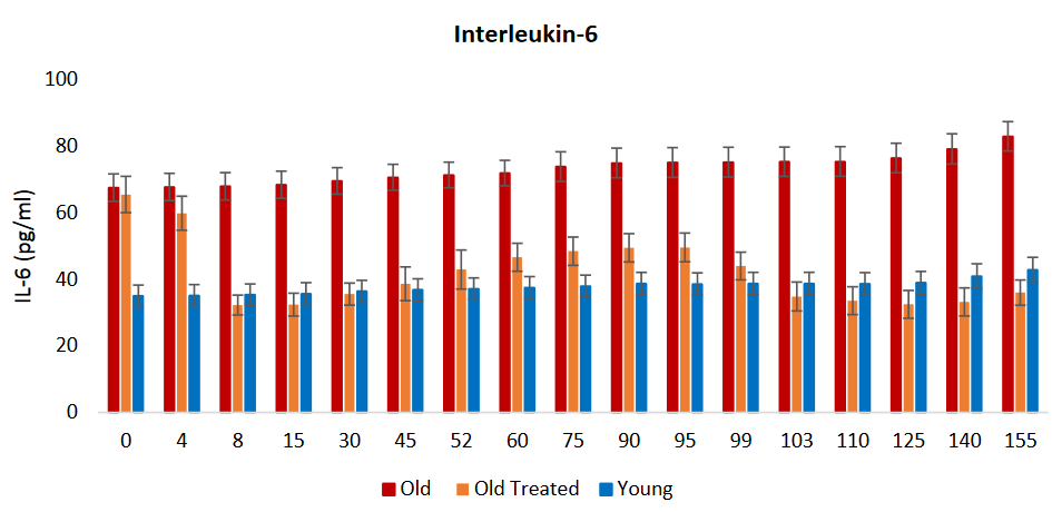
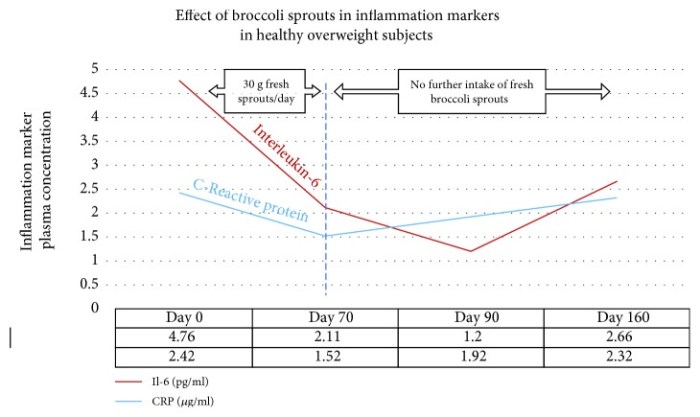
“In a recent study using 30 grams of fresh broccoli sprouts incorporated daily into diet, two key inflammatory cytokines were measured at four time points in forty healthy overweight [BMI 24.9 – 29.9] people. Levels of both interleukin-6 (Il-6) and C-reactive protein (CRP) declined over the 70 days during which sprouts were ingested.

These biomarkers were measured again at day 90, wherein it was found that Il-6 continued to decline, whereas CRP climbed again. When the final measurement was taken at day 160, CRP, although climbing, had not returned to its baseline value. Il-6 remained significantly below baseline level at day 160.

Sprouts contained approximately 51 mg (117 μmol) GRN [glucoraphanin], and plasma and urinary SFN metabolites were measured to confirm that SFN had been produced when sprouts were ingested.”


From a clinical trial perspective, glucoraphanin dosage was “1.67 (GR) μmol/kg BW.” This wasn’t accurate, however. It was assumed into existence by:

“In cases where authors did not indicate dosage in μmol/kg body weight (BW), we have made those calculations using a priori assumption of a 70 kg BW.”

117 μmol / 1.67 μmol/kg = 70 kg.

This study provided overweight subjects’ mean weight in its Table 1 as “85.8 ± 16.7 kg.” So its actual average glucoraphanin dosage per kg body weight was 117 μmol / 85.8 kg = 1.36 μmol/kg. Was making an accurate calculation too difficult?

A clinical trial perspective included this study in Section “3.2. Clinical Studies with Broccoli-Based Preparations: Efficacy” subsection “3.2.8. Diabetes, Metabolic Syndrome, and Related Disorders.” This was somewhat misleading, as it was grouped with studies such as a 2012 Iranian Effects of broccoli sprout with high sulforaphane concentration on inflammatory markers in type 2 diabetic patients: A randomized double-blind placebo-controlled clinical trial (not freely available).

A commercial perspective pointed out substantial differences between these two studies:

“Where the study described above by Lopez-Chillon et al. investigated healthy overweight people to assess effects of SFN-yielding broccoli sprout homogenate on biomarkers of inflammation, Mirmiran et al. in 2012 had used a SFN-yielding supplement in T2DM patients. Although the data are not directly comparable, the latter study using the powdered supplement resulted in significant lowering of Il-6, hs-CRP, and TNF-α over just 4 weeks.

It is not possible to further compare the two studies due to vastly different time periods over which each was conducted.”


The commercial perspective impressed as more balanced than the clinical trial perspective. The clinical trial perspective also had an undisclosed conflict of interest!

A. The clinical trial perspective:

  • Effectively promoted one commercial product whose supplier was associated with a coauthor;
  • Downgraded several other commercial products; and
  • Tried to shift responsibility for the lack of “minimally effective doses in humans” to commercial products with:

    “Only recently have there been attempts to define minimally effective doses in humans – an outcome made possible by the development of consistently formulated, stable, bioavailable broccoli-derived preparations.”

But unless four years previous is “recently,” using commercial products to excuse slow research progress can be dismissed. A coauthor of the clinical trial perspective was John Hopkins’ lead researcher for a November 2015 Sulforaphane Bioavailability from Glucoraphanin-Rich Broccoli: Control by Active Endogenous Myrosinase, which commended “high quality, commercially available broccoli supplements” per:

“We have now discontinued making BSE [broccoli sprout extract], because there are several high quality, commercially available broccoli supplements on the market.”

The commercial perspective didn’t specifically mention any commercial products.

B. The commercial perspective didn’t address taste, which may be a consumer acceptance problem.

C. The commercial perspective provided practical dosage recommendations, reflecting their consumer orientation. These recommendations didn’t address how much sulforaphane is suitable for healthy people, though.

The clinical trial perspective will eventually have to make practical dosage recommendations after they stop dodging their audience – which includes clinicians trying to apply clinical trial data – with unhelpful statements such as:

“Reporting of administered dose of glucoraphanin and/or sulforaphane is a poor measure of the bioavailable / bioactive dose of sulforaphane.”

How practical was their “internal dose” recommendation for non-researcher readers?


Here’s what I’m doing to answer how much sulforaphane is suitable for healthy people.

I’d like to posthumously credit my high school literature teachers Dorothy Jasiecki and Martin Obrentz for this post’s compare-and-contrast approach. They both required their students to read at least two books monthly, then minimally handwrite a 3-page (single-spaced) paper comparing and contrasting those books.

Each monthly assignment was individualized so that students couldn’t undo the assignment’s purpose – to think for yourself – with parasitical collaboration. This former practice remains a good measure of intentional dumbing-down of young people, the intent of which has become clearer.

You can see from these linked testimonials that their approach was in a bygone era, back when some teachers considered a desired outcome of public education to be that each individual learned to think for themself. My younger brother contributed:

“I can still remember everything Mr. Obrentz ever assigned for me to read. He was the epitome of what a teacher should be.”

The epigenetics of perinatal stress

This 2019 McGill review discussed long-lasting effects of perinatal stress:

“Epigenetic processes are involved in embedding the impact of early-life experience in the genome and mediating between social environments and later behavioral phenotypes. Since these phenotypes are apparent a long time after early experience, changes in gene expression programming must be stable.

Although loss of methylation in a promoter is necessary for expression, it is not sufficient. Demethylation removes a barrier for expression, but expression might be realized at the right time or context when needed factors or signals are present.

DNA methylation anticipates future transcriptional response to triggers. Comparing steady-state expression with DNA methylation does not capture the full meaning and scope of regulatory roles of differential methylation.

A model for epigenetic programming by early life stress:

  1. Perinatal stress perceived by the brain triggers release of glucocorticoids (GC) from the adrenal in the mother prenatally or the newborn postnatally.
  2. GC activate nuclear glucocorticoid receptors across the body, which epigenetically program (demethylate) genes that are targets of GR in brain and white blood cells (WBC).
  3. Demethylation events are insufficient for activation of these genes. A brain specific factor (TF) is required for expression and will activate low expression of the gene in the brain but not in blood.
  4. During adulthood a stressful event transiently triggers a very high level of expression of the GR regulated gene specifically in the brain.

Horizontal arrow, transcription; circles, CpG sites; CH3 in circles, methylated sites; empty circles, unmethylated CpG sites; horizon[t]al curved lines, mRNA.”

Review points discussed:

  • “Epigenetic marks are laid down and maintained by enzymes that either add or remove epigenetic modifications and are therefore potentially reversible in contrast to genetic changes.
  • Response to early life stress and maternal behavior is also not limited to the brain and involves at least the immune system as well.
  • The placenta is also impacted by maternal social experience and early life stress.
  • Most studies are limited to peripheral tissues such as saliva and white blood cells, and relevance to brain physiology and pathology is uncertain.
  • Low absolute differences in methylation seen in most human behavioral EWAS raise questions about their biological significance.

  • Although post-mortem studies examine epigenetic programming in physiologically relevant tissues, they represent only a final and single stage that does not capture dynamic evolution of environments and epigenetic programming in living humans.”

https://www.ncbi.nlm.nih.gov/pmc/articles/PMC6952743/ “The epigenetics of perinatal stress”


Other reviewers try to ignore times when we were all fetuses and newborns. For example, in the same journal issue was a Boston review of PTSD that didn’t mention anything about earliest times of human lives! Those reviewers speculated around this obvious gap on their way to being paid by NIH.

Why would researchers ignore perinatal stress events that prime humans for later-life PTSD? Stress generally has a greater impact on fetuses and newborns than on infants, and a greater impact on infants than on adults.

Using oxytocin receptor gene methylation to pursue an agenda

A pair of 2019 Virginia studies involved human mother/infant subjects:

“We show that OXTRm [oxytocin receptor gene DNA methylation] in infancy and its change is predicted by maternal engagement and reflective of behavioral temperament.”

https://www.ncbi.nlm.nih.gov/pmc/articles/PMC6795517 “Epigenetic dynamics in infancy and the impact of maternal engagement”

“Infants with higher OXTRm show enhanced responses to anger and fear and attenuated responses to happiness in right inferior frontal cortex, a region implicated in emotion processing through action-perception coupling.

Infant fNIRS [functional near-infrared spectroscopy] is limited to measuring responses from cerebral cortex. It is unknown whether OXTR is expressed in the cerebral cortex during prenatal and early postnatal human brain development.”

https://www.sciencedirect.com/science/article/pii/S187892931830207X “Epigenetic modification of the oxytocin receptor gene is associated with emotion processing in the infant brain”


Both studies had weak disclosures of limitations on their findings’ relevance and significance. The largest non-disclosed contrary finding was from the 2015 Early-life epigenetic regulation of the oxytocin receptor gene:

These results suggest that:

  • Blood Oxtr DNA methylation may reflect early experience of maternal care, and
  • Oxtr methylation across tissues is highly concordant for specific CpGs, but
  • Inferences across tissues are not supported for individual variation in Oxtr methylation.

That rat study found that blood OXTR methylation of 25 CpG sites couldn’t accurately predict the same 25 CpG sites’ OXTR methylation in each subject’s hippocampus, hypothalamus, and striatum (which includes the nucleus accumbens) brain areas. Without significant effects in these limbic system structures, there couldn’t be any associated behavioral effects.

But CpG site associations and correlations were deemed good in the two current studies because they cited:

“Recent work in prairie voles has found that both brain- and blood-derived OXTRm levels at these sites are negatively associated with gene expression in the brain and highly correlated with each other.”

https://www.sciencedirect.com/science/article/pii/S0306453018306103 “Early nurture epigenetically tunes the oxytocin receptor”

The 2018 prairie vole study – which included several of the same researchers as the two current studies – found four nucleus accumbens CpG sites that had high correlations to humans. Discarding one of these CpG sites allowed their statistics package to make a four-decimal place finding:

“The methylation state of the blood was also associated with the level of transcription in the brain at three of the four CpG sites..whole blood was capable of explaining 94.92% of the variance in Oxtr DNA methylation and 18.20% of the variance in Oxtr expression.”

Few limitations on the prairie vole study findings were disclosed. Like the two current studies, there wasn’t a limitation section that placed research findings into suitable contexts. So readers didn’t know researcher viewpoints on items such as:

  • What additional information showed that 3 of the 30+ million human CpGs accurately predicted specific brain OXTR methylation and expression from saliva OXTR methylation?
  • What additional information demonstrated how “measuring responses from cerebral cortex” although “it is unknown whether OXTR is expressed in the cerebral cortex” provided detailed and dependable estimates of limbic system CpG site OXTR methylation and expression?
  • Was the above 25-CpG study evidence considered?

Further contrast these three studies with a typical, four-point, 285-word limitation section of a study like Prenatal stress heightened adult chronic pain. The word “limit” appeared 6 times in that pain study, 3 times in the current fNIRS study, and 0 times in the current maternal engagement and cited prairie vole studies.

Frank interpretations of one’s own study findings to acknowledge limitations is one way researchers can address items upfront that will be questioned anyway. Such analyses also indicate a goal to advance science.

Prenatal stress heightened adult chronic pain

This 2019 McGill rodent study found:

Prenatal stress exacerbates pain after injury. Analysis of mRNA expression of genes related to epigenetic regulation and stress responses in the frontal cortex and hippocampus, brain structures implicated in chronic pain, showed distinct sex and region-specific patterns of dysregulation.

In general, mRNA expression was most frequently altered in the male hippocampus and effects of prenatal stress were more prevalent than effects of nerve injury. Recent studies investigating chronic pain-related pathology in the hippocampus in humans and in rodent models demonstrate functional abnormalities in the hippocampus, changes in associated behavior, and decreases in adult hippocampal neurogenesis.

The change in expression of epigenetic- and stress-related genes is not a consequence of nerve injury but rather precedes nerve injury, consistent with the hypothesis that it might play a causal role in modulating the phenotypic response to nerve injury. These findings demonstrate the impact of prenatal stress on behavioral sensitivity to a painful injury.

Decreased frontal mRNA expression of BDNF and BDNF IV in male offspring following neuropathic pain or prenatal stress respectively. Relative mRNA expression of other stress-related genes (GR17, FKBP5) and epigenetic-related genes (DNMTs, TETs, HDACs, MBDs, MeCP2) in male offspring.

A drastic decrease in expression of HDAC1 was observed in all groups compared to sham-control animals. CCI: chronic constriction injury.”


The study’s design was similar to the PRS (prenatal restraint stress) model, except that the PRS procedure covered gestational days 11 to 21 (birth):

“Prenatal stress was induced on Embryonic days 13 to 17 by restraining the pregnant dams in transparent cylinder with 5 mm water, under bright light exposure, 3 times per day for 45 min.”

None of the French, Italian, and Swiss PRS studies were cited.

The limitation section included:

  1. “Although our study shows significant changes in expression of epigenetic enzymes, it didn’t examine the impact of these changes on genes that are epigenetically regulated by this machinery or their involvement in intensifying pain responses.
  2. The current study is limited by the focus on changes in gene expression which do not necessarily correlate with changes in protein expression.
  3. Another limitation of this study is the inability to distinguish the direct effects of stress in utero vs. changes in the dam’s maternal behavior due to stress during pregnancy; cross-fostering studies are needed to address this issue.
  4. Functional experiments that involve up and down regulation of epigenetic enzymes in specific brain regions are required to establish a causal role for these processes in chronic pain.”

What do you think about possible human applicability of this study’s “effects of prenatal stress were more prevalent than effects of nerve injury” finding?

Are there any professional therapeutic frameworks that instruct trainees to recognize that if a person’s mother was stressed while pregnant, their prenatal experiences could cause more prevalent biological and behavioral effects than a recent injury?

https://www.sciencedirect.com/science/article/pii/S0166432819315219 “Prenatal maternal stress is associated with increased sensitivity to neuropathic pain and sex-specific changes in supraspinal mRNA expression of epigenetic- and stress-related genes in adulthood” (not freely available)

An epigenetic clock review by committee

This 2019 worldwide review of epigenetic clocks was a semi-anonymous mishmash of opinions, facts, hypotheses, unwarranted extrapolations, and beliefs. Diversity of viewpoints among the 21 coauthors wasn’t evident.

1. Citations of coauthors’ works seemed excessive, and they apologized for omissions. However:

  • Challenge 5 was titled “Single-cell analysis of aging changes and disease” and
  • Table 1 “Major biological and analytic issues with epigenetic DNA methylation clocks” had single-cell analysis as the Proposed solution to five Significant issues.

Yet studies such as High-Resolution Single-Cell DNA Methylation Measurements Reveal Epigenetically Distinct Hematopoietic Stem Cell Subpopulations were unmentioned.

2. Some coauthors semi-anonymously expressed faith that using current flawed methodologies in the future – only more thoroughly, with newer equipment, etc. – would yield better results. If all 21 coauthors were asked their viewpoints of Proposed solutions to the top three Significant issues of epigenetic clocks, what would they emphasize when quoted?

3. Techniques were praised:

“Given the precision with which DNA methylation clock age can be estimated and evolving measures of biological, phenotype-, and disease-related age (e.g., PhenoAge, GrimAge)..”

Exactly why these techniques have at times produced inexplicable results wasn’t examined, though. Two examples:

  • In Reversal of aging and immunosenescent trends, Levine PhenoAge methodology estimated that the 51-65 year old subjects’ biological ages at the beginning of the study averaged 17.5 years less than their chronological age. Comparing that to Horvath average biological age of 3.95 years less raised the question: exactly why did PhenoAge show such a large difference?
  • The paper mentioned GrimAge methodology findings about “smoking-related changes.” But it didn’t explain why GrimAge methylation findings most closely associated with smoking history also accurately predicted future disease risk with non-smokers.

Eluding explanations for these types of findings didn’t help build confidence in methodologies.

4. A more readable approach to review by committee could have coauthors – in at least one section – answer discussion questions, as Reversing epigenetic T cell exhaustion did with 18 experts.

https://genomebiology.biomedcentral.com/articles/10.1186/s13059-019-1824-y “DNA methylation aging clocks: challenges and recommendations”

A review of fetal adverse events

This 2019 Australian review subject was fetal adversities:

“Adversity during the perinatal period is a significant risk factor for the development of neurodevelopmental disorders long after the causative event. Despite stemming from a variety of causes, perinatal compromise appears to have similar effects on the developing brain, thereby resulting in behavioural disorders of a similar nature.

These behavioural disorders occur in a sex‐dependent manner, with males affected more by externalizing behaviours such as attention deficit hyperactivity disorder (ADHD) and females by internalizing behaviours such as anxiety. The term ‘perinatal compromise’ serves as an umbrella term for intrauterine growth restriction, maternal immune activation, prenatal stress, early life stress, premature birth, placental dysfunction, and perinatal hypoxia.

The above conditions are associated with imbalanced excitatory-inhibitory pathways resulting from reduced GABAergic signalling. Methylation of the GAD1/GAD67 gene, which encodes the key glutamate‐to‐GABA synthesizing enzyme Glutamate Decarboxylase 1, resulting in increased levels of glutamate is one epigenetic mechanism that may account for a tendency towards excitation in disorders such as ADHD.

The posterior cerebellum’s role in higher executive functioning is becoming well established due to its connections with the prefrontal cortex, association cortices, and limbic system. It is now suggested that disruptions to cerebellar development, which can occur due to late gestation compromises such as preterm birth, can have a major impact on the region of the brain to which it projects.

Activation of the maternal hypothalamic-pituitary adrenal (HPA) axis and placental protection. Psychological stress is perceived by the maternal HPA axis, which stimulates cortisol release from the maternal adrenal gland.

High levels of maternal cortisol are normally prevented from reaching the fetus by the 11β-hydroxysteroid dehydrogenase 2 (HSD11B2) enzyme, which converts cortisol to the much less active cortisone. Under conditions of high maternal stress, this protective mechanism can be overwhelmed, with the gene encoding the enzyme becoming methylated, which reduces its expression allowing cortisol to cross the placenta and reach the fetus.”


The reviewers extrapolated many animal study findings to humans, although most of their own work was with guinea pigs. The “suggest” and “may” qualifiers were used often – 22 and 37 times, respectively. More frequent use of the “appears,” “hypothesize,” “propose,” and “possible” terms was justified.

As a result, many reviewed items such as the above graphic and caption should be viewed as hypothetical for humans rather than reflecting solid evidence from quality human studies.

The reviewers focused on the prenatal (before birth) period more than the perinatal (last trimester of pregnancy to one month after birth) period. There were fewer mentions of birth and early infancy adversities.

https://onlinelibrary.wiley.com/doi/abs/10.1111/jne.12814 “Perinatal compromise contributes to programming of GABAergic and Glutamatergic systems leading to long-term effects on offspring behaviour” (not freely available)

Organismal aging and cellular senescence

I’ll curate this 2019 German review through its figures:

“With the discovery of beneficial aspects of cellular senescence and evidence of senescence being not limited to replicative cellular states, a redefinition of our comprehension of aging and senescence appears scientifically overdue.

Figure 1. Current determinants and relevant open questions, marking the processes of aging and senescence as discussed in the text. Aspects represented in green are considered as broadly accepted or scientifically consolidated. Novel aspects that are yet unproven, or are under debate, are highlighted in red.

SASP = senescence-associated secretory phenotype. AASP = putative aging-associated secretory phenotype as suggested in the text.

Figure 2. Theories on the causality and purpose of aging. Graphically summarized are four contrasting concepts crystallized from current evidence addressing the inductive driving force of aging. Apart from a stochastic deleteriome, there are arguments for a pseudo-programmed, programmed or at least partially programmed nature of aging.

Figure 3. Comparative representation of the aging and senescence processes highlighting different levels of interaction and putative sites of interventions.

(1) As discussed in the text, causative mechanisms of aging are still not well understood, however, multiple factors including genetic, epigenetic and stress-related effects seem to have an orchestrated role in the progression of aging. Senescence on the other hand, is seen as a programmed response to different kinds of stressors, which proceed in defined stages. Whether, in analogy, aging also follows a defined program or sequential stages is not known.

(2) Senescence involves autocrine and paracrine factors, which are responsible for a ‘seno-infection’ or bystander effect in neighboring cells. There is currently no direct evidence for a similar factor composition propagating the aging process via a kind of ‘gero-infection’.

(3) Accumulation of senescent cells has been described as a hallmark of aging; however, whether they are a causative factor or a consequence of tissue and organismal aging is still unknown. As discussed in the text, it appears possible that aging and senescence mutually influence each other through positive feedback at this level, leading to accelerated tissue damage and aging.

(4,5) Clearance of senescent or aging cells might constitute putative targets for interventional approaches aimed to reduce or reverse the impact of aging and improve cell and tissue homeostasis by inducing a ‘rejuvenation’ process.

Figure 4. Pathological and beneficial functions of aging and senescence, according to current knowledge. In red are represented pathological consequences and in green beneficial functions of aging and senescence.

The impact of aging has mainly been described at the organismal level, since a complete cellular functional profile has not yet been established. Accordingly, whether beneficial consequences of the aging process exist at the cellular level is unclear.”


The assertion of Figure 3 (2) that:

“There is currently no direct evidence for a similar factor composition propagating the aging process via a kind of ‘gero-infection.”

was shown to be false in Reevaluate findings in another paradigm:

“It was demonstrated that increased aging occurred as a result of lack of gonadotropin-releasing hormone and that increased lifespan resulted from its provision during aging.

In this manner:

  1. Aging of hypothalamic microglia leads to
  2. Aging of the hypothalamus, which leads to
  3. Aging elsewhere in the body.

So here we have a multi-level interaction:

  1. Activation of NF-κB leads to
  2. Cellular aging, leading to
  3. A diminished production of GnRH, which then
  4. Acts (through cells with a receptor for it, or indirectly as a result of changes to GnRH-receptor-possessing cells) to decrease lifespan.

So the age state of hypothalamic cells, at least with respect to NF-κB activation, is communicated to other cells via reduced output of GnRH.”


The reviewers’ position on Figure 2 was:

“In our view, recent evidence that

  • Senescence is based on an unterminated developmental growth program and the finding that
  • The concept of post-mitotic senescence requires the activation of expansion, or ‘growth’ factors as a second hit,

favor the assumption that aging underlies a grating of genetic determination similarly to what is summarized above under the pseudo-programmed causative approach.”

Their position on Figure 4’s beneficial effects of aging began with the sentence:

“If we assume that aging already starts before birth, it can be considered simply a developmental stage, required to complete the evolutionary program associated with species-intrinsic biological functions such as reproduction, survival, and selection.”

Cited studies included:

https://www.mdpi.com/2073-4409/8/11/1446 “Dissecting Aging and Senescence-Current Concepts and Open Lessons”

Do genes or maternal environments shape fetal brains?

This 2019 Singapore human study used Diffusion Tensor Imaging on 5-to-17-day old infants to find:

“Our findings showed evidence for region-specific effects of genotype and GxE on individual differences in human fetal development of the hippocampus and amygdala. Gene x Environment models outcompeted models containing genotype or environment only, to best explain the majority of measures but some, especially of the amygdaloid microstructure, were best explained by genotype only.

Models including DNA methylation measured in the neonate umbilical cords outcompeted the Gene and Gene x Environment models for the majority of amygdaloid measures and minority of hippocampal measures. The fact that methylation models outcompeted gene x environment models in many instances is compatible with the idea that DNA methylation is a product of GxE.

A genome-wide association study of SNP [single nucleotide polymorphism] interactions with the prenatal environments (GxE) yielded genome wide significance for 13 gene x environment models. The majority (10) explained hippocampal measures in interaction with prenatal maternal mental health and SES [socioeconomic status]. The three genome-wide significant models predicting amygdaloid measures, explained right amygdala volume in interaction with maternal depression.

The transcription factor CUX1 was implicated in the genotypic variation interaction with prenatal maternal health to shape the amygdala. It was also a central node in the subnetworks formed by genes mapping to the CpGs in neonatal umbilical cord DNA methylation data associating with both amygdala and hippocampus structure and substructure.

Our results implicated the glucocorticoid receptor (NR3C1) in population variance of neonatal amygdala structure and microstructure.

Estrogen in the hippocampus affects learning, memory, neurogenesis, synapse density and plasticity. In the brain testosterone is commonly aromatized to estradiol and thus the estrogen receptor mediates not only the effects of estrogen, but also that of testosterone.”

https://onlinelibrary.wiley.com/doi/full/10.1111/gbb.12576 “Neonatal amygdalae and hippocampi are influenced by genotype and prenatal environment, and reflected in the neonatal DNA methylome” (not freely available)

A drug that countered effects of a traumatizing mother

This 2019 US rodent study concerned transmitting poor maternal care to the next generation:

“The quality of parental care received during development profoundly influences an individual’s phenotype, including that of maternal behavior. Infant experiences with a caregiver have lifelong behavioral consequences.

Maternal behavior is a complex behavior requiring the recruitment of multiple brain regions including the nucleus accumbens, bed nucleus of the stria terminalis, ventral tegmental area, prefrontal cortex, amygdala, and medial preoptic area. Dysregulation within this circuitry can lead to altered or impaired maternal responsiveness.

We administered zebularine, a drug known to alter DNA methylation, to dams exposed during infancy to the scarcity-adversity model of low nesting resources, and then characterized the quality of their care towards their offspring.

  1. We replicate that dams with a history of maltreatment mistreat their own offspring.
  2. We show that maltreated-dams treated with zebularine exhibit lower levels of adverse care toward their offspring.
  3. We show that administration of zebularine in control dams (history of nurturing care) enhances levels of adverse care.
  4. We show altered methylation and gene expression in maltreated dams normalized by zebularine.

These findings lend support to the hypothesis that epigenetic alterations resulting from maltreatment causally relate to behavioral outcomes.

Maternal behavior is an intergenerational behavior. It is important to establish the neurobiological underpinnings of aberrant maternal behavior and explore treatments that can improve maternal behavior to prevent the perpetuation of poor maternal care across generations.”


The study authors demonstrated intergenerational epigenetic effects, and missed an opportunity to also investigate transgenerational epigenetically inherited effects. They cited reference 60 for the first part of the above quotation, but the cited reviewer misused the transgenerational term by applying it to grand-offspring instead of the great-grand-offspring.

There were resources available to replicate the study authors’ previous findings, which didn’t show anything new. Why not use such resources to uncover evidence even more applicable to humans by extending experiments to great-grand-offspring that would have no potential germline exposure to the initial damaging cause?

Could a study design similar to A limited study of parental transmission of anxiety/stress-reactive traits have been integrated? That study’s thorough removal of parental behavior would be an outstanding methodology to confirm by falsifiability whether parental behavior is both an intergenerational and a transgenerational epigenetic inheritance mechanism.

Rodent great-grand-offspring can be studied in < 9 months. It takes > 50 years for human studies to reach the great-grand-offspring transgenerational generation.

  • Why not attempt to “prevent the perpetuation of poor maternal care across generations?”
  • Isn’t it a plausible hypothesis that humans “with a history of maltreatment mistreat their own offspring?”
  • Isn’t it worth the extra effort to extend animal research to investigate this unfortunate chain?

https://www.nature.com/articles/s41598-019-46539-4 “Pharmacological manipulation of DNA methylation normalizes maternal behavior, DNA methylation, and gene expression in dams with a history of maltreatment”

A better method of measuring neurogenesis

One of the references cited in Linking adult neurogenesis to Alzheimer’s disease was https://www.nature.com/articles/s41591-019-0375-9 “Adult hippocampal neurogenesis is abundant in neurologically healthy subjects and drops sharply in patients with Alzheimer’s disease” (not freely available).

This 2019 Spanish human study used improved techniques to find:

“Adult hippocampal neurogenesis (AHN), confers an unparalleled degree of plasticity to the entire hippocampal circuitry. Direct evidence of AHN in humans has remained elusive. Determining whether new neurons are continuously incorporated into the human dentate gyrus (DG) during physiological and pathological aging is a crucial question with outstanding therapeutic potential.

By combining human brain samples obtained under tightly controlled conditions and state-of-the-art tissue processing methods, we identified thousands of immature neurons in the DG of neurologically healthy human subjects up to the ninth decade of life. These neurons exhibited variable degrees of maturation along differentiation stages of AHN. In sharp contrast, the number and maturation of these neurons progressively declined as AD advanced.

These results demonstrate the persistence of AHN during both physiological and pathological aging in humans and provide evidence for impaired neurogenesis as a potentially relevant mechanism underlying memory deficits in AD that might be amenable to novel therapeutic strategies.”


The control group was 13 neurologically healthy deceased people aged 43 to 87. The AD group was 45 deceased people, distributed among the six Braak stages of the pathology, aged 52 to 97.