Aging as an unintended consequence

The coauthors of 2018’s The epigenetic clock theory of aging reviewed progress that’s been made todate in understanding epigenetic clock mechanisms.

1. Proven DNA methylation features of epigenetic clocks:

  1. “Methylation of cytosines is undoubtedly a binary event.
  2. The increase in epigenetic age is contributed by changes of methylation profiles in a very small percent of cells in a population.
  3. The clock ticks extremely fast in early post-natal years and much slower after puberty.
  4. Clock CpGs have specific locations in the genome.
  5. It applies to prenatal biological samples and embryonic stem cells.

While consistency with all the five attributes does not guarantee veracity of a model, inconsistency with any one will signal the unlikely validity of a hypothesis.”

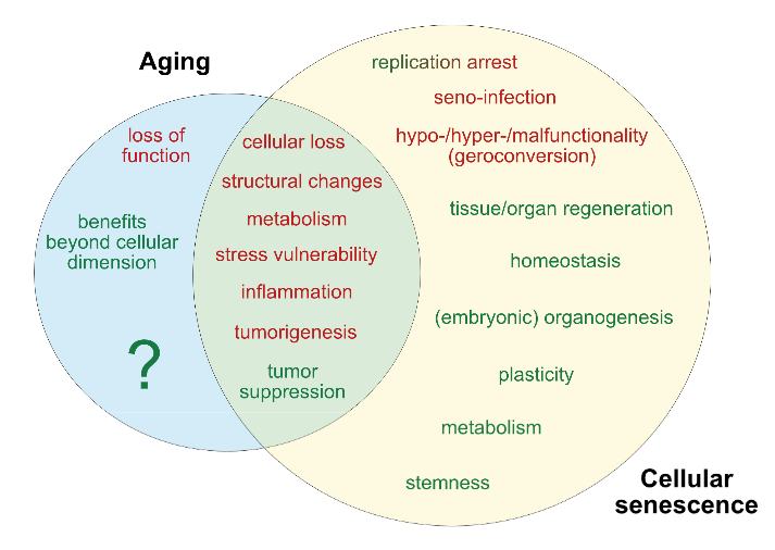
2. Regarding what epigenetic clocks don’t measure:

“The effects of

  • Telomere maintenance,
  • Cellular senescence,
  • DNA damage signaling,
  • Terminal differentiation and
  • Cellular proliferation

have all been tested and found to be unrelated to epigenetic ageing.”

3. Regarding cyclical features:

Both the epigenetic and circadian clocks are present in all cells of the body, but their ticking rates are regulated. Both these clocks lose synchronicity when cells are isolated from tissues and grown in vitro.

These similarities compel one to ponder potential links between them.”

This was among the points that Linear thinking about biological age clocks missed.

4. The reviewers discussed 3 of the 5 treatment elements in Reversal of aging and immunosenescent trends:

“It is not known at this stage whether the rejuvenating effect is mediated through the regeneration of the thymus or a direct effect of the treatment modality on the body. Also, it is not known if the effect is mediated by all three compounds or one or two of them.

What we know at this stage does not allow the formation of general principles regarding the impact of hormones on epigenetic age, but their involvement in development and maintenance of the body argue that they do indeed have a very significant impact on the epigenetic clock.”

Not sure why they omitted 3000 IU vitamin D and 50 mg zinc, especially since:

“It is not known if the effect is mediated by all three [five] compounds or one or two of them.”

5. They touched on the specialty of Aging as a disease researchers with:

“Muscle stem cells isolated from mice were epigenetically much younger independently of the ages of the tissue / animal from which they were derived.

The proliferation and differentiation of muscle stem cells cease upon physical maturation. These activities are initiated in adult muscles only in response to injury.

6. The reviewers agreed with those researchers in the Conclusion:

“Epigenetic ageing begins from very early moments after the embryonic stem cell stage and continues uninterrupted through the entire lifespan. The significance of this is profound as the question of why we age has been attributed to many different things, most commonly to ‘wear-and-tear.’

The ticking of the epigenetic clock from the embryonic state challenges this perspective and supports the notion that ageing is an unintended consequence of processes that are necessary for

  • The development of the organism and
  • Tissue homeostasis thereafter.”


https://journals.sagepub.com/doi/10.1177/1535370220918329 “Current perspectives on the cellular and molecular features of epigenetic ageing” (not freely available)

Broccoli sprouts oppose effects of advanced glycation end products (AGEs)

This 2020 Australian/UK review subject was AGEs:

“AGEs are formed during cooking and food processing or produced endogenously as a consequence of metabolism. Deleterious effects of AGEs are underpinned by their ability to trigger mechanisms well known to elicit metabolic dysfunction, including activation of inflammatory pathways, oxidative stress and impaired mitochondrial oxidative metabolism. They have been widely implicated in complications of diabetes affecting cardiovascular health, the nervous system, eyes and kidneys.

Reactive carbonyl groups are constantly being produced via normal metabolism and when production overrides detoxification, AGEs accumulate. AGE formation may take several days or weeks to complete in the body.

Factors affecting AGE content of food depends on composition of protein, fat, and sugar and types of processing and cooking methods employed, predominantly on temperature and duration of preparation. Circulating free-AGEs concentrations are a good marker for dietary AGE intake while plasma protein-bound AGEs better represent endogenously produced AGEs.

Receptor for Advanced glycation end products (RAGE) signals via transcription factor NF-kB increasing gene expression of inflammatory mediators and production of ROS (reactive oxygen species).”

https://onlinelibrary.wiley.com/doi/abs/10.1002/mnfr.201900934 “The Role of Dietary Advanced Glycation End Products (AGEs) in Metabolic Dysfunction” (not freely available)


Let’s use the Australian 2019 Sulforaphane: Its “Coming of Age” as a Clinically Relevant Nutraceutical in the Prevention and Treatment of Chronic Disease as a reference for how sulforaphane may counter effects of AGEs:

1. “Activation of inflammatory pathways”

“Antioxidants in general and glutathione in particular can be depleted rapidly under conditions of oxidative stress, and this can signal inflammatory pathways associated with NF-κB. SFN [sulforaphane] has been shown to inhibit NF-κB in endothelial cells.

Two key inflammatory cytokines were measured at four time points in forty healthy overweight people [our model clinical trial, Effects of long-term consumption of broccoli sprouts on inflammatory markers in overweight subjects]. Levels of both interleukin-6 (Il-6) and C-reactive protein (CRP) declined over the 70 days during which sprouts were ingested. These biomarkers were measured again at day 90, wherein it was found that Il-6 continued to decline, whereas CRP climbed again. When the final measurement was taken at day 160, CRP, although climbing, had not returned to its baseline value. Il-6 remained significantly below the baseline level at day 160.”

OMCL2019-2716870.010

2. “When production overrides detoxification”

“SFN significantly activates Nrf2 and as such has the potential to modulate the expression of genes associated with redox balance, inflammation, detoxification, and antimicrobial capacity, all key components of the upstream cellular defence processes.

Toxins presented to Phase 1 enzymes produce intermediate compounds which are sometimes more toxic to cells than the initial toxin. It is therefore important that Phase 2 is sufficiently active that intermediate products cannot accumulate in the cellular environment.

As a monofunctional inducer, SFN has been described as an ideal detoxifier, as its effect on Phase 1 is minimal compared with its significant activity on Phase 2.”

3. “Oxidative stress”

“As a mediator for amplification of the mammalian defence system against various stressors, Nrf2 sits at the interface between our prior understanding of oxidative stress and endogenous mechanisms cells use to deal with it. Diseases known to be underpinned by oxidative stress are proving to be more responsive to amplification of cellular defences via Nrf2 activation than by administration of direct-acting antioxidant supplements.

SFN, with absolute bioavailability of around 80%, is capable of increasing several endogenous antioxidant compounds via transcription factor Nrf2.”

4. “Complications of diabetes affecting cardiovascular health, the nervous system, eyes and kidneys”

“Nrf2 is ubiquitously expressed with highest concentrations (in descending order) in the kidney, muscle, lung, heart, liver, and brain. Nrf2 was shown to prevent endothelial cells from exhibiting a proinflammatory state. Nrf2 is required for protection against glucose-induced oxidative stress and cardiomyopathy in the heart.

Well in excess of 500 genes have been identified as being activated by SFN via the Nrf2/ARE [Antioxidant Response Element] pathway, and it is likely that this underestimates the number as others are being discovered. Of available SFN clinical trials associated with genes induced via Nrf2 activation, many demonstrate a linear dose-response. More recently, it has become apparent that SFN can behave hormetically with different effects responsive to different doses.

It [sulforaphane] is not only a potent Nrf2 inducer but also highly bioavailable so that modest practical doses can produce significant clinical responses. Other Nrf2 activators [shown in the above image] not only lack potency but also lack the bioavailability to be considered as significant intracellular Nrf2 activators.”


As mentioned in Changing an inflammatory phenotype with broccoli sprouts, per the above bolded part of section 3, I stopped taking N-acetyl-cysteine, the precursor to our endogenous antioxidant glutathione. I stopped taking curcumin last year due to no noticeable effects, probably because of its poor bioavailability. I may soon stop taking more vitamin E than the RDA, and β-carotene.

I changed my diet last summer to reduce AGEs, with mild effects. I expect stronger effects from also daily eating 60 grams of 3-day-old broccoli sprouts that yield 27 mg of sulforaphane after microwaving.

Forcing people to learn helplessness

Learned helplessness is a proven animal model. Its reliably-created phenotype is often the result of applying chronic unpredictable stress.

As we’re finding out worldwide, forcing humans to learn helplessness works in much the same way, with governments imposing what amounts to martial law. Never mind that related phenotypes and symptoms include:

  • “Social defeat
  • Social avoidance behavior
  • Irritable bowel syndrome
  • Depression
  • Anxiety
  • Anhedonia
  • Increased hypothalamic-pituitary-adrenal (HPA)-axis sensitivity
  • Visceral hypersensitivity” [1]

Helplessness is both a learned behavior and a cumulative set of experiences. Animal models demonstrate that these phenotypes usually continue on throughout the subjects’ entire lifespans.

Will the problems caused in humans by humans be treated by removing the causes? Or will the responses be approaches such as drugs to treat the symptoms?


A major difference between our current situation and the situation depicted below is that during communism, most people didn’t really trust or believe what the authorities, newspapers, television, and radio said:

Image from Prague’s Memorial to the Victims of Communism


[1] 2014 GABAB(1) receptor subunit isoforms differentially regulate stress resilience curated in If research provides evidence for the causes of stress-related disorders, why only focus on treating the symptoms?

The epigenetics of perinatal stress

This 2019 McGill review discussed long-lasting effects of perinatal stress:

“Epigenetic processes are involved in embedding the impact of early-life experience in the genome and mediating between social environments and later behavioral phenotypes. Since these phenotypes are apparent a long time after early experience, changes in gene expression programming must be stable.

Although loss of methylation in a promoter is necessary for expression, it is not sufficient. Demethylation removes a barrier for expression, but expression might be realized at the right time or context when needed factors or signals are present.

DNA methylation anticipates future transcriptional response to triggers. Comparing steady-state expression with DNA methylation does not capture the full meaning and scope of regulatory roles of differential methylation.

A model for epigenetic programming by early life stress:

  1. Perinatal stress perceived by the brain triggers release of glucocorticoids (GC) from the adrenal in the mother prenatally or the newborn postnatally.
  2. GC activate nuclear glucocorticoid receptors across the body, which epigenetically program (demethylate) genes that are targets of GR in brain and white blood cells (WBC).
  3. Demethylation events are insufficient for activation of these genes. A brain specific factor (TF) is required for expression and will activate low expression of the gene in the brain but not in blood.
  4. During adulthood a stressful event transiently triggers a very high level of expression of the GR regulated gene specifically in the brain.

Horizontal arrow, transcription; circles, CpG sites; CH3 in circles, methylated sites; empty circles, unmethylated CpG sites; horizon[t]al curved lines, mRNA.”

Review points discussed:

  • “Epigenetic marks are laid down and maintained by enzymes that either add or remove epigenetic modifications and are therefore potentially reversible in contrast to genetic changes.
  • Response to early life stress and maternal behavior is also not limited to the brain and involves at least the immune system as well.
  • The placenta is also impacted by maternal social experience and early life stress.
  • Most studies are limited to peripheral tissues such as saliva and white blood cells, and relevance to brain physiology and pathology is uncertain.
  • Low absolute differences in methylation seen in most human behavioral EWAS raise questions about their biological significance.

  • Although post-mortem studies examine epigenetic programming in physiologically relevant tissues, they represent only a final and single stage that does not capture dynamic evolution of environments and epigenetic programming in living humans.”

https://www.ncbi.nlm.nih.gov/pmc/articles/PMC6952743/ “The epigenetics of perinatal stress”


Other reviewers try to ignore times when we were all fetuses and newborns. For example, in the same journal issue was a Boston review of PTSD that didn’t mention anything about earliest times of human lives! Those reviewers speculated around this obvious gap on their way to being paid by NIH.

Why would researchers ignore perinatal stress events that prime humans for later-life PTSD? Stress generally has a greater impact on fetuses and newborns than on infants, and a greater impact on infants than on adults.

Clearing out the 2019 queue of interesting papers

I’m clearing out the below queue of 27 studies and reviews I’ve partially read this year but haven’t taken the time to curate. I have a pesky full-time job that demands my presence elsewhere during the day. :-\

Should I add any of these back in? Let’s be ready for the next decade!


Early life

https://link.springer.com/article/10.1007/s12035-018-1328-x “Early Behavioral Alterations and Increased Expression of Endogenous Retroviruses Are Inherited Across Generations in Mice Prenatally Exposed to Valproic Acid” (not freely available)

https://www.sciencedirect.com/science/article/pii/S0166432818309392 “Consolidation of an aversive taste memory requires two rounds of transcriptional and epigenetic regulation in the insular cortex” (not freely available)

https://www.nature.com/articles/s41380-018-0265-4 “Intergenerational transmission of depression: clinical observations and molecular mechanisms” (not freely available)

mother

https://www.ncbi.nlm.nih.gov/pmc/articles/PMC6454089/ “Epigenomics and Transcriptomics in the Prediction and Diagnosis of Childhood Asthma: Are We There Yet?”

https://www.ncbi.nlm.nih.gov/pmc/articles/PMC6628997/Placental epigenetic clocks: estimating gestational age using placental DNA methylation levels”

https://www.ncbi.nlm.nih.gov/pmc/articles/PMC6770436/ “Mismatched Prenatal and Postnatal Maternal Depressive Symptoms and Child Behaviours: A Sex-Dependent Role for NR3C1 DNA Methylation in the Wirral Child Health and Development Study”

https://www.sciencedirect.com/science/article/pii/S0889159119306440 “Environmental influences on placental programming and offspring outcomes following maternal immune activation”

https://academic.oup.com/mutage/article-abstract/34/4/315/5581970 “5-Hydroxymethylcytosine in cord blood and associations of DNA methylation with sex in newborns” (not freely available)

https://physoc.onlinelibrary.wiley.com/doi/full/10.1113/JP278270 “Paternal diet impairs F1 and F2 offspring vascular function through sperm and seminal plasma specific mechanisms in mice”

https://onlinelibrary.wiley.com/doi/full/10.1111/nmo.13751 “Sex differences in the epigenetic regulation of chronic visceral pain following unpredictable early life stress” (not freely available)

https://www.ncbi.nlm.nih.gov/pmc/articles/PMC6811979/ “Genome-wide DNA methylation data from adult brain following prenatal immune activation and dietary intervention”

https://link.springer.com/article/10.1007/s00702-019-02048-2miRNAs in depression vulnerability and resilience: novel targets for preventive strategies”


Later life

https://www.ncbi.nlm.nih.gov/pmc/articles/PMC6543991/ “Effect of Flywheel Resistance Training on Balance Performance in Older Adults. A Randomized Controlled Trial”

https://www.mdpi.com/2411-5142/4/3/61/htm “Eccentric Overload Flywheel Training in Older Adults”

https://www.nature.com/articles/s41577-019-0151-6 “Epigenetic regulation of the innate immune response to infection” (not freely available)

https://link.springer.com/chapter/10.1007/978-981-13-6123-4_1 “Hair Cell Regeneration” (not freely available)

https://www.ncbi.nlm.nih.gov/pmc/articles/PMC6422915/Histone Modifications as an Intersection Between Diet and Longevity”

https://www.sciencedirect.com/science/article/abs/pii/S0306453019300733 “Serotonin transporter gene methylation predicts long-term cortisol concentrations in hair” (not freely available)

https://www.sciencedirect.com/science/article/abs/pii/S0047637419300338 “Frailty biomarkers in humans and rodents: Current approaches and future advances” (not freely available)

https://onlinelibrary.wiley.com/doi/full/10.1111/pcn.12901 “Neural mechanisms underlying adaptive and maladaptive consequences of stress: Roles of dopaminergic and inflammatory responses

https://www.ncbi.nlm.nih.gov/pmc/articles/PMC6627480/ “In Search of Panacea—Review of Recent Studies Concerning Nature-Derived Anticancer Agents”

https://www.sciencedirect.com/science/article/abs/pii/S0028390819303363 “Reversal of oxycodone conditioned place preference by oxytocin: Promoting global DNA methylation in the hippocampus” (not freely available)

https://www.futuremedicine.com/doi/10.2217/epi-2019-0102 “Different epigenetic clocks reflect distinct pathophysiological features of multiple sclerosis”

https://www.ncbi.nlm.nih.gov/pmc/articles/PMC6834159/ “The Beige Adipocyte as a Therapy for Metabolic Diseases”

https://www.sciencedirect.com/science/article/abs/pii/S8756328219304077 “Bone adaptation: safety factors and load predictability in shaping skeletal form” (not freely available)

https://www.nature.com/articles/s41380-019-0549-3 “Successful treatment of post-traumatic stress disorder reverses DNA methylation marks” (not freely available)

https://www.sciencedirect.com/science/article/abs/pii/S0166223619301821 “Editing the Epigenome to Tackle Brain Disorders” (not freely available)

Using oxytocin receptor gene methylation to pursue an agenda

A pair of 2019 Virginia studies involved human mother/infant subjects:

“We show that OXTRm [oxytocin receptor gene DNA methylation] in infancy and its change is predicted by maternal engagement and reflective of behavioral temperament.”

https://www.ncbi.nlm.nih.gov/pmc/articles/PMC6795517 “Epigenetic dynamics in infancy and the impact of maternal engagement”

“Infants with higher OXTRm show enhanced responses to anger and fear and attenuated responses to happiness in right inferior frontal cortex, a region implicated in emotion processing through action-perception coupling.

Infant fNIRS [functional near-infrared spectroscopy] is limited to measuring responses from cerebral cortex. It is unknown whether OXTR is expressed in the cerebral cortex during prenatal and early postnatal human brain development.”

https://www.sciencedirect.com/science/article/pii/S187892931830207X “Epigenetic modification of the oxytocin receptor gene is associated with emotion processing in the infant brain”


Both studies had weak disclosures of limitations on their findings’ relevance and significance. The largest non-disclosed contrary finding was from the 2015 Early-life epigenetic regulation of the oxytocin receptor gene:

These results suggest that:

  • Blood Oxtr DNA methylation may reflect early experience of maternal care, and
  • Oxtr methylation across tissues is highly concordant for specific CpGs, but
  • Inferences across tissues are not supported for individual variation in Oxtr methylation.

That rat study found that blood OXTR methylation of 25 CpG sites couldn’t accurately predict the same 25 CpG sites’ OXTR methylation in each subject’s hippocampus, hypothalamus, and striatum (which includes the nucleus accumbens) brain areas. Without significant effects in these limbic system structures, there couldn’t be any associated behavioral effects.

But CpG site associations and correlations were deemed good in the two current studies because they cited:

“Recent work in prairie voles has found that both brain- and blood-derived OXTRm levels at these sites are negatively associated with gene expression in the brain and highly correlated with each other.”

https://www.sciencedirect.com/science/article/pii/S0306453018306103 “Early nurture epigenetically tunes the oxytocin receptor”

The 2018 prairie vole study – which included several of the same researchers as the two current studies – found four nucleus accumbens CpG sites that had high correlations to humans. Discarding one of these CpG sites allowed their statistics package to make a four-decimal place finding:

“The methylation state of the blood was also associated with the level of transcription in the brain at three of the four CpG sites..whole blood was capable of explaining 94.92% of the variance in Oxtr DNA methylation and 18.20% of the variance in Oxtr expression.”

Few limitations on the prairie vole study findings were disclosed. Like the two current studies, there wasn’t a limitation section that placed research findings into suitable contexts. So readers didn’t know researcher viewpoints on items such as:

  • What additional information showed that 3 of the 30+ million human CpGs accurately predicted specific brain OXTR methylation and expression from saliva OXTR methylation?
  • What additional information demonstrated how “measuring responses from cerebral cortex” although “it is unknown whether OXTR is expressed in the cerebral cortex” provided detailed and dependable estimates of limbic system CpG site OXTR methylation and expression?
  • Was the above 25-CpG study evidence considered?

Further contrast these three studies with a typical, four-point, 285-word limitation section of a study like Prenatal stress heightened adult chronic pain. The word “limit” appeared 6 times in that pain study, 3 times in the current fNIRS study, and 0 times in the current maternal engagement and cited prairie vole studies.

Frank interpretations of one’s own study findings to acknowledge limitations is one way researchers can address items upfront that will be questioned anyway. Such analyses also indicate a goal to advance science.

A review of fetal adverse events

This 2019 Australian review subject was fetal adversities:

“Adversity during the perinatal period is a significant risk factor for the development of neurodevelopmental disorders long after the causative event. Despite stemming from a variety of causes, perinatal compromise appears to have similar effects on the developing brain, thereby resulting in behavioural disorders of a similar nature.

These behavioural disorders occur in a sex‐dependent manner, with males affected more by externalizing behaviours such as attention deficit hyperactivity disorder (ADHD) and females by internalizing behaviours such as anxiety. The term ‘perinatal compromise’ serves as an umbrella term for intrauterine growth restriction, maternal immune activation, prenatal stress, early life stress, premature birth, placental dysfunction, and perinatal hypoxia.

The above conditions are associated with imbalanced excitatory-inhibitory pathways resulting from reduced GABAergic signalling. Methylation of the GAD1/GAD67 gene, which encodes the key glutamate‐to‐GABA synthesizing enzyme Glutamate Decarboxylase 1, resulting in increased levels of glutamate is one epigenetic mechanism that may account for a tendency towards excitation in disorders such as ADHD.

The posterior cerebellum’s role in higher executive functioning is becoming well established due to its connections with the prefrontal cortex, association cortices, and limbic system. It is now suggested that disruptions to cerebellar development, which can occur due to late gestation compromises such as preterm birth, can have a major impact on the region of the brain to which it projects.

Activation of the maternal hypothalamic-pituitary adrenal (HPA) axis and placental protection. Psychological stress is perceived by the maternal HPA axis, which stimulates cortisol release from the maternal adrenal gland.

High levels of maternal cortisol are normally prevented from reaching the fetus by the 11β-hydroxysteroid dehydrogenase 2 (HSD11B2) enzyme, which converts cortisol to the much less active cortisone. Under conditions of high maternal stress, this protective mechanism can be overwhelmed, with the gene encoding the enzyme becoming methylated, which reduces its expression allowing cortisol to cross the placenta and reach the fetus.”


The reviewers extrapolated many animal study findings to humans, although most of their own work was with guinea pigs. The “suggest” and “may” qualifiers were used often – 22 and 37 times, respectively. More frequent use of the “appears,” “hypothesize,” “propose,” and “possible” terms was justified.

As a result, many reviewed items such as the above graphic and caption should be viewed as hypothetical for humans rather than reflecting solid evidence from quality human studies.

The reviewers focused on the prenatal (before birth) period more than the perinatal (last trimester of pregnancy to one month after birth) period. There were fewer mentions of birth and early infancy adversities.

https://onlinelibrary.wiley.com/doi/abs/10.1111/jne.12814 “Perinatal compromise contributes to programming of GABAergic and Glutamatergic systems leading to long-term effects on offspring behaviour” (not freely available)

Organismal aging and cellular senescence

I’ll curate this 2019 German review through its figures:

“With the discovery of beneficial aspects of cellular senescence and evidence of senescence being not limited to replicative cellular states, a redefinition of our comprehension of aging and senescence appears scientifically overdue.

Figure 1. Current determinants and relevant open questions, marking the processes of aging and senescence as discussed in the text. Aspects represented in green are considered as broadly accepted or scientifically consolidated. Novel aspects that are yet unproven, or are under debate, are highlighted in red.

SASP = senescence-associated secretory phenotype. AASP = putative aging-associated secretory phenotype as suggested in the text.

Figure 2. Theories on the causality and purpose of aging. Graphically summarized are four contrasting concepts crystallized from current evidence addressing the inductive driving force of aging. Apart from a stochastic deleteriome, there are arguments for a pseudo-programmed, programmed or at least partially programmed nature of aging.

Figure 3. Comparative representation of the aging and senescence processes highlighting different levels of interaction and putative sites of interventions.

(1) As discussed in the text, causative mechanisms of aging are still not well understood, however, multiple factors including genetic, epigenetic and stress-related effects seem to have an orchestrated role in the progression of aging. Senescence on the other hand, is seen as a programmed response to different kinds of stressors, which proceed in defined stages. Whether, in analogy, aging also follows a defined program or sequential stages is not known.

(2) Senescence involves autocrine and paracrine factors, which are responsible for a ‘seno-infection’ or bystander effect in neighboring cells. There is currently no direct evidence for a similar factor composition propagating the aging process via a kind of ‘gero-infection’.

(3) Accumulation of senescent cells has been described as a hallmark of aging; however, whether they are a causative factor or a consequence of tissue and organismal aging is still unknown. As discussed in the text, it appears possible that aging and senescence mutually influence each other through positive feedback at this level, leading to accelerated tissue damage and aging.

(4,5) Clearance of senescent or aging cells might constitute putative targets for interventional approaches aimed to reduce or reverse the impact of aging and improve cell and tissue homeostasis by inducing a ‘rejuvenation’ process.

Figure 4. Pathological and beneficial functions of aging and senescence, according to current knowledge. In red are represented pathological consequences and in green beneficial functions of aging and senescence.

The impact of aging has mainly been described at the organismal level, since a complete cellular functional profile has not yet been established. Accordingly, whether beneficial consequences of the aging process exist at the cellular level is unclear.”


The assertion of Figure 3 (2) that:

“There is currently no direct evidence for a similar factor composition propagating the aging process via a kind of ‘gero-infection.”

was shown to be false in Reevaluate findings in another paradigm:

“It was demonstrated that increased aging occurred as a result of lack of gonadotropin-releasing hormone and that increased lifespan resulted from its provision during aging.

In this manner:

  1. Aging of hypothalamic microglia leads to
  2. Aging of the hypothalamus, which leads to
  3. Aging elsewhere in the body.

So here we have a multi-level interaction:

  1. Activation of NF-κB leads to
  2. Cellular aging, leading to
  3. A diminished production of GnRH, which then
  4. Acts (through cells with a receptor for it, or indirectly as a result of changes to GnRH-receptor-possessing cells) to decrease lifespan.

So the age state of hypothalamic cells, at least with respect to NF-κB activation, is communicated to other cells via reduced output of GnRH.”


The reviewers’ position on Figure 2 was:

“In our view, recent evidence that

  • Senescence is based on an unterminated developmental growth program and the finding that
  • The concept of post-mitotic senescence requires the activation of expansion, or ‘growth’ factors as a second hit,

favor the assumption that aging underlies a grating of genetic determination similarly to what is summarized above under the pseudo-programmed causative approach.”

Their position on Figure 4’s beneficial effects of aging began with the sentence:

“If we assume that aging already starts before birth, it can be considered simply a developmental stage, required to complete the evolutionary program associated with species-intrinsic biological functions such as reproduction, survival, and selection.”

Cited studies included:

https://www.mdpi.com/2073-4409/8/11/1446 “Dissecting Aging and Senescence-Current Concepts and Open Lessons”

Do genes or maternal environments shape fetal brains?

This 2019 Singapore human study used Diffusion Tensor Imaging on 5-to-17-day old infants to find:

“Our findings showed evidence for region-specific effects of genotype and GxE on individual differences in human fetal development of the hippocampus and amygdala. Gene x Environment models outcompeted models containing genotype or environment only, to best explain the majority of measures but some, especially of the amygdaloid microstructure, were best explained by genotype only.

Models including DNA methylation measured in the neonate umbilical cords outcompeted the Gene and Gene x Environment models for the majority of amygdaloid measures and minority of hippocampal measures. The fact that methylation models outcompeted gene x environment models in many instances is compatible with the idea that DNA methylation is a product of GxE.

A genome-wide association study of SNP [single nucleotide polymorphism] interactions with the prenatal environments (GxE) yielded genome wide significance for 13 gene x environment models. The majority (10) explained hippocampal measures in interaction with prenatal maternal mental health and SES [socioeconomic status]. The three genome-wide significant models predicting amygdaloid measures, explained right amygdala volume in interaction with maternal depression.

The transcription factor CUX1 was implicated in the genotypic variation interaction with prenatal maternal health to shape the amygdala. It was also a central node in the subnetworks formed by genes mapping to the CpGs in neonatal umbilical cord DNA methylation data associating with both amygdala and hippocampus structure and substructure.

Our results implicated the glucocorticoid receptor (NR3C1) in population variance of neonatal amygdala structure and microstructure.

Estrogen in the hippocampus affects learning, memory, neurogenesis, synapse density and plasticity. In the brain testosterone is commonly aromatized to estradiol and thus the estrogen receptor mediates not only the effects of estrogen, but also that of testosterone.”

https://onlinelibrary.wiley.com/doi/full/10.1111/gbb.12576 “Neonatal amygdalae and hippocampi are influenced by genotype and prenatal environment, and reflected in the neonatal DNA methylome” (not freely available)

Transgenerational epigenetic inheritance of thyroid hormone sensitivity

My 500th curation is a 2019 Portuguese human study of Azorean islanders:

“This study demonstrates a transgenerational epigenetic inheritance in humans produced by exposure to high TH [thyroid hormone] in fetal life, in the absence of maternal influences secondary to thyrotoxicosis. The inheritance is along the male line.

The present work took advantage of the relatively frequent occurrence of fetal exposure to high TH levels in the Azorean island of São Miguel. This is the consequence of a missense mutation in the THRB gene causing the amino-acid replacement R243Q, resulting in reduced affinity of the TH receptor beta (TRβ) for TH and thus RTHβ.

Its origin has been traced to a couple who lived at the end of the 19th century. F0 represented the third generation and F3 the sixth and seventh generation descendant.”


These researchers provided the first adequately evidenced human transgenerational epigenetic inheritance study! However, the lead sentence in its Abstract wasn’t correct:

“Evidence for transgenerational epigenetic inheritance in humans is still controversial, given the requirement to demonstrate persistence of the phenotype across three generations.”

Although found in this study, there is no “requirement to demonstrate persistence of the phenotype.” Observing the same phenotype in each generation is NOT required for human transgenerational epigenetic inheritance to exist!

Animal transgenerational studies have shown that epigenetic inheritance mechanisms may both express different phenotypes for each generation:

and entirely skip a phenotype in one or more generations!

  • Transgenerational pathological traits induced by prenatal immune activation found a F2 and F3 generation phenotype of impaired sociability, abnormal fear expression and behavioral despair – effects that weren’t present in the F1 offspring;
  • The transgenerational impact of Roundup exposure “Found negligible impacts of glyphosate on the directly exposed F0 generation, or F1 generation offspring pathology. In contrast, dramatic increases in pathologies in the F2 generation grand-offspring, and F3 transgenerational great-grand-offspring were observed.” (a disease phenotype similarly skipped the first offspring generation);
  • Epigenetic transgenerational inheritance mechanisms that lead to prostate disease “There was also no increase in prostate histopathology in the directly exposed F1 or F2 generation.” (a prostate disease phenotype skipped the first two male offspring generations before it was observed in the F3 male offspring); and
  • Epigenetic transgenerational inheritance of ovarian disease “There was no increase in ovarian disease in direct fetal exposed F1 or germline exposed F2 generation. The F3 generation can have disease while the F1 and F2 generations do not, due to this difference in the molecular mechanisms involved.” (an ovarian disease phenotype similarly skipped the first two female offspring generations before it was observed in the F3 female offspring).

Details of epigenetic inheritance mechanisms were provided in Another important transgenerational epigenetic inheritance study. Mechanisms from fetal exposure to the fungicide vinclozolin were compared with mechanisms from fetal DDT exposure, and summarized as:

The fetal exposure initiates a developmental cascade of aberrant epigenetic programming, and does NOT simply induce a specific number of DMRs [DNA methylation regions] that are maintained throughout development.

I emailed references to the studies in the first five above curations to the current study’s corresponding coauthor. They replied “What is the mechanism for the transgenerational inheritance you describe?” and my reply included a link to the sixth curation’s study.

Are there still other transgenerational epigenetically inherited effects due to fetal exposure to high thyroid hormone levels?

https://www.liebertpub.com/doi/full/10.1089/thy.2019.0080 “Reduced Sensitivity to Thyroid Hormone as a Transgenerational Epigenetic Marker Transmitted Along the Human Male Line”

Perinatal stress and sex differences in circadian activity

This 2019 French/Italian rodent study used the PRS model to investigate its effects on circadian activity:

“The aim of this study was to explore the influence of PRS on the circadian oscillations of gene expression in the SCN [suprachiasmatic nucleus of the hypothalamus] and on circadian locomotor behavior, in a sex-dependent manner.

Research on transcriptional rhythms has shown that more than half of all genes in the human and rodent genome follow a circadian pattern. We focused on genes belonging to four functional classes, namely the circadian clock, HPA axis stress response regulation, signaling and glucose metabolism in male and female adult PRS rats.

Our findings provide evidence for a specific profile of dysmasculinization induced by PRS at the behavioral and molecular level, thus advocating the necessity to include sex as a biological variable to study the set-up of circadian system in animal models.”

“There was a clear-cut effect of sex on the effect of PRS on the levels of activity:

  • During the period of lower activity (light phase), both CONT and PRS females were more active than males. During the light phase, PRS increased activity in males, which reached levels of CONT females.
  • More interestingly, during the period of activity (dark phase), male PRS rats were more active than male CONT rats. In contrast, female PRS rats were less active than CONT females.
  • During the dark phase, CONT female rats were less active than CONT male rats.

The study presented evidence for sex differences in circadian activity of first generation offspring that was caused by stress experienced by the pregnant mother:

“Exposure to gestational stress and altered maternal behavior programs a life-long disruption in the reactive adaptation such as:

  •  A hyperactive response to stress and
  • A defective feedback of the hypothalamus-pituitary-adrenal (HPA) axis together with
  • Long-lasting modifications in stress/anti-stress gene expression balance in the hippocampus.”

It would advance science if these researchers carried out experiments to two more generations to investigate possible transgenerational epigenetic inheritance of effects caused by PRS. What intergenerational and transgenerational effects would they possibly find by taking a few more months and extending research efforts to F2 and F3 generations? Wouldn’t these findings likely help humans?


One aspect of the study was troubling. One of the marginally-involved coauthors was funded by the person described in How one person’s paradigms regarding stress and epigenetics impedes relevant research. Although no part of the current study was sponsored by that person, there were three gratuitous citations of their work.

All three citations were reviews. Unlike study researchers, reviewers aren’t bound to demonstrate evidence from tested hypotheses. Reviewers are free to:

  • Express their beliefs as facts;
  • Over/under emphasize study limitations; and
  • Disregard and misrepresent evidence as they see fit.

Fair or not, comparisons of reviews with Cochrane meta-analyses of the same subjects consistently show the extent of reviewers’ biases. Reviewers also aren’t obligated to make post-publication corrections for their errors and distortions.

As such, reviews can’t be cited for reliable evidence. Higher-quality studies that were more relevant and recent than a 1993 review could have elucidated points.

Sucking up to the boss and endorsing their paradigm was predictable. Since that coauthor couldn’t constrain themself to funder citations only in funder studies, it was the other coauthors’ responsibilities to edit out unnecessary citations.

https://www.frontiersin.org/articles/10.3389/fnmol.2019.00089/full “Perinatal Stress Programs Sex Differences in the Behavioral and Molecular Chronobiological Profile of Rats Maintained Under a 12-h Light-Dark Cycle”

A drug that countered effects of a traumatizing mother

This 2019 US rodent study concerned transmitting poor maternal care to the next generation:

“The quality of parental care received during development profoundly influences an individual’s phenotype, including that of maternal behavior. Infant experiences with a caregiver have lifelong behavioral consequences.

Maternal behavior is a complex behavior requiring the recruitment of multiple brain regions including the nucleus accumbens, bed nucleus of the stria terminalis, ventral tegmental area, prefrontal cortex, amygdala, and medial preoptic area. Dysregulation within this circuitry can lead to altered or impaired maternal responsiveness.

We administered zebularine, a drug known to alter DNA methylation, to dams exposed during infancy to the scarcity-adversity model of low nesting resources, and then characterized the quality of their care towards their offspring.

  1. We replicate that dams with a history of maltreatment mistreat their own offspring.
  2. We show that maltreated-dams treated with zebularine exhibit lower levels of adverse care toward their offspring.
  3. We show that administration of zebularine in control dams (history of nurturing care) enhances levels of adverse care.
  4. We show altered methylation and gene expression in maltreated dams normalized by zebularine.

These findings lend support to the hypothesis that epigenetic alterations resulting from maltreatment causally relate to behavioral outcomes.

Maternal behavior is an intergenerational behavior. It is important to establish the neurobiological underpinnings of aberrant maternal behavior and explore treatments that can improve maternal behavior to prevent the perpetuation of poor maternal care across generations.”


The study authors demonstrated intergenerational epigenetic effects, and missed an opportunity to also investigate transgenerational epigenetically inherited effects. They cited reference 60 for the first part of the above quotation, but the cited reviewer misused the transgenerational term by applying it to grand-offspring instead of the great-grand-offspring.

There were resources available to replicate the study authors’ previous findings, which didn’t show anything new. Why not use such resources to uncover evidence even more applicable to humans by extending experiments to great-grand-offspring that would have no potential germline exposure to the initial damaging cause?

Could a study design similar to A limited study of parental transmission of anxiety/stress-reactive traits have been integrated? That study’s thorough removal of parental behavior would be an outstanding methodology to confirm by falsifiability whether parental behavior is both an intergenerational and a transgenerational epigenetic inheritance mechanism.

Rodent great-grand-offspring can be studied in < 9 months. It takes > 50 years for human studies to reach the great-grand-offspring transgenerational generation.

  • Why not attempt to “prevent the perpetuation of poor maternal care across generations?”
  • Isn’t it a plausible hypothesis that humans “with a history of maltreatment mistreat their own offspring?”
  • Isn’t it worth the extra effort to extend animal research to investigate this unfortunate chain?

https://www.nature.com/articles/s41598-019-46539-4 “Pharmacological manipulation of DNA methylation normalizes maternal behavior, DNA methylation, and gene expression in dams with a history of maltreatment”

Our brains are shaped by our early environments

This 2019 McGill paper reviewed human and animal studies on brain-shaping influences from the fetal period through childhood:

“In neonates, regions of the methylome that are highly variable across individuals are explained by the genotype alone in 25 percent of cases. The best explanation for 75 percent of variably methylated regions is the interaction of genotype with different in utero environments.

A meta-analysis including 45,821 individuals with attention-deficit/hyperactivity disorder and 9,207,363 controls suggests that conditions such as preeclampsia, Apgar score lower than 7 at 5 minutes, breech/transverse presentations, and prolapsed/nuchal cord – all of which involve some sort of poor oxygenation during delivery – are significantly associated with attention-deficit/hyperactivity disorder. The dopaminergic system seems to be one of the brain systems most affected by perinatal hypoxia-ischemia.

Exposure to childhood trauma activates the stress response systems and dysregulates serotonin transmission that can adversely impact brain development. Smaller cerebral, cerebellar, prefrontal cortex, and corpus callosum volumes were reported in maltreated young people as well as reduced hippocampal activity.

Environmental enrichment has a series of beneficial effects associated with neuroplasticity mechanisms, increasing hippocampal volume, and enhancing dorsal dentate gyrus-specific differences in gene expression. Environmental enrichment after prenatal stress decreases depressive-like behaviors and fear, and improves cognitive deficits.”


The reviewers presented strong evidence until the Possible Factors for Reversibility section, which ended with the assertion:

“All these positive environmental experiences mentioned in this section could counterbalance the detrimental effects of early life adversities, making individuals resilient to brain alterations and development of later psychopathology.”

The review’s penultimate sentence recognized that research is seldom done on direct treatments of causes:

“The cross-sectional nature of most epigenetic studies and the tissue specificity of the epigenetic changes are still challenges.”

Cross-sectional studies won’t provide definitive data on cause-and-effect relationships.

The question yet to be examined is: How can humans best address these early-life causes to ameliorate their lifelong effects?

https://onlinelibrary.wiley.com/doi/full/10.1111/dmcn.14182 “Early environmental influences on the development of children’s brain structure and function” (not freely available)

An hour of the epigenetic clock

Starting the fifth year of this blog with a 2018 presentation by the founder of the epigenetic clock method describing the state of the art up through July 2018. The webinar was given on the release day of The epigenetic clock now includes skin study.


Segments before the half-hour mark provide an introduction to the method and several details about the concurrently-released study. The Q&A section starts a little before the hour mark.

Epigenetic factors affecting female rat sexual behavior

This 2018 Baltimore/Montreal rodent study found:

“If sexually naïve females have their formative sexually rewarding experiences paired with the same male, they will recognize that male and display mate-guarding behavior towards him in the presence of a female competitor. Female rats that display mate-guarding behavior also show enhanced activation of oxytocin and vasopressin neurons in the supraoptic and paraventricular hypothalamic nucleus.

We examined the effect of a lysine-specific demethylase-1 inhibitor to block the action of demethylase enzymes and maintain the methylation state of corresponding genes. Female rats treated with the demethylase inhibitor failed to show any measure of mate guarding, whereas females treated with vehicle displayed mate guarding behavior. Demethylase inhibitor treatment also blocked the ability of familiar male cues to activate oxytocin and vasopressin neurons, whereas vehicle-treated females showed this enhanced activation.”

General principles and their study-specific illustrations were:

Histone modifications are a key element in gene regulation through chromatin remodeling. Histone methylation / demethylation does not have straightforward transcriptional outcomes as do other histone modifications, like acetylation, which is almost invariably associated with transcriptional activation.

What is of vital importance in regards to histone methylation / demethylation is the pattern of methylation that is established. Patterns of methylation incorporate both methylated and demethylated residues, and are what ultimately play a role in transcriptional outcomes.

In the present study, inhibiting LSD1 demethylase enzymes disrupted the ability of cells to properly establish histone methylation / demethylation patterns, thus creating a deficit in the cells’ ability to transcribe the gene products necessary for the enhanced induction of OT, AVP, and the subsequent mate-guarding behaviors we observed. This study is the first to demonstrate a definitive role of epigenetic histone modifications in a conditioned sexual response.”

https://www.sciencedirect.com/science/article/pii/S0031938418303421 “Inhibition of lysine-specific demethylase enzyme disrupts sexually conditioned mate guarding in the female rat” (not freely available)