Trained immunity responses to bacterial infections

This 2019 Swiss rodent study investigated immune responses to five types of bacterial infections:

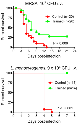
“The innate immune system recalls a challenge to adapt to a secondary challenge, a phenomenon called trained immunity. Trained immunity protected mice from a large panel of clinically relevant bacterial pathogens inoculated systematically and locally to induce peritonitis, enteritis and pneumonia.

Induction of trained immunity remodeled bone marrow and blood cellular compartments, providing efficient barriers against bacterial infections. Protection was remarkably broad when considering the pathogens and sites of infection tested.

We are running experiments to delineate the length of protection conferred by trained immunity. Trained immunity is most typically induced with β-glucan.

Mice were injected with methicillin-resistant Staphylococcus aureus (MRSA). Trained mice survived better than control mice (31% vs. 0% survival) and had 10-fold less bacteria in blood 2 days post-infection.

Mice were challenged with a lethal dose of Listeria monocytogenes. Most strikingly, all trained mice survived infection while all control mice died within 5 days. Bacteria were not detected in blood collected from trained mice 2 and 3 days post-infection.”

https://academic.oup.com/jid/advance-article/doi/10.1093/infdis/jiz692/5691195 “Trained immunity confers broad-spectrum protection against bacterial infections”


One of the coauthors also published:

Epigenetic inheritance and microRNAs

This 2019 Canadian rodent study found:

“Folic acid (FA) supplementation mitigates sperm miRNA profiles transgenerationally following in utero paternal exposure to POPs [persistent organic pollutants]. Across the F1 – F4 generations, sperm miRNA profiles were less perturbed with POPs + FA compared to sperm from descendants of dams treated with POPs alone..and only in F1 sperm.

The POPs mixture represents the pollutant composition found in Ringed seal blubber of Northern Quebec which is a traditional food of Inuit people in that region.

F0 founder dams were gavaged with the POPs mixture corresponding to 500 µg PCBs/kg body weight or corn oil (CTRL) thrice weekly and were fed the AIN-93G diet containing either 2 mg/kg (1X) or 6 mg/kg (3X) of FA ad libitum. Treatments were only administered to F0 founder dams for 9 weeks in total; 5 weeks before mating to untreated males at postnatal day 90 and until parturition. Subsequent lineages, F1 through F4, were neither exposed to POPs nor 3X FA – instead they received 1X FA diet ad libitum.”


Folic acid’s mechanisms weren’t clear:

“The protective role of FA supplementation in the F1 sperm may be partly explained by its antioxidant activity if the miRNA changes are caused by oxidative stress induced by POPs exposure. If, however, the miRNA changes in POPs exposed sperm are due to an altered methylation capacity or dysregulated nucleotide synthesis or mutations, then the increased availability of methyl groups provided by FA supplementation may mitigate the POPs effect by supporting DNA repair through nucleotide synthesis. Additional studies of the interaction between POPs and FA are required.”

Epigenetic inheritance mechanisms were also unclear:

“It remains puzzling how environmentally perturbed paternal miRNAs can persist across multiple generations. To become heritable, parts of the sperm chromatin must escape reprogramming, leading to the possibility that sperm miRNA profiles are subsequently modified by environmental factors. There are clear examples of sperm DNA methylation that escape reprogramming and histones can be involved.”

The study may have produced more clarity had its design investigated DNA methylation as Epigenetic transgenerational inheritance extends to the great-great-grand offspring did. That study also had an intercross breeding scheme with the populations for the F1 – F3 generations before an outcross for the F4 generation because:

“An intercross within the exposure lineage population (with no sibling or cousin breeding to avoid inbreeding artifacts) provides the optimal phenotypes (i.e. pathology) and germline epigenetic alterations.”

Which breeding scheme do you think would more fairly represent the humans of this study? I’d guess that intercross would – if all Inuits eat Ringed seal blubber and have children with other Inuits.

https://academic.oup.com/eep/article/5/4/dvz024/5677505 “Folic acid supplementation reduces multigenerational sperm miRNA perturbation induced by in utero environmental contaminant exposure”

Prenatal stress heightened adult chronic pain

This 2019 McGill rodent study found:

“Prenatal stress exacerbates pain after injury. Analysis of mRNA expression of genes related to epigenetic regulation and stress responses in the frontal cortex and hippocampus, brain structures implicated in chronic pain, showed distinct sex and region-specific patterns of dysregulation.

In general, mRNA expression was most frequently altered in the male hippocampus and effects of prenatal stress were more prevalent than effects of nerve injury. Recent studies investigating chronic pain-related pathology in the hippocampus in humans and in rodent models demonstrate functional abnormalities in the hippocampus, changes in associated behavior, and decreases in adult hippocampal neurogenesis.

The change in expression of epigenetic- and stress-related genes is not a consequence of nerve injury but rather precedes nerve injury, consistent with the hypothesis that it might play a causal role in modulating the phenotypic response to nerve injury. These findings demonstrate the impact of prenatal stress on behavioral sensitivity to a painful injury.

Decreased frontal mRNA expression of BDNF and BDNF IV in male offspring following neuropathic pain or prenatal stress respectively. Relative mRNA expression of other stress-related genes (GR17, FKBP5) and epigenetic-related genes (DNMTs, TETs, HDACs, MBDs, MeCP2) in male offspring.

A drastic decrease in expression of HDAC1 was observed in all groups compared to sham-control animals. CCI: chronic constriction injury.”


The study’s design was similar to the PRS (prenatal restraint stress) model, except that the PRS procedure covered gestational days 11 to 21 (birth):

“Prenatal stress was induced on Embryonic days 13 to 17 by restraining the pregnant dams in transparent cylinder with 5 mm water, under bright light exposure, 3 times per day for 45 min.”

None of the French, Italian, and Swiss PRS studies were cited.

The limitation section included:

  1. “Although our study shows significant changes in expression of epigenetic enzymes, it didn’t examine the impact of these changes on genes that are epigenetically regulated by this machinery or their involvement in intensifying pain responses.
  2. The current study is limited by the focus on changes in gene expression which do not necessarily correlate with changes in protein expression.
  3. Another limitation of this study is the inability to distinguish the direct effects of stress in utero vs. changes in the dam’s maternal behavior due to stress during pregnancy; cross-fostering studies are needed to address this issue.
  4. Functional experiments that involve up and down regulation of epigenetic enzymes in specific brain regions are required to establish a causal role for these processes in chronic pain.”

What do you think about possible human applicability of this study’s “effects of prenatal stress were more prevalent than effects of nerve injury” finding?

Are there any professional therapeutic frameworks that instruct trainees to recognize that if a person’s mother was stressed while pregnant, their prenatal experiences could cause more prevalent biological and behavioral effects than a recent injury?

https://www.sciencedirect.com/science/article/pii/S0166432819315219 “Prenatal maternal stress is associated with increased sensitivity to neuropathic pain and sex-specific changes in supraspinal mRNA expression of epigenetic- and stress-related genes in adulthood” (not freely available)

A transgenerational view of the rise in obesity

This 2019 Washington State University rodent study found epigenetically inherited transgenerational effects in great-grand offspring due to their great-grandmothers’ toxicant exposures during pregnancy:

“Previous studies found an increased susceptibility to obesity in F3 generation rats ancestrally exposed to the pesticide DDT, and an increase in a lean phenotype in the F3 generation rats ancestrally exposed to the herbicide atrazine. The present study investigated whether there were common DMR [differential DNA methylated region] and associated genes between the control, DDT, and atrazine lineage male and female adipocytes in order to identify potential novel gene pathways modulated by DNA methylation.

Comparison of epigenetic alterations indicated that there were substantial overlaps between the different treatment lineage groups for both the lean and obese phenotypes. Novel correlated genes and gene pathways associated with DNA methylation were identified, and may aid in the discovery of potential therapeutic targets for metabolic diseases such as obesity.

Given that the first widespread [DDT] exposures to gestating human females started in the 1950s, the majority of the subsequent F3 generation are adults today. Ancestral exposures to environmental toxicants like DDT may have had a role in the dramatic rise in obesity rates worldwide.”


This same research group noted in Transgenerational diseases caused by great-grandmother DDT exposure:

“DDT was banned in the USA in 1973, but it is still recommended by the World Health Organization for indoor residual spray. India is by far the largest consumer of DDT worldwide.

India has experienced a 5-fold increase of type II diabetes over the last three decades with a predisposition to obesity already present at birth in much of the population. Although a large number of factors may contribute to this increased incidence of obesity, the potential contribution of ancestral toxicant exposures in the induction of obesity susceptibility requires further investigation.”

https://www.tandfonline.com/doi/full/10.1080/21623945.2019.1693747 “Adipocyte epigenetic alterations and potential therapeutic targets in transgenerationally inherited lean and obese phenotypes following ancestral exposures”

Epigenetic transgenerational inheritance extends to the great-great-grand offspring

This 2019 rodent study by the Washington State University labs of Dr. Michael Skinner continued to F4 generation great-great-grand offspring, and demonstrated that epigenetic inheritance mechanisms are similar to imprinted genes:

“Epigenetic transgenerational inheritance potentially impacts disease etiology, phenotypic variation, and evolution. An increasing number of environmental factors from nutrition to toxicants have been shown to promote the epigenetic transgenerational inheritance of disease.

Imprinted genes are a special class of genes since their DNA methylation patterns are unchanged over the generation and are not affected by the methylation erasure occurring early in development. The transgenerational epigenetic alterations in the germline appear to be permanently reprogrammed like imprinted genes, and appear protected from this DNA methylation erasure and reprogramming at fertilization in the subsequent generations. Similar to imprinted genes, the epigenetic transgenerational germline epimutations appear to have a methylation erasure in the primordial germ cells involving an epigenetic molecular memory.

Comparison of the transgenerational F3 generation, with the outcross to the F4 generation through the paternal or maternal lineages, allows an assessment of parent-of-origin transmission of disease or pathology. Observations provided examples of the following:

  1. Pathology that required combined contribution of both paternal and maternal alleles to promote disease [testis and ovarian disease];
  2. Pathology that is derived from the opposite sex allele such as father to daughter [kidney disease] or mother to son [prostate disease];
  3. Pathology that is derived from either parent-of-origin alleles independently [obesity];
  4. Pathology that is transmitted within the same sex, such as maternal to daughter [mammary tumor development]; and
  5. Pathology that is observed only following a specific parent-of-origin outcross [both F4 male obesity and F4 female kidney disease in the vinclozolin lineage].”

https://www.sciencedirect.com/science/article/pii/S0012160619303471 “Epigenetic transgenerational inheritance of parent-of-origin allelic transmission of outcross pathology and sperm epimutations”


This study showed that epigenetically inherited legacies extend to the fifth generation. Do any of us know our ancestors’ medical histories back to our great-great-grandparents?

Will toxicologists take their jobs seriously, catch up to current science, and investigate possible effects in at least the F3 generation that had no direct toxicant exposure?

Maternal obesity causes heart disease in every offspring generation

This 2019 St. Louis rodent study found:

“We hypothesized that maternal obesity induces cardiac mitochondrial dysfunction in the offspring via transgenerational inheritance of abnormal oocyte mitochondria. All F1 to F3 descendants bred via the female in each generation were nonobese and demonstrated cardiac mitochondrial abnormalities.

Contrary to our hypothesis, male F1 also transmitted these effects to their offspring, ruling out maternal mitochondria as the primary mode of transmission. We conclude that transmission of obesity-induced effects in the oocyte nucleus rather than abnormal mitochondria underlie transgenerational inheritance of cardiac mitochondrial defects in descendants of obese females.”


For some reason, the researchers didn’t cite any of Dr. Michael Skinner’s research on epigenetic transgenerational inheritance. Their time, efforts, and resources would have been more productive had they used Dr. Skinner’s studies – such as the 2018 Epigenetic transgenerational inheritance of ovarian disease – as guides.

A podcast with the researchers is available here.

https://www.physiology.org/doi/abs/10.1152/ajpheart.00013.2019 “Maternal High-Fat, High-Sucrose Diet Induces Transgenerational Cardiac Mitochondrial Dysfunction Independent of Maternal Mitochondrial Inheritance” (not freely available)

Emotional responses and BDNF methylation

This 2019 German human study found:

“A critical role of BDNF [brain-derived neurotrophic factor] methylation in human amygdala response to negative emotional stimuli, whereby:

  • High BDNF methylation rates were for the first time shown to be associated with a high reactivity in the amygdala; and
  • High BDNF methylation and high amygdala reactivity were associated with low novelty seeking.

There was no interaction or main effect of the Val66Met polymorphism on amygdala reactivity.

Our data adds evidence to the hypothesis that epigenetic modifications of BDNF can result in an endophenotype associated with anxiety and mood disorders. However, since correlations do not prove causality:

  • A direct link between human BDNF mRNA/protein levels, methylation, amygdala reactivity and psychiatric disorders is still missing, demanding further research.
  • Determining the underlying directions of the relations between BDNF methylation, amygdala reactivity, and NS [novelty seeking] cannot be accomplished based on our data and must await further research.

The fact that our results mainly involve the right amygdala is in line with previous studies. Recent reviews suggest a general right hemisphere dominance for all kinds of emotions, and, more specifically, a critical role of the right amygdala in the early assessment of emotional stimuli.

The experimental fMRI paradigm utilized a face‐processing task (faces with anger or fear expressions), alternating with a sensorimotor control task. Harm avoidance, novelty seeking, and reward dependence were measured using the Tridimensional Personality Questionnaire.”

https://onlinelibrary.wiley.com/doi/full/10.1002/hbm.24825 “The role of BDNF methylation and Val 66 Met in amygdala reactivity during emotion processing”

May you be the hero who solves your own problems

This 2019 Germany/US review subject was the failure of psychotherapy and pharmacotherapy:

“Each mental disorder raises its own host of issues. However, recent evidence across multiple meta-analyses on key mental disorders provides an overarching picture of limited benefits for both psychotherapy and pharmacotherapy.

Some differences for specific disorders are not strong enough to weaken the overall impression that a dead end has been reached in the treatment of mental disorders. For this reason, a paradigm shift seems to be required.”


Investigate the above linked Primal Therapy category to figure out what you could do for yourself. Follow the below review link for reasons to avoid treatments that waste your one precious life.

https://www.cambridge.org/core/journals/psychological-medicine/article/toward-a-paradigm-shift-in-treatment-and-research-of-mental-disorders/FDE68FF26E946276A334FA90ACE28D9F/core-reader “Toward a paradigm shift in treatment and research of mental disorders”

Transgenerational epigenetic inheritance of thyroid hormone sensitivity

My 500th curation is a 2019 Portuguese human study of Azorean islanders:

“This study demonstrates a transgenerational epigenetic inheritance in humans produced by exposure to high TH [thyroid hormone] in fetal life, in the absence of maternal influences secondary to thyrotoxicosis. The inheritance is along the male line.

The present work took advantage of the relatively frequent occurrence of fetal exposure to high TH levels in the Azorean island of São Miguel. This is the consequence of a missense mutation in the THRB gene causing the amino-acid replacement R243Q, resulting in reduced affinity of the TH receptor beta (TRβ) for TH and thus RTHβ.

Its origin has been traced to a couple who lived at the end of the 19th century. F0 represented the third generation and F3 the sixth and seventh generation descendant.”


These researchers provided the first adequately evidenced human transgenerational epigenetic inheritance study! However, the lead sentence in its Abstract wasn’t correct:

“Evidence for transgenerational epigenetic inheritance in humans is still controversial, given the requirement to demonstrate persistence of the phenotype across three generations.”

Although found in this study, there is no “requirement to demonstrate persistence of the phenotype.” Observing the same phenotype in each generation is NOT required for human transgenerational epigenetic inheritance to exist!

Animal transgenerational studies have shown that epigenetic inheritance mechanisms may both express different phenotypes for each generation:

and entirely skip a phenotype in one or more generations!

  • Transgenerational pathological traits induced by prenatal immune activation found a F2 and F3 generation phenotype of impaired sociability, abnormal fear expression and behavioral despair – effects that weren’t present in the F1 offspring;
  • The transgenerational impact of Roundup exposure “Found negligible impacts of glyphosate on the directly exposed F0 generation, or F1 generation offspring pathology. In contrast, dramatic increases in pathologies in the F2 generation grand-offspring, and F3 transgenerational great-grand-offspring were observed.” (a disease phenotype similarly skipped the first offspring generation);
  • Epigenetic transgenerational inheritance mechanisms that lead to prostate disease “There was also no increase in prostate histopathology in the directly exposed F1 or F2 generation.” (a prostate disease phenotype skipped the first two male offspring generations before it was observed in the F3 male offspring); and
  • Epigenetic transgenerational inheritance of ovarian disease “There was no increase in ovarian disease in direct fetal exposed F1 or germline exposed F2 generation. The F3 generation can have disease while the F1 and F2 generations do not, due to this difference in the molecular mechanisms involved.” (an ovarian disease phenotype similarly skipped the first two female offspring generations before it was observed in the F3 female offspring).

Details of epigenetic inheritance mechanisms were provided in Another important transgenerational epigenetic inheritance study. Mechanisms from fetal exposure to the fungicide vinclozolin were compared with mechanisms from fetal DDT exposure, and summarized as:

“The fetal exposure initiates a developmental cascade of aberrant epigenetic programming, and does NOT simply induce a specific number of DMRs [DNA methylation regions] that are maintained throughout development.“

I emailed references to the studies in the first five above curations to the current study’s corresponding coauthor. They replied “What is the mechanism for the transgenerational inheritance you describe?” and my reply included a link to the sixth curation’s study.

Are there still other transgenerational epigenetically inherited effects due to fetal exposure to high thyroid hormone levels?

https://www.liebertpub.com/doi/full/10.1089/thy.2019.0080 “Reduced Sensitivity to Thyroid Hormone as a Transgenerational Epigenetic Marker Transmitted Along the Human Male Line”

Preliminary findings from a senolytics clinical trial

This 2019 US human clinical trial reported preliminary results. See Reanalysis of findings from a senolytics clinical trial for strikeout changes.

“Senescent cells, which can release factors that cause inflammation and dysfunction, the senescence-associated secretory phenotype (SASP), accumulate with ageing and at etiological sites in multiple chronic diseases. Senolytics, including the combination of Dasatinib and Quercetin (D + Q), selectively eliminate senescent cells by transiently disabling pro-survival networks that defend them against their own apoptotic environment.

Since the target of senolytics is senescent cells, these drugs do not need to be continuously present in the circulation in the same way as drugs whose mechanism of action is to occupy a receptor, modulate an enzyme, or act on a particular biochemical pathway, at least in mice. Intermittently administering D + Q effectively circumvents any potential off-target effects due to continuous receptor occupancy or modulation of an enzyme or biochemical pathway.

To test whether intermittent D + Q is effective in targeting senescent cells in humans, we administered a single 3 day course of oral D + Q and assayed senescent cell abundance 11 days after the last dose in subjects with DKD [diabetic kidney disease], the most common cause of end-stage kidney failure and which is characterized by increased senescent cell burden.

In this interim report of findings, we found the single brief course of D + Q:

  • Attenuated adipose tissue and skin senescent cell burden,
  • Decreased resulting adipose tissue macrophage accumulation,
  • Enhanced adipocyte progenitor replicative potential, and
  • Reduced key circulating SASP factors.”

gr2_lrg.jpg

“In adipose tissue D + Q significantly reduced raw numbers of:

  • p16INK4A+ cells by 35%;
  • p21CIP1+ cells by 17%;
  • SAβgal+ cells by 62%;
  • CD68+ macrophages by 28%; and
  • Crown-like structures by 86%.”

https://www.ebiomedicine.com/article/S2352-3964(19)30591-2/fulltext “Senolytics decrease senescent cells in humans: Preliminary report from a clinical trial of Dasatinib plus Quercetin in individuals with diabetic kidney disease”


In a referenced 2019 rodent study by many of the same researchers:

“We also found that even Q alone can prevent high fat diet-induced increases in markers of senescence, renal fibrosis, decreases in renal oxygenation, and increased creatinine in mice, although Q alone did not prevent insulin resistance.”

The rodent study’s 50 mg/kg quercetin dose scaled human-equivalent dose would be (0.081 x 50 mg) = 13.3 mg/kg. This was 375% higher than a 1,000 mg/75 kg quercetin dose (clinical trial participants’ weights weren’t disclosed.)

https://onlinelibrary.wiley.com/doi/full/10.1111/acel.12950 “Targeting senescent cells alleviates obesity‐induced metabolic dysfunction”

Developmental disorders and the epigenetic clock

This 2019 UK/Canada/Germany human study investigated thirteen developmental disorders to identify genes that changed aspects of the epigenetic clock:

“Sotos syndrome accelerates epigenetic aging [+7.64 years]. Sotos syndrome is caused by loss-of-function mutations in the NSD1 gene, which encodes a histone H3 lysine 36 (H3K36) methyltransferase.

This leads to a phenotype which can include:

  • Prenatal and postnatal overgrowth,
  • Facial gestalt,
  • Advanced bone age,
  • Developmental delay,
  • Higher cancer predisposition, and, in some cases,
  • Heart defects.

Many of these characteristics could be interpreted as aging-like, identifying Sotos syndrome as a potential human model of accelerated physiological aging.

This research will shed some light on the different processes that erode the human epigenetic landscape during aging and provide a new hypothesis about the mechanisms behind the epigenetic aging clock.”

“Proposed model that highlights the role of H3K36 methylation maintenance on epigenetic aging:

  • The H3K36me2/3 mark allows recruiting de novo DNA methyltransferases DNMT3A (in green) and DNMT3B (not shown).
  • DNA methylation valleys (DMVs) are conserved genomic regions that are normally found hypomethylated.
  • During aging, the H3K36 methylation machinery could become less efficient at maintaining the H3K36me2/3 landscape.
  • This would lead to a relocation of de novo DNA methyltransferases from their original genomic reservoirs (which would become hypomethylated) to other non-specific regions such as DMVs (which would become hypermethylated and potentially lose their normal boundaries),
  • With functional consequences for the tissues.”

The researchers improved methodologies of several techniques:

  1. “Previous attempts to account for technical variation have used the first 5 principal components estimated directly from the DNA methylation data. However, this approach potentially removes meaningful biological variation. For the first time, we have shown that it is possible to use the control probes from the 450K array to readily correct for batch effects in the context of the epigenetic clock, which reduces the error associated with the predictions and decreases the likelihood of reporting a false positive.
  2. We have confirmed the suspicion that Horvath’s model underestimates epigenetic age for older ages and assessed the impact of this bias in the screen for epigenetic age acceleration.
  3. Because of the way that the Horvath epigenetic clock was trained, it is likely that its constituent 353 CpG sites are a low-dimensional representation of the different genome-wide processes that are eroding the epigenome with age. Our analysis has shown that these 353 CpG sites are characterized by a higher Shannon entropy when compared with the rest of the genome, which is dramatically decreased in the case of Sotos patients.”

https://genomebiology.biomedcentral.com/articles/10.1186/s13059-019-1753-9 “Screening for genes that accelerate the epigenetic aging clock in humans reveals a role for the H3K36 methyltransferase NSD1”

Too cheap for clinical trials

Let’s compare and contrast a 2019 meta-analysis and a 2017 review of using acetyl-L-carnitine to treat diabetic neuropathy.

A 2019 Brazilian meta-analysis Acetyl‐L‐carnitine for the treatment of diabetic peripheral neuropathy of four previous trials stated:

  • “The risk of bias was high in both trials of different ALC doses and low in the other two trials.
  • No included trial measured the proportion of participants with at least moderate (30%) or substantial (50%) pain relief.
  • At doses greater than 1500 mg/day, ALC reduced pain more than placebo. This subgroup analysis should be viewed with caution as the evidence was even less certain than the overall analysis, which was already of very low certainty.
  • The placebo-controlled studies did not measure functional impairment and disability scores.
  • No study used validated symptom scales.
  • Two studies were funded by the manufacturer of ALC and the other two studies had at least one co-author who was a consultant for an ALC manufacturer.

Authors’ conclusions:

  • We are very uncertain whether ALC causes a reduction in pain after 6 to 12 months treatment in people with DPN, when compared with placebo, as the evidence is sparse and of low certainty.
  • Data on functional and sensory impairment and symptoms are lacking, or of very low certainty.
  • The evidence on adverse events is too uncertain to make any judgements on safety.”

A 2017 Italian review Effects of acetyl-L-carnitine in diabetic neuropathy and other geriatric disorders stated:

“A long history of diabetes mellitus and increasing age are associated with the onset of diabetic neuropathy, a painful and highly disabling complication with a prevalence peaking at 50% among elderly diabetic patients. The management of diabetic neuropathy is extremely difficult: in addition to the standard analgesics used for pain control, common treatments include opioids, anticonvulsants, antidepressants, and local anesthetics, alone or in combination. Such therapies still show a variable, often limited efficacy, however.

Many patients do not spontaneously report their symptoms to physicians, but, if asked, they often describe having experienced a persistent and non-abating pain for many years. The prevalence of painful symptoms is just as high in patients with mild neuropathy as in those with more advanced DPN.

Through the donation of acetyl groups, ALC exerts a positive action on mitochondrial energy metabolism. ALC has cytoprotective, antioxidant, and antiapoptotic effects in the nervous system.

ALC has also been proposed for the treatment of other neurological and psychiatric diseases, such as mood disorders and depression, dementia, Alzheimer’s disease, and Parkinson’s disease, given that synaptic energy states and mitochondrial dysfunctions are core factors in their pathogenesis. Compared to other treatments, ALC is safe and extremely well tolerated.

In nerve injury, the mGlu2 receptor overexpressed by ALC binds the glutamate, reducing its concentration in the synapses with an analgesic effect. ALC may improve nerve regeneration and damage repair after primary nerve trauma.”


Where will the money come from to realize what the 2017 review promised, as well as provide what the 2019 meta-analysis required?

Do we prefer the current “limited efficacy” treatments of “opioids, anticonvulsants, antidepressants, and local anesthetics?”

Who will initiate clinical trials of a multiple of the normal dietary supplement dose (500 mg at $.25 a day, retail)? How profitable is a product whose hypothetical effective dosage for diabetic neuropathy (3000 mg) sells for only $1.50 a day?

Perinatal stress and sex differences in circadian activity

This 2019 French/Italian rodent study used the PRS model to investigate its effects on circadian activity:

“The aim of this study was to explore the influence of PRS on the circadian oscillations of gene expression in the SCN [suprachiasmatic nucleus of the hypothalamus] and on circadian locomotor behavior, in a sex-dependent manner.

Research on transcriptional rhythms has shown that more than half of all genes in the human and rodent genome follow a circadian pattern. We focused on genes belonging to four functional classes, namely the circadian clock, HPA axis stress response regulation, signaling and glucose metabolism in male and female adult PRS rats.

Our findings provide evidence for a specific profile of dysmasculinization induced by PRS at the behavioral and molecular level, thus advocating the necessity to include sex as a biological variable to study the set-up of circadian system in animal models.”

“There was a clear-cut effect of sex on the effect of PRS on the levels of activity:

  • During the period of lower activity (light phase), both CONT and PRS females were more active than males. During the light phase, PRS increased activity in males, which reached levels of CONT females.
  • More interestingly, during the period of activity (dark phase), male PRS rats were more active than male CONT rats. In contrast, female PRS rats were less active than CONT females.
  • During the dark phase, CONT female rats were less active than CONT male rats.

The study presented evidence for sex differences in circadian activity of first generation offspring that was caused by stress experienced by the pregnant mother:

“Exposure to gestational stress and altered maternal behavior programs a life-long disruption in the reactive adaptation such as:

  •  A hyperactive response to stress and
  • A defective feedback of the hypothalamus-pituitary-adrenal (HPA) axis together with
  • Long-lasting modifications in stress/anti-stress gene expression balance in the hippocampus.”

It would advance science if these researchers carried out experiments to two more generations to investigate possible transgenerational epigenetic inheritance of effects caused by PRS. What intergenerational and transgenerational effects would they possibly find by taking a few more months and extending research efforts to F2 and F3 generations? Wouldn’t these findings likely help humans?


One aspect of the study was troubling. One of the marginally-involved coauthors was funded by the person described in How one person’s paradigms regarding stress and epigenetics impedes relevant research. Although no part of the current study was sponsored by that person, there were three gratuitous citations of their work.

All three citations were reviews. Unlike study researchers, reviewers aren’t bound to demonstrate evidence from tested hypotheses. Reviewers are free to:

  • Express their beliefs as facts;
  • Over/under emphasize study limitations; and
  • Disregard and misrepresent evidence as they see fit.

Fair or not, comparisons of reviews with Cochrane meta-analyses of the same subjects consistently show the extent of reviewers’ biases. Reviewers also aren’t obligated to make post-publication corrections for their errors and distortions.

As such, reviews can’t be cited for reliable evidence. Higher-quality studies that were more relevant and recent than a 1993 review could have elucidated points.

Sucking up to the boss and endorsing their paradigm was predictable. Since that coauthor couldn’t constrain themself to funder citations only in funder studies, it was the other coauthors’ responsibilities to edit out unnecessary citations.

https://www.frontiersin.org/articles/10.3389/fnmol.2019.00089/full “Perinatal Stress Programs Sex Differences in the Behavioral and Molecular Chronobiological Profile of Rats Maintained Under a 12-h Light-Dark Cycle”
Caloric restriction’s epigenetic effects

This 2019 US review subject was caloric restriction (CR) without malnutrition:

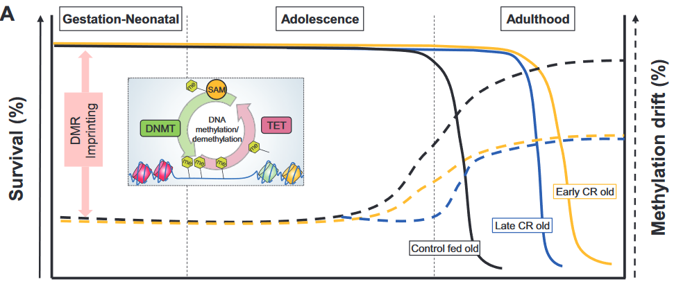
“Cellular adaptation that occurs in response to dietary patterns can be explained by alterations in epigenetic mechanisms such as DNA methylation, histone modifications, and microRNA. Epigenetic reprogramming of the underlying chronic low-grade inflammation by CR can lead to immuno-metabolic adaptations that enhance quality of life, extend lifespan, and delay chronic disease onset.

Short- and long-term CRs produce significant changes in different tissues and across species, in some animal models even with sex-specific effects. Early CR onset may cause a different and even an opposite effect on physiological outcomes in animal models such as body weight.”

https://academic.oup.com/advances/article-abstract/10/3/520/5420411 “Epigenetic Regulation of Metabolism and Inflammation by Calorie Restriction” (not freely available)


1. The review didn’t present evidence to equate survival (left axis) with methylation drift (right axis) per the above graphic. Methylation drift should point in the opposite direction of survival, if anything.

2. No mention was made of the epigenetic clock method of measuring age acceleration, although it’s been available since 2013 and recent diet studies have used it. The sole citation of an age acceleration study was from 2001, which was unacceptable for a review published in 2019.

3. The review provided many cellular-level details about the subject. However, organism-level areas weren’t sufficiently evidenced:

A. Arguments for an effect usually include explanations for no effect as well as for opposite effects. The reviewers didn’t provide direct evidence for why, if caloric restriction extended lifespan, caloric overabundance produced shorter lifespans.

B. Caloric restriction evidence was presented as if only it was responsible for organism-level effects. Other mechanisms may have been involved.

An example of such a mechanism was demonstrated in a 2007 rodent study Reduced Oxidant Stress and Extended Lifespan in Mice Exposed to a Low Glycotoxin Diet which compared two 40%-calorie-restricted diets.

The calories and composition of both diets were identical. However, advanced glycation end product (AGE) levels were doubled in standard chow because heating temperatures were “sufficiently high to inadvertently cause standard mouse chow to be rich in oxidant AGEs.”

The study found that a diet with lower chow heating temperatures increased lifespan and health span irrespective of caloric restriction!

  • The low-AGE calorie-restricted diet group lived an average of 15% longer (>20 human equivalent years) than the CR group.
  • 40% of the low-AGE calorie-restricted diet group were still alive when the last CR group member died.
  • The CR group also had significantly more: 1) oxidative stress damage; 2) glucose and insulin metabolism problems; and 3) kidney, spleen, and liver injuries.

A drug that countered effects of a traumatizing mother

This 2019 US rodent study concerned transmitting poor maternal care to the next generation:

“The quality of parental care received during development profoundly influences an individual’s phenotype, including that of maternal behavior. Infant experiences with a caregiver have lifelong behavioral consequences.

Maternal behavior is a complex behavior requiring the recruitment of multiple brain regions including the nucleus accumbens, bed nucleus of the stria terminalis, ventral tegmental area, prefrontal cortex, amygdala, and medial preoptic area. Dysregulation within this circuitry can lead to altered or impaired maternal responsiveness.

We administered zebularine, a drug known to alter DNA methylation, to dams exposed during infancy to the scarcity-adversity model of low nesting resources, and then characterized the quality of their care towards their offspring.

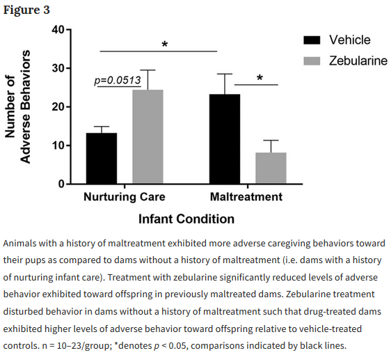
  1. We replicate that dams with a history of maltreatment mistreat their own offspring.
  2. We show that maltreated-dams treated with zebularine exhibit lower levels of adverse care toward their offspring.
  3. We show that administration of zebularine in control dams (history of nurturing care) enhances levels of adverse care.
  4. We show altered methylation and gene expression in maltreated dams normalized by zebularine.

These findings lend support to the hypothesis that epigenetic alterations resulting from maltreatment causally relate to behavioral outcomes.

Maternal behavior is an intergenerational behavior. It is important to establish the neurobiological underpinnings of aberrant maternal behavior and explore treatments that can improve maternal behavior to prevent the perpetuation of poor maternal care across generations.”


The study authors demonstrated intergenerational epigenetic effects, and missed an opportunity to also investigate transgenerational epigenetically inherited effects. They cited reference 60 for the first part of the above quotation, but the cited reviewer misused the transgenerational term by applying it to grand-offspring instead of the great-grand-offspring.

There were resources available to replicate the study authors’ previous findings, which didn’t show anything new. Why not use such resources to uncover evidence even more applicable to humans by extending experiments to great-grand-offspring that would have no potential germline exposure to the initial damaging cause?

Could a study design similar to A limited study of parental transmission of anxiety/stress-reactive traits have been integrated? That study’s thorough removal of parental behavior would be an outstanding methodology to confirm by falsifiability whether parental behavior is both an intergenerational and a transgenerational epigenetic inheritance mechanism.

Rodent great-grand-offspring can be studied in < 9 months. It takes > 50 years for human studies to reach the great-grand-offspring transgenerational generation.

  • Why not attempt to “prevent the perpetuation of poor maternal care across generations?”
  • Isn’t it a plausible hypothesis that humans “with a history of maltreatment mistreat their own offspring?”
  • Isn’t it worth the extra effort to extend animal research to investigate this unfortunate chain?

https://www.nature.com/articles/s41598-019-46539-4 “Pharmacological manipulation of DNA methylation normalizes maternal behavior, DNA methylation, and gene expression in dams with a history of maltreatment”