Eat broccoli sprouts for DIM

This 2019 Spanish human study ran in parallel with Our model clinical trial for Changing to a youthful phenotype with broccoli sprouts. I’ll focus on the aspect of diindolylmethane (DIM) from eating broccoli sprouts:

“The aim of this study is to evaluate the effect of gender or hormonal status (menopause) on the bioavailability of broccoli sprouts in different cohorts of overweight adult subjects: men, non-menopausal women and post-menopausal women.

3,3′-diindolylmethane (DIM) was detected and quantified in all volunteers. It increased significantly during broccoli [sprouts] ingestion in men. However, a steady decrease of its urinary concentration was observed in post-menopausal women that was significant at day 50. No significant changes were observed in premenopausal women. Albeit this different behaviour, no significant differences between the three groups were detected by the different statistical tests performed.

High increases observed in SFN-metabolites in the three cohorts confirm that the fresh product is a good source of bioactive compounds bioavailable in the organism. We detected high amounts of 3,3-DIM in urine samples, which can be related to the metabolism of glucobrassicin derivatives from our broccoli sprouts.

Post-menopausal women seem to metabolize isothiocyanates in a greater extension. Hormonal status and differences in gut microbiota may influence the bioavailability of isothiocyanates from broccoli sprouts but more studies are needed to support this statement.”

https://www.sciencedirect.com/science/article/abs/pii/S1756464619303147 “Bioavailability of broccoli sprouts in different human overweight populations” (not freely available)


“Post-menopausal women seem to metabolize isothiocyanates in a greater extension. A steady decrease of its [DIM] urinary concentration was observed in post-menopausal women that was significant at day 50.”

Subjects ate 30 grams of broccoli super sprouts every day through Day 35, then stopped, and were measured again at Day 50. The only example of measurements where Day 35 was less than Day 0 was postmenopausal women metabolizing more DIM.

That Day 35 data point didn’t have an asterisk next to it to indicate a statistically significant decrease. But the overweight postmenopausal women group’s next Day 50 significant “steady decrease” finding supported an interpretation that eating broccoli sprouts supplied them with DIM that they especially needed.

Regarding the huge percentage changes above, our model clinical trial found in a longer time frame:

The decrease in IL-6 levels was significantly related to the increase in 24 h urine SFN [sulforaphane] levels. In case of C-reactive protein, the decrease was significantly related to the increases in 24 h urine SFN-NAC [SFN-N-acetylcysteine] and SFN-CYS [SFN-cysteine].

I’ll guess that these parallel trial subjects also experienced similar benefits from eating broccoli  sprouts every day for five weeks. See Day 70 results from Changing to a youthful phenotype with broccoli sprouts for another guess that even shorter time frames would be effective.


Broccoli super sprout indolic compounds were as follows:

indolic glucosinolates

Assuming that only glucobrassicin is a precursor to DIM, subjects’ DIM bioavailability can be calculated as μmol DIM / 21.61 μmol. For example, overweight postmenopausal women Day 35 average of 0.5544 μmol DIM that ranged from 0.1771 to 0.8034 μmol DIM represented an average 2.57% DIM bioavailability with a range of 0.82% to 3.72% DIM bioavailability.

See Part 2 for DIM follow-up.

Day 70 results from Changing to a youthful phenotype with broccoli sprouts

Here are my Day 70 measurements* to follow up Our model clinical trial for Changing to a youthful phenotype with broccoli sprouts, which had these findings:


Keep in mind that I’m not in the population represented by the clinical trial sample:

  1. My chronological age is above their inclusion range;
  2. My BMI is below their inclusion range; and
  3. I take supplements and meet other exclusion criteria.

I also didn’t take Day 0 measurements.

June 2019 BMI: 24.8

June 2020 BMI: 22.4

2020 IL-6: 1.0 pg / ml. See Part 2 of Rejuvenation therapy and sulforaphane for comparisons.

2020 C-reactive protein: < 1 mg / l.

IL-6 2020

2019 and 2020 No biological age measurements. Why aren’t epigenetic clocks standard and affordable?


I’ve made four lifestyle “interventions” since last summer:

  1. In July 2019 I started to reduce my consumption of advanced glycation end products after reading Dr. Vlassara’s AGE-Less Diet: How a Chemical in the Foods We Eat Promotes Disease, Obesity, and Aging and the Steps We Can Take to Stop It.
  2. In September I started non-prescription daily treatments of Vitamin D, zinc, and DHEA per clinical trial Reversal of aging and immunosenescent trends.
  3. Also in September, I started non-prescription intermittent quercetin treatments of Preliminary findings from a senolytics clinical trial.
  4. I started eating broccoli sprouts every day eleven weeks ago.

1. Broccoli sprouts oppose effects of advanced glycation end products (AGEs) provided examples of Items 1 and 4 interactions.

2. Two examples of Item 2 treatment interactions with Item 4 are in Reversal of aging and immunosenescent trends with sulforaphane:

  • “The effects of the combined treatment with BSE [broccoli sprout extract] and zinc were always greater than those of single treatments.” [Zinc and broccoli sprouts – a winning combination]
  • “Vitamin D administration decreased tumor incidence and size, and the co-administration with SFN [sulforaphane] magnified the effects. The addition of SFN decreased the activity of histone deacetylase and increased autophagy.”

3. How broccoli sprout compounds may complement three supplements I take was in a 2020 review Central and Peripheral Metabolic Defects Contribute to the Pathogenesis of Alzheimer’s Disease: Targeting Mitochondria for Diagnosis and Prevention:

“The nutrients benefit mitochondria in four ways, by:

  • Ameliorating oxidative stress, for example, lipoic acid;
  • Activating phase II enzymes that improve antioxidant defenses, for example, sulforaphane;
  • Enhancing mitochondrial remodeling, for example, acetyl-l-carnitine; and
  • Protecting mitochondrial enzymes and/or stimulating mitochondrial enzyme activities, for example, enzyme cofactors, such as B vitamins and coenzyme Q10 .

In addition to using mitochondrial nutrients individually, the combined use of mitochondrial nutrients may provide a better strategy for mitochondrial protection.”

The review provided a boatload of mitochondrial multifactorial analyses for Alzheimer’s. But these analyses didn’t include effective mitochondrial treatments of ultimate aging causes. I didn’t see evidence of why, after fifteen years of treating mitochondrial effects with supplements, treating one more effect could account for my Week 9 vastly different experiences.


I nod to An environmental signaling paradigm of aging explanations. Its Section 10 reviewed IL-6, C-reactive protein, senescence, and NF-κB in terms of feedback loops, beginning with:

“It is clear that the increasing number of senescent cells depends on the post-adult developmental stage rather than chronological age. The coincidence that these processes result in particular forms of impairment in old age does not seem to be random as it is present in all mammals, and may be causative of many aspects of aging.”

A derived hypothesis: After sufficient strength and duration, broccoli sprout compounds changed my signaling environment, with appreciable effects beginning in Week 9.

I offered weak supporting evidence in Upgrade your brain’s switchboard with broccoli sprouts where a study’s insufficient one week duration of an insufficient daily 17.3 mg sulforaphane dosage still managed to change a blood antioxidant that may have changed four thalamus-brain-area metabolites. For duration and weight comparisons, I doubled my daily amount of broccoli seeds from one to two tablespoons just before Week 6 (Day 35), and from that point onward consumed a estimated 52 mg sulforaphane with microwaving 3-day-old broccoli sprouts every day.

Maybe a promised “In a submitted study, we will report that peripheral GSH levels may be correlated with cognitive functions” will provide stronger evidence? I’m not holding my breath for relevant studies because:

  • There wouldn’t be potential payoffs for companies to study any broccoli sprout compound connections with research areas such as aging, migraines, etc. Daily clinically-relevant broccoli sprout dosages can be grown for < $500 a year.
  • Sponsors would have to change paradigms, a very-low-probability event. They’d have to explain why enormous resources dedicated to current frameworks haven’t produced effective long-term treatments.

What long-term benefits could be expected if I continue eating broccoli sprouts every day?

The longest relevant clinical trial I’ve seen – referenced in Part 2 of Reversal of aging and immunosenescent trends with sulforaphane – was twelve weeks. Part 2 also provided epigenetic clock examples of changes measured after 9 months, which accelerated from there to the 12-month end-of-trial point.

Reviewing clinical trials of broccoli sprouts and their compounds pointed out:

“Biomarkers of effect need more time than biomarkers of exposure to be influenced by dietary treatment.”


A contrary argument: Perhaps people don’t require long durations to effectively change their signaling environments?

I apparently didn’t start eating an effective-for-me daily broccoli sprouts dosage until Day 35, when I changed from one to two tablespoons of broccoli seeds a day. If so, Weeks 6 through 8 may account for my substantial responses during Week 9.

  • Could eating broccoli sprouts every day for four weeks dramatically change a person’s signaling environment?
  • Do you have four weeks and $38 to find out? Two tablespoons of broccoli seeds = 21.4 g x 30 days = .642 kg or 1.42 lbs.

This is what twice-a-day one-tablespoon starting amounts of broccoli seeds look like through three days:


Maintaining the sprouting process hasn’t been a big effort compared with the benefits.

In the absence of determinative evidence, I’ll continue eating broccoli sprouts every day. Several areas of my annual physical have room for improvements. Extending my four lifestyle “interventions” a few more months may also provide hints toward inadequately researched connections.

* Results may not be extrapolatable to other people, to any specific condition, etc.

Our model clinical trial for Changing to a youthful phenotype with broccoli sprouts

The further I get into a daily regimen of eating broccoli sprouts for ten weeks, the more I appreciate “Effects of long-term consumption of broccoli sprouts on inflammatory markers in overweight subjects.”

“This study represents an advance in intervention studies as the broccoli sprouts were included in a daily dietary pattern in quantities that reflect a real consumption. The hypothesis of our research is that broccoli sprouts are able to reduce the inflammatory status in overweight subjects due to their content in phytochemicals, mainly glucosinolates.

Total concentration of aliphatic glucosinolates was 80.50 mg/30 gf.w. This concentration was two-fold higher than indolic glucosinolates. Volunteers consumed an average of 51 mg (117 μmol) and 20 mg (42 μmol) of glucoraphanin and neoglucobrassicin, respectively, on a daily basis, during the 70 days of the dietary intervention. Considering an amount of GRA [glucoraphanin] of 117 μmol by serving, a 4% on average was metabolized through mercapturic acid pathway.

No significant changes were observed in weight and BMI. By contrast, body fat mass slightly decreased significantly after 70 days of broccoli [sprout] consumption and returned to basal levels at day 90, a state that was maintained until day 160.

The decrease in IL-6 levels was significantly related to the increase in 24 h urine SFN [sulforaphane] levels. In case of C-reactive protein, the decrease was significantly related to the increases in 24 h urine SFN-NAC [SFN-N-acetylcysteine] and SFN-CYS [SFN-cysteine].

The possible synergistic interaction of both SFN and 3,30-DIM and the isothiocyanates erucin and sulforaphane are interconvertible, so that the anti-inflammatory effects observed with broccoli sprouts intake are likely due to the combined effects of all the hydrolysis products of glucosinolates.

https://www.sciencedirect.com/science/article/abs/pii/S0261561418301183 (Not freely available, better format) and https://researchonline.lshtm.ac.uk/id/eprint/4647168/ (freely available)


Modifications I’ve made to the clinical trial’s protocols include:

  1. I start new broccoli sprout batches twice a day with one tablespoon of seeds per A pair of broccoli sprout studies.
  2. I eat 131 grams daily as calculated in Estimating daily consumption of broccoli sprout compounds.
  3. Per 3-day-old broccoli sprouts have the optimal yields, I consume broccoli sprouts when they’re 3 days old. The clinical trial subjects ate broccoli sprouts that were at least a week old.
  4. I immerse 3-day-old broccoli sprouts in 100 ml distilled water, then microwave them on 1000W full power for 35 seconds to ≤ 60°C (140°F) per Microwave broccoli to increase sulforaphane levels.
  5. Per Enhancing sulforaphane content, after microwaving I transfer broccoli sprouts to a strainer, and allow further myrosinase hydrolization of glucoraphanin and other glucosinolates into sulforaphane and other healthy compounds.

I use the above studies as guides to create broccoli sprout hydrolysis compounds just before eating them. I don’t depend on my metabolism to create sulforaphane, indole-3-carbinol, erucin, and other hydrolysis compounds as did the clinical trial. But then again, those subjects ate super sprouts:

“We used the elicitor methyl jasmonate (MeJA) by priming the seeds as well as by spraying daily over the cotyledons from day 4-7 of germination. We observed that MeJA at concentrations of 250 μmol act as stressor in the plant and enhances the biosynthesis of the phytochemicals glucosinolates.

Compared to control plants without MeJA treatment, the content of compounds as the aliphatic glucosinolate glucoraphanin was enhanced up to a 70% and similar increases were observed with glucoiberin or glucobrassicin. In this way, we improved the content of these health-promoting compounds.”


I’ve referenced our model clinical trial in 15 previous blog posts. They are, in date descending order:

  1. A pair of broccoli sprout studies
  2. Reversal of aging and immunosenescent trends with sulforaphane
  3. A hair color anecdote
  4. Week 7 of Changing to a youthful phenotype with broccoli sprouts
  5. Part 2 of Rejuvenation therapy and sulforaphane
  6. A rejuvenation therapy and sulforaphane
  7. Week 6 of Changing an inflammatory phenotype with broccoli sprouts
  8. Week 3 of Changing an inflammatory phenotype with broccoli sprouts
  9. Broccoli sprouts oppose effects of advanced glycation end products (AGEs)
  10. Reviewing clinical trials of broccoli sprouts and their compounds
  11. Understanding a clinical trial’s broccoli sprout amount
  12. Week 2 of Changing an inflammatory phenotype with broccoli sprouts
  13. Changing an inflammatory phenotype with broccoli sprouts
  14. Growing a broccoli sprouts Victory Garden
  15. How much sulforaphane is suitable for healthy people?

Uses of the lymphocytes-to-monocytes ratio

To follow up a presentation topic of Part 2 of Reversal of aging and immunosenescent trends with sulforaphane, here are a few papers no earlier than 2015 that address the ratio of lymphocytes to monocytes (LMR), or its reciprocal MLR. Because inquiring minds want to know. 🙂

  1. Monocyte heterogeneity and functions in cancer

    “The ratio of lymphocytes to monocytes has emerged as a prognostic factor, including for B cell lymphoma, colorectal cancer, lung cancer, and ovarian cancer. For example, in patients with stage III colon cancer, a higher lymphocyte to monocyte ratio was associated with increased time to recurrence and overall survival.”

  2. Distinct Transcriptional and Anti-Mycobacterial Profiles of Peripheral Blood Monocytes Dependent on the Ratio of Monocytes: Lymphocytes

    “Our observation of monocyte functional and transcriptional differences dependent on the ML ratio (but on neither constituent alone) suggests that qualitative differences in monocytes are better reflected by the ML ratio than by monocyte counts alone, potentially explaining epidemiologic associations of the ratio. The ML ratio was associated with mycobacterial growth in vitro (β = 2.23, SE 0.91, p = 0.02). The significant enrichment of interferon signalling we found supports a common role for type I and II interferons in altering the ML ratio and monocyte function sufficiently to explain altered disease course, consistent with the central role of interferons in mycobacterial and inflammatory diseases. In humans, myeloid-biased HSC accumulate with age and explain the relative increase in myeloid cells in blood with age. Therefore changes in ML ratio in blood are likely a marker of changes in the frequency of lineage-biased HSC.”

  3. Monocyte–lymphocyte ratio is a valuable predictor for diabetic nephropathy in patients with type 2 diabetes

    “T2D patients without diabetic-related complications had higher MLR than control patients. MLR was significantly higher in DN patients than in T2D patients without diabetic-related complications.”

  4. Monocyte lymphocyte ratio predicts the new-onset of chronic kidney disease: A cohort study

    “Increased baseline MLR is strongly associated with the risk of new-onset CKD in people with normal or near-normal kidney function at baseline. Inflammatory markers such as interleukin are difficult to be measured by primary medical care. Therefore, search for simpler inflammatory markers to predict the risk of CKD. MLR represent[s] the state of balance between inflammatory activators and inflammatory regulators. The higher the ratio, the greater the imbalance, the more severe the inflammatory response and the stronger the immune suppression. In addition to increasing the risk of new-onset CKD, our study found MLR was positively related to inflammatory factors, such as leukocytes, neutrophils, NLR, PLR and platelet distribution width. In addition, MLR was positively correlated with age, blood pressure and BMI. However, there was no significant correlation between MLR and fasting plasma glucose in non-diabetic participants. A total of 11280 participants (6592 male and 4688 female) were enrolled in this longitudinal study.”


A LMR of 5 and a MLR of 0.2 are easy-to-measure heuristics, adequate for screening people. These ratios can be used along with many other measurements as starting points to investigate underlying causes.

Item 1 described how LMR also has prognostic value for cancers. The other studies used MLR as a biomarker for the future course of inflammatory diseases per:

“The higher the ratio, the greater the imbalance, the more severe the inflammatory response and the stronger the immune suppression.”


I’d seen the below presentation graphic several times since September 2019. My reaction was “Oh, that’s interesting” each time.

On Friday I understood it: This was what resetting your internal environment looked like.

Did my paradigm change? Yes, among other things, and all of that allowed me to see.

An environmental signaling paradigm of aging provided evidence up through 2015 for its hypothesis and framework. Its treatments’ capabilities to “reset to different age-phenotypes will be tested as the 2020 study underlying A rejuvenation therapy and sulforaphane is tested.

Caution is warranted before getting carried away with ratio analyses of a 9-subject pilot study. Are hormone ratios useful in explaining health? Behavior? Neurobiology? Anything? recommended:

“Analysis of the individual variables offers more information and a more accurate picture of the underlying relationships.

Ratios should either be analyzed with non-parametric techniques, or be log-transformed before parametric statistical methods are applied.”

There was monocyte but not lymphocyte data in the clinical trial’s supplementary material.

A review of sulforaphane and aging

This 2019 Mexican review stated:

“We describe some of the molecular and physical characteristics of SFN, its mechanisms of action, and the effects that SFN treatment induces in order to discuss its relevance as a ‘miraculous’ drug to prevent aging and neurodegeneration. SFN has been shown to modulate several cellular pathways in order to activate diverse protective responses, which might allow avoiding cancer and neurodegeneration as well as improving cellular lifespan and health span.

NF-κB is in charge of inflammatory response regulation. Under basal conditions, NF-κB is sequestrated into the cytosol by IκB, but when pro-inflammatory ligands bind to its receptors, the IKK protein family phosphorylates IκB to degrade it via proteasome, so NF-κB is able to translocate into the nucleus and transcript several inflammatory mediators. Sulforaphane is capable to inhibit IκB phosphorylation and NF-κB nuclear translocation.

SFN upregulated Nrf2 expression by reducing DNA demethylation levels of the Nrf2 promoter. In another model using the triple-transgenic mouse model of Alzheimer’s disease (3 × Tg-AD), the use of SFN regulates the expression of the Brain-derived neurotrophic factor (BDNF) via HDAC inhibition, thus increasing H3 and H4 acetylation on the BDNF promoter. Enhancing BDNF expression as an effect of SFN treatment increased the neuronal content of several synaptic molecules like MAP 2, synaptophysin, and PSD-95 in primary cortical neurons of 3 × Tg-AD.”

https://www.ncbi.nlm.nih.gov/pmc/articles/PMC6885086/ “Sulforaphane – role in aging and neurodegeneration”


I came across this review while searching PubMed for sulforaphane commonalities with presentation topics in Part 2 of Reversal of aging and immunosenescent trends with sulforaphane. The review outlined some aging aspects and presented relevant sulforaphane studies. Others such as eye and muscle decline weren’t addressed.

Since sulforaphane’s “a ‘miraculous’ drug” in the Abstract, I expected but didn’t see corresponding excitement in the review body. Just phrases like “it is known” and non-specific “more research is needed.”

Other papers published after this review were found by a PubMed “sulforaphane signal aging” search:


Part 2 of Reversal of aging and immunosenescent trends with sulforaphane

Reversal of aging and immunosenescent trends with sulforaphane covered only the first 13 minutes of a super informative presentation by the lead researcher of clinical trial Reversal of aging and immunosenescent trends.  Commonalities with sulforaphane research were found by PubMed searches of sulforaphane and each presentation topic, and used a 1/1/2015 publication date cutoff.

Continuing presentation topics from the 13:40 mark:

Cancer

Lymphocyte/monocyte ratio

CD38 monocytes

  • NQO1-induced activation of AMPK contributes to cancer cell death by oxygen-glucose deprivation

    “NQO1 plays a key role in AMPK-induced cancer cell death in OGD through the CD38/cADPR/RyR/Ca2+/CaMKII signaling pathway. Expression of NQO1 is elevated by hypoxia/reoxygenation or inflammatory stresses through nuclear accumulation of the NQO1 transcription factor, Nrf2 (NFE2-related factor 2). Activation of the cytoprotective Nrf2 antioxidant pathway by sulforaphane protects immature neurons and astrocytes from death caused by exposure to combined hypoxia and glucose deprivation.”

Thymus – no recent sulforaphane studies

Renal function

  • Rapid and Sustainable Detoxication of Airborne Pollutants by Broccoli Sprout Beverage: Results of a Randomized Clinical Trial in China

    “Rapid and sustained, statistically significant increases in levels of excretion of glutathione-derived conjugates of benzene (61%), acrolein (23%), but not crotonaldehyde were found in those receiving broccoli sprout beverage compared with placebo. Excretion of benzene-derived mercapturic acid was higher in participants who were GSTT1-positive compared to the null genotype, irrespective of study arm assignment. Measures of sulforaphane metabolites in urine indicated that bioavailability did not decline over the 12-week daily dosing period. Intervention with broccoli sprouts enhances detoxication of some airborne pollutants, and may provide a frugal means to attenuate their associated long-term health risks.”

Hair rejuvenation

Epigenetic clocks – There are no sulforaphane studies that use epigenetic clocks, although broccoli compounds have epigenetic effects on aging, as reviewed in 2019:

  • Sulforaphane – role in aging and neurodegeneration

    “SFN has been shown to modulate several cellular pathways in order to activate diverse protective responses, which might allow avoiding cancer and neurodegeneration as well as improving cellular lifespan and health span.”


Both biomarker (Lymphocyte / monocyte ratio) and epigenetic clock (GrimAge) measurements done 6 months after the clinical trial ended suggested trial subjects’ aging phenotypes had been reset:

An environmental signaling paradigm of aging explained:

“Apart from being slowed down or sped up, the body clock can also be reset. Organisms, organs, and their cells can be reset to different age-phenotypes depending on their environment.

This is not so much a principle as an application of principle that the environment determines age-phenotype.”

There wouldn’t be a potential payoff for a company to study any broccoli compound / aging connections. People can achieve clinically relevant, daily doses of broccoli sprouts for < $500 a year.

What sponsor would be interested enough to put sulforaphane research on the clock?

Presentation topics are continued in Uses of the lymphocytes to monocytes ratio and A review of sulforaphane and aging. This pilot trial’s follow-on clinical trial was updated in The next phase of reversing aging and immunosenescent trends.

Reversal of aging and immunosenescent trends with sulforaphane

Sulforaphane research findings have commonalities with a super informative presentation by the lead researcher of clinical trial Reversal of aging and immunosenescent trends. I did a PubMed search of sulforaphane and each presentation topic, and used a 1/1/2015 publication date cutoff.

Presentation topics through the first 13 minutes were:

Thymus – no recent sulforaphane studies

Treatments

PSA

C-reactive protein and IL-6

Bone marrow fat – no recent studies

T cells

PD-1 / PD-L1

Treatment cost

I estimate the annual cost of the non-prescription treatments of the clinical trial to be $100. The estimated annual cost of eating broccoli sprouts every day is < $500 for the broccoli seeds.

broccoli seed label

The above image isn’t an endorsement although it’s what I’ve used. It’s buzzword marketing to put “sprouts” and “sulforaphane” but not “seeds” on the label of a broccoli seeds package. For another thing, broccoli sprouts don’t “abound with phytochemical sulforaphane.”

Repeating a point from Estimating daily consumption of broccoli sprout compounds, broccoli seeds and sprouts contain little or no sulforaphane. They have glucoraphanin and myrosinase enzyme which are structurally separated. Disturbing their cells mixes the two, and the enzyme hydrolyzes glucoraphanin and other glucosinolates into sulforaphane and other healthy compounds.

Continue presentation topic commonalities with sulforaphane research at A pair of broccoli sprout studies and Part 2 of Reversal of aging and immunosenescent trends with sulforaphane.

Do broccoli sprouts treat migraines?

While rereading a review in Eat broccoli sprouts today, it occurred to me that I haven’t needed to take migraine medicine during the 9 weeks I’ve been eating broccoli sprouts every day. Since 14 weeks of lockdown overlap this period, it’s also possible that I’ve avoided triggering conditions. I look at brightly-lit screens all day, but don’t have cold air blowing on my head that’s the other half of my most common triggering condition.

I started having intermittent ~monthly episodes about ten years ago. I wouldn’t take sumatriptan unless I have a half-day-long headache that doesn’t respond to acetaminophen. It stops a headache from turning into a 3-day-long migraine.

I went over to PubMed and did a “sulforaphane migraine” search, which turned up exactly 1 (!!) result. A 2016 Chinese rodent study Activation of the nuclear factor E2-related factor 2/anitioxidant response element alleviates the nitroglycerin-induced hyperalgesia in rats found:

“Activation of the Nrf2/ARE pathway inhibited the activation of TGVS [trigeminovascular system] and prevented the induction of hyperalgesia. Sulforaphane might therefore be an effective agent for hyperalgesia.”

Plausible conclusion. Nitroglycerin definitely jolts a monster headache.

Two of the eleven papers citing this study were:


There wouldn’t be any potential payoff for a company to be interested in studying a sulforaphane-migraine connection. What sponsor would be interested enough to double the number of studies in this area?

See Part 2 of Do broccoli sprouts treat migraines? for a follow up.

Enhancing sulforaphane content

This 2020 Chinese study experimented with enhancing sulforaphane content of broccoli florets in a range of conditions:

“For direct water blanching at 60°C, sulforaphane yield increased with treatment time from 1698.0 ± 121.9 μmol per kg DW (0 min) to 2833.3 ± 118.6 μmol per kg DW (1 min) and then steadily decreased to the lowest value of 2345.8 ± 57.7 μmol per kg DW for 5 min.

The sulforaphane yield of broccoli after 5 min thermal treatment at 65 °C was even lower than the value obtained for raw broccoli. The reason could be leaching of glucoraphanin into blanching water coupled with partial inactivation of myrosinase resulting in low yield of sulforaphane.

For direct water blanching, the best treatment temperature for maximizing sulforaphane yield was 60 °C.

Sulforaphane yield depends on the relative activity of myrosinase and ESP in the broccoli matrix and 3 min treatment at 65 °C during in-pack processing in this study was found to be the best condition that favours conversion into sulforaphane instead of sulforaphane nitrile. This indicates that the condition favours inactivation of ESP to a larger extent while maintaining sufficient myrosinase activity resulting in optimal conversion into sulforaphane.

Under this condition, it seems that all extractable glucoraphanin is converted to sulforaphane assuming 1 to 1 conversion, since glucoraphanin content of broccoli samples were determined to be 3141.2 μmol per kg DW whereas the sulforaphane yield was 3983 μmol per kg DW. The slightly higher sulforaphane yield than would be predicted from the level of glucoraphanin in raw broccoli requires further investigation.”

https://pubs.rsc.org/en/content/articlehtml/2020/fo/c9fo02089f “Mild heat combined with lactic acid fermentation: a novel approach for enhancing sulforaphane yield in broccoli puree”


1. The study presented evidence for kitchen practices:

  • Per the above graphic’s point a, I’ve changed to let broccoli sprout heating continue for 1 minute after microwaving to achieve up to but no more than 60°C (140°F). This allows further myrosinase hydrolization of glucoraphanin into sulforaphane. My practice had been to immediately cool them down, which was suboptimal point c on the 60°C line. I still transfer broccoli sprouts to a strainer immediately after microwaving.
  • The 60°C cliff finding of Microwave broccoli to increase sulforaphane levels was confirmed by this study’s 65°C direct blanching finding.

2. I didn’t view this study’s in-pack or lactic acid bacteria fermentation findings as having practical kitchen use. Maybe it’s a cultural difference?

3. Poor performance at 65°C after 5 minutes was partially attributed to “leaching of glucoraphanin into the blanching water.” But poor 65°C performance was evident at the 1 minute point compared with good 60°C performance.

“Partial inactivation of myrosinase” at 65°C was more likely to be the dominant factor.

4. Regarding:

“The slightly higher sulforaphane yield than would be predicted from the level of glucoraphanin in raw broccoli requires further investigation.”

The microwaving study author was on a productive investigation track with:

“Microwave irradiation might help to release more conjugated forms of glucosinolates and then get hydrolyzed by released myrosinase.”

That track developed in part from finding that broccoli florets microwaved on full power to 60°C increased glucoraphanin past control (raw) levels:

“The control GLR amount was 2.18 µmol/g DW, while the HL60 GLR amount was 2.78 µmol/g DW.”

Not to mention the coincident 1,114% increase in sulforaphane content of ordinary broccoli purchased at a grocery store!


I arrived at this study through it being referenced in the enjoyable 2020 Spanish review Functional Ingredients From Brassicaceae Species: Overview and Perspectives. Those reviewers noted that this study’s 2019 predecessor Fermentation for enhancing the bioconversion of glucoraphanin into sulforaphane and improve the functional attributes of broccoli puree (not freely available) found:

“Preferential formation of SFN-nitrile (less potential as inducer of phase II detoxification enzymes than SFN) instead of SFN.”

Part 3 of Rejuvenation therapy and sulforaphane

Part 1 focused on the study’s clinical biomarkers. Part 2 highlighted its epigenetic clocks. Now we’ll look at rejuvenation of cognitive function.

Charts for this study’s most relevant human aging applications – measured by the new human-rat relative biological age clock – were in supplementary data due to combining study untreated tissue samples into clock training data. Reanalyses showed:

“Using the final version of the epigenetic clocks, we find that treatment effects become even more significant especially for the hypothalamus.”

Human-rat relative clock percentages of rejuvenation were:

  • “Blood 70.6%
  • Liver 79.4%
  • Heart 61.6%
  • Hypothalamus 20.9%”

The Discussion section addressed hypothalamus rejuvenation:

“Why does plasma fraction treatment not reduce brain epigenetic age by the same magnitude as it does other organs? We can only begin to address this question after having first understood what epigenetic aging entails.

As it stands, our knowledge in this area remains limited, but it is nevertheless clear that:

  1. Epigenetic aging is distinct from the process of cellular senescence and telomere attrition,
  2. Several types of tissue stem cells are epigenetically younger than non-stem cells of the same tissue,
  3. A considerable number of age-related methylation sites, including some clock CpGs, are proximal to genes whose proteins are involved in the process of development,
  4. Epigenetic clocks are associated with developmental timing, and
  5. Relate to an epigenomic maintenance system.

Collectively, these features indicate that epigenetic aging is intimately associated with the process of development and homeostatic maintenance of the body post-maturity.

  • While most organs of the body turnover during the lifetime of the host, albeit at different rates, the brain appears at best to do this at a very much slower rate.
  • While most tissues harbor stem cells that are necessary for replenishment and turnover, stem cells in adult brain have only been detected in a defined and very limited area of the subventricular zone, olfactory bulb (in rats), hippocampus and hypothalamic proliferative region.

As such, if plasma fraction treatment’s rejuvenating effect is:

  • Mediated through the process of development and
  • Involves tissue stem cells

then its effect on epigenetic age of the brain would appear to be modest, which indeed it does.

It is to be noted however, that improving brain function does not depend on neurogenesis as much as it does on synapse formation and factors such as NMDA receptors, which decline in density with age.

Assessment of plasma fraction treatment on cognitive function (learning and memory). Rats were subjected to Barnes maze test – nine consecutive days of test where the time (in seconds) required by rats to find the escape hole (latency) was recorded and plotted. Error bars depict 2 standard errors.

Within a month of plasma fraction treatment, rats exhibited significantly reduced latency to escape, i.e., they learned and remembered better. After the second month, treated rats began with a slightly reduced latency period compared to untreated old rats, and once again, they learned much faster than the latter.

By the third month, it was clear that treated rats remembered the maze much better than untreated ones even from the first day of test as their latency period was significantly reduced. By the end of the test period, their latency was similar to that of young rats. This feature was sustained and repeated in the fourth month.”

Not sure why there’s a 62-day gap between “Second month” and “Third month.” Maybe it had something to do with “First month” starting 10 days after the first treatment and “Third month” similarly starting 13 days after the second treatment?


Many of us know older people who lived well past their time of good cognitive function:

  • We see how they’re helpless and dependent; and
  • We see how others take advantage of them in their morbidity phase, where healthspan stops increasing but lifespan continues.

We can make personal plans for that day, sure. But let’s also put some urgency into applying this study’s new human-rat relative biological age clock, and make:

“A step change in aging research. Although conservation of aging mechanism could be equally deduced from the existence of multiple individual clocks for other mammals (mouse, dog), the single formula of the human-rat clock that is equally applicable to both species effectively demonstrates this fact.”

Part 2 of Rejuvenation therapy and sulforaphane

A rejuvenation therapy and sulforaphane focused on the study’s clinical biomarkers and not its biological age measurements. This Part 2 curation of the study highlights its epigenetic clocks because:

“While clinical biomarkers have obvious advantages (being indicative of organ dysfunction or disease), they are neither sufficiently mechanistic nor proximal to fundamental mechanisms of aging to serve as indicators of them. It has long been recognized that epigenetic changes are one of several primary hallmarks of aging.

DNA methylation (DNAm) epigenetic clocks capture aspects of biological age. The discrepancy between DNAm age and chronological age (term as ‘epigenetic age acceleration’) is predictive of all-cause mortality. Pathologies and conditions that are associated with epigenetic age acceleration includes, but are not limited to, cognitive and physical functioning, centenarian status, Down syndrome, HIV infection, obesity, and early menopause.

The [new] human-rat clocks apply to both species. The two human-rat pan-tissue clocks are distinct, by way of measurement parameters. One estimates absolute age (in units of years), while the other estimates relative age, which is the ratio of chronological age to maximum lifespan; with values between 0 and 1. This ratio allows alignment and biologically meaningful comparison between species with very different lifespan (rat and human), which is not afforded by mere measurement of absolute age.

Relative age estimation was made using the formula: Relative age = Age / maxLifespan where the maximum lifespan for rats and humans were set to 3.8 years and 122.5 years, respectively.”

From Supplementary Table 3, old control and old treatment subjects were males 109 weeks old, 55% of their maximum lifespan (109 / 197.6). Young control subjects were males 30 weeks old, 15% of their maximum lifespan.

The money charts for this study’s human aging applications – measured by the new human-rat relative biological age clock – were buried in Supplementary Figure 12, bar plots M through P:

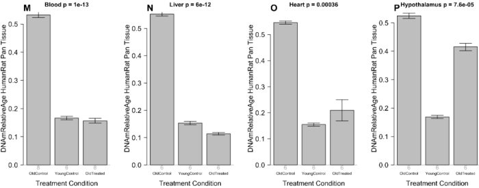
“Human-rat clock measure of relative age defined as age/maximum species lifespan. Each bar-plot reports the mean value and one standard error.”

From Supplementary Table 8, the percentages of rejuvenation for the above bar plots, calculated as “(100 * (1 – Old Treated / Old Control)” were:

  • “Blood 70.6%
  • Liver 79.4%
  • Heart 61.6%
  • Hypothalamus 20.9%”

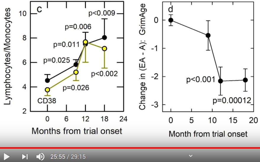
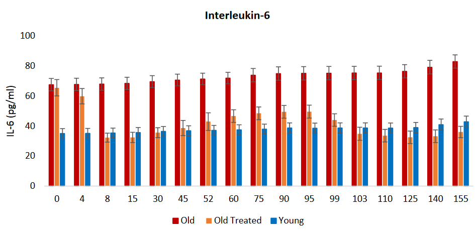
Let’s return to clinical biomarkers for comparison purposes. The current study measured pro-inflammatory cytokine IL-6 blood plasma levels at every time point, but didn’t publish numbers. Bar plots and narrative were:

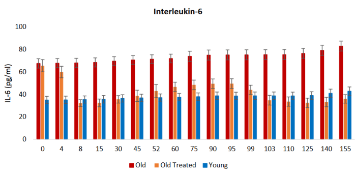
“Inflammation is an important response that helps protect the body, but excess inflammation especially in terms of duration of this response can have very detrimental effects instead. This occurs when inflammation fails to subside and persists indefinitely; a condition referred to as chronic inflammation, which for reasons not well-understood, increases with age and is associated with a multitude of conditions and pathologies.

The levels of two of the most reliable and common biomarkers of chronic inflammation, interleukin 6 (IL-6) and tumor necrosis factor α (TNF-α), are found to be considerably higher in old rats, and these were very rapidly diminished, within days by plasma fraction treatment, to comparable levels with those of young rats. This was especially stark with IL-6.

In time, the levels of these inflammatory factors began to rise gradually, but they were once again very effectively reduced following the second administration of the plasma fraction on the 95th day.”

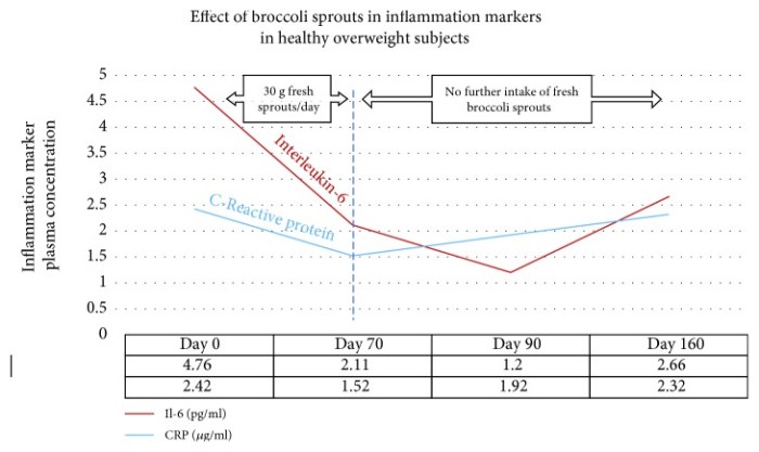
Let’s compare the above IL-6 graphic with IL-6 concentration improvements of our 2018 model clinical trial, Effects of long-term consumption of broccoli sprouts on inflammatory markers in overweight subjects, calculated as (100 * (1 – Day _ mean / Day 0 mean):

Mean pg/ml | % improvement | Period | Broccoli sprout consumption

  • 4.594 | 0% | Day 0 | “One week before the beginning of the intervention period, subjects were asked to avoid the consumption of Brassica vegetables (broccoli, radish, cauliflower, Brussel sprouts, mustards, among others) and their derived products.”
  • 1.748 | 62.0% | Day 0 to 70 | Subjects ate 30 g raw broccoli sprouts every day, and stopped eating them after Day 70.
  • 0.896 | 80.5% | Day 0 to 90 | “After the intervention period, a follow-up recovery period for all subjects continued for another 90 days with no ingestion of broccoli sprouts.”
  • 2.170 | 52.8% | Day 0 to 160 | Subjects had not eaten broccoli sprouts after Day 70.

Results between the studies were similar in that:

  1. IL-6 levels improved during early treatments through rat Day 8 and human Day 70, respectively.
  2. IL-6 levels continued decreasing shortly after treatments for 7 days (through rat Day 15) and 20 days (through human Day 90), respectively.
  3. IL-6 levels rose after rat Day 15 and human Day 90, respectively, but were still significantly below Day 0 values at rat Day 95 and human Day 160.

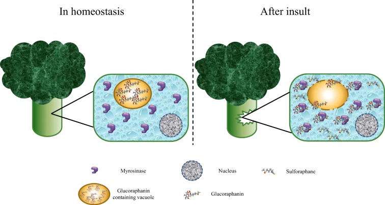
The current study measured Nrf2 but didn’t publish numbers. Bar plots and narrative were:

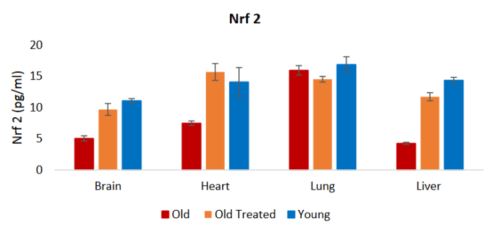
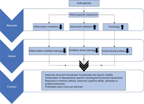
“The reduction of these inflammation markers is consistent with the profile of the nuclear factor erythroid 2-like 2 protein (Nrf2), which plays a major role in resolving inflammation, in part by inhibiting the expression of IL-6 and TNF-α. Nrf2 also induces the expression of antioxidants that neutralizes ROS [reactive oxygen species], which is also a significant feature in inflammation.”

A PubMed search on “nrf2 sulforaphane human” didn’t turn up relevant 2020 human in vivo studies. I disregarded reviews, cancer studies, disproven hypotheses, and other compounds listed in the below graphic.

I won’t repeat the entire Nrf2 section from the Part 1 curation, just one graphic and paragraph:

It [sulforaphane] is not only a potent Nrf2 inducer but also highly bioavailable [around 80%], so that modest practical doses can produce significant clinical responses. Other Nrf2 activators [shown in the above image] not only lack potency but also lack the bioavailability to be considered as significant intracellular Nrf2 activators.”


As noted in Reviewing clinical trials of broccoli sprouts and their compounds, there are no sulforaphane clinical trials that also use epigenetic clocks. Broccoli sprouts and their compounds’ effects on human aging is an area that hasn’t drawn attention and funding.

What effects may broccoli sprout compounds have on human aging? With this new human-rat relative biological age clock, researchers can get reliable answers from rat studies, with human clinical trials needed only to confirm those findings!

As rejuvenation research continues, what could people do easily, cheaply, and today for our long-term selves? Don’t know about the hypothalamus, but our blood, liver, and heart biological ages may decrease as we reduce inflammation and oxidative stress by eating broccoli sprouts.

I’m at a similar percentage of species maximum lifespan as were the study’s treated subjects. It’s my choice as to what my healthspan will be.

There isn’t evidence today to definitively say that changing my inflammatory phenotype with broccoli sprouts has had / will have rejuvenation effects on biological ages of my cells, organs, and body. But if eating broccoli sprouts every day not only reduces chronic inflammation and oxidative stress as expected, but also makes me younger, I could probably learn to live with that. 🙂

Continued with Part 3 of Rejuvenation therapy and sulforaphane.

An environmental signaling paradigm of aging

To follow up A rejuvenation therapy and sulforaphane, the study’s lead laboratory researcher – Dr. Harold Katcher – provided evidence for an environmental signaling paradigm of aging in this 2015 paper:

“The age-phenotype of a cell or organ depends on its environment and not its history.

Organ dysfunction is not the cause of aging, but is the result of its milieu. Therefore, the aged milieu is the cause. Though it has been thought that the aging immune system is the cause of aging, it can seen to be the result of aging.

The systemic milieu of an organism sets the age-phenotype of its cells, tissues and organs. Cells and organs secrete factors into blood, which are determined by the age-phenotype and repair-states of those cells and organs. The presence and concentrations of these blood-borne factors determine the age-phenotype of cells and organs.

Here we must be a bit more speculative. Changes in concentrations of factors present in blood, rather than their presence or absence, determines age-phenotype.

Interactions between disparate levels of the body’s hierarchy establish a consensus age-phenotype for cells and organs, and this largely occurs via the bloodstream. There appear to be positive factors that promote youthful age-phenotypes and negative factors that promote the aged phenotypes.

We readily consider development as a ‘program’, and it seems clear that we must consider post-adult development as ‘programmed’ as well. But if there is a program it is neither in genes nor chromatin, but in interaction of complex, interconnected systems spanning hierarchical levels.

If these aforementioned principles are correct, it should be easy to verify. If so, whole organism rejuvenation might require little more than:

  • Changing concentrations of all age-determining molecules of the bloodstream and various stem cell niche environments to youthful levels;
  • For a time sufficient to cause rejuvenation at the cellular level.

Once cells start secreting factors appropriate to their new, younger age-phenotypes, cognate changes should propagate through hierarchical levels.

The analogy to workings of a mechanical clock is not very exact. ‘Gears’ represent individual aging clocks, both cellular and organic (shown at different levels within the mechanism) which interact, ultimately resulting in organismic age, i.e. ‘body clock’, represented by the ‘hour hand’ (no minute hand is shown).

In mammals, readout of the clock corresponds to age-related composition of blood plasma. In this model, moving the hour hand backwards should result in a turning back of composite clocks as well – a result obtained when induction to pluripotence is used to reset cellular clocks.

Apart from being slowed down or sped up, the body clock can also be reset. Organisms, organs, and their cells can be reset to different age-phenotypes depending on their environment.

We know that old transplanted tissues and organs can regain function and live for the entire life of the younger host at least in rodents. We must suppose that age-phenotype changes must have taken place at the cellular level to allow this.

Rejuvenation cannot be explained on the basis that aging represents accumulation of irreparable cellular damage.

None of these principles are rigorously established as such, but all are supported by experimental evidence.”

http://www.eurekaselect.com/130538/article “Towards an Evidence-based Model of Aging”


Here are some of his responses to comments on the blog post that first curated his current research:

“We’ve (scientists), spent the past 70 years trying to definitively prove the commonsense ‘wear and tear’ theories and have not succeeded. So I tried something different, looking at results of experiments.

This is not based on ‘theory’ (say mitochondrial aging or ‘wear and tear’) but on experimental evidence. Theory comes in explaining our results, not achieving them. There is a theory becoming clear, one very different from the commonsense view of ‘wear and tear’ aging.

We haven’t examined immune response. All that we know for sure is that chronic inflammation of aging stopped. I can definitively say that chronic inflammation due to aging can be reversed with factors present in young blood.

There are amazing things that Big Pharma won’t touch as there’s not enough profit in them (they can’t be patented). So I guess we’re somewhat the same, but we know what to do and have proven it – for us, it’s not money. However, money allows you to do things.

Being 75 myself puts a time-frame around the project. We plan to propose its use for diseases of aging – eventually, everyone will use it. It will end up changing humanity. As people already seem to have too much free time to begin with, what will people do with those extra years they will be given?”


Sections 3 “Aging Manifestations that Have Hitherto Been Proposed as the Causes of Aging are the Consequences of Aging” and 10 “Several Factors ‘Conspire’ to Promote Inflammation in Old Mammalian Bodies, Inflammation Leads to Several Diseases of Aging and Perhaps to Aging Itself” were especially informative.

The former section discussed cells that were capable of making repairs but didn’t make repairs, with aging being the consequence of this behavior. The latter reviewed topics such as senescence, IL-6, NF-κB, and C-reactive protein in terms of feedback loops.

See Reevaluate findings in another paradigm for comparisons of Section 6 with another view of hypothalamic aging.

A rejuvenation therapy and sulforaphane

The founder of the epigenetic clock methodology with the coauthor of Aging as an unintended consequence released a 2020 rodent study “Reversing age: dual species measurement of epigenetic age with a single clock” at https://www.biorxiv.org/content/10.1101/2020.05.07.082917v1.full.pdf:

“We employed six clocks to investigate the rejuvenation effects of a plasma fraction treatment in different rat tissues. Two of these epigenetic clocks apply to both humans and rats.

The treatment more than halved the epigenetic ages of blood, heart, and liver tissue. A less pronounced, but statistically significant, rejuvenation effect could be observed in the hypothalamus.

The treatment was accompanied by progressive improvement in the function of these organs as ascertained through numerous biochemical/physiological biomarkers and behavioral responses to assess cognitive functions. Cellular senescence, which is not associated with epigenetic aging, was also considerably reduced in vital organs.

Plasma fraction treatment consists of two series of intravenous injections of plasma fraction. Rats were injected four times on alternate days for 8 days. A second identical series of injections were administered 95 days later. In its entirety, the experiment lasted 155 days.

Overall, this study demonstrates that a plasma-derived treatment markedly reverses aging according to epigenetic clocks and benchmark biomarkers of aging.”

The study hasn’t been peer reviewed, so can’t be viewed yet as conclusive. Given that researchers’ single-most valuable asset is their reputations, though, will the findings have major revisions?


I was alerted to the study by Josh Mitteldorf’s blog post Age Reduction Breakthrough, who did his usual excellent curation:

“Most of the explosion in aging research (and virtually all the venture capital startups) are looking to treat aging at the cellular level. Their paradigm is that aging is an accumulation of molecular damage, and they see their job as engineering of appropriate repair mechanisms.

The truth, as Katcher [the lead lab researcher] understands it, is that, to a large extent, aging is coordinated system-wide via signal molecules in the blood. The problem is that there are thousands of constituents represented in tiny concentrations in blood plasma, but conveying messages that cells read. Which of these are responsible for aging?

The two-species clock[s] was [were] a significant innovation, a first bridge for translating results from an animal model into their probable equivalent in humans. Besides the methylation clock[s], the paper presents evidence of rejuvenation by many other measures. For example:

  • IL-6, a marker of inflammation, was restored to low youthful levels;
  • Glutathione (GSH), superoxide dismutase (SOD), and other antioxidants were restored to higher youthful levels;
  • In tests of cognitive function (Barnes maze), treated rats scored better than old rats, but not as well as young rats.;
  • Blood triglycerides were brought down to youthful levels;
  • HDL cholesterol rose to youthful levels; and
  • Blood glucose fell toward youthful levels.

These results bring together three threads that have been gaining credibility over the last decade. Mutually reinforcing, the three have a strength that none of them could offer separately.

  1. The root cause of aging is epigenetic progression = changes in gene expression over a lifetime.
  2. Methylation patterns in nuclear DNA are not merely a marker of aging, but its primary source. Thus aging can be reversed by reprogramming DNA methylation.
  3. Information about the body’s age state is transmitted system-wide via signal molecules in the blood. Locally, tissues respond to these signals and adopt a young or an old cellular phenotype as they are directed.”

Several of these aging measurements are also positively affected by sulforaphane. Using Sulforaphane: Its “Coming of Age” as a Clinically Relevant Nutraceutical in the Prevention and Treatment of Chronic Disease as a reference:

1. “Chronic inflammation”

“Antioxidants in general and glutathione in particular can be depleted rapidly under conditions of oxidative stress, and this can signal inflammatory pathways associated with NF-κB. SFN [sulforaphane] has been shown to inhibit NF-κB in endothelial cells.

Two key inflammatory cytokines were measured at four time points in forty healthy overweight people [our model clinical trial, Effects of long-term consumption of broccoli sprouts on inflammatory markers in overweight subjects]. The levels of both interleukin-6 (Il-6) and C-reactive protein (CRP) declined over the 70 days during which the sprouts were ingested. These biomarkers were measured again at day 90, wherein it was found that Il-6 continued to decline, whereas CRP climbed again. When the final measurement was taken at day 160, CRP, although climbing, had not returned to its baseline value. Il-6 remained significantly below the baseline level at day 160.”

OMCL2019-2716870.010

2. “Oxidative stress”

“As a mediator for amplification of the mammalian defence system against various stressors, Nrf2 [nuclear factor erythroid 2-related factor 2] sits at the interface between our prior understanding of oxidative stress and the endogenous mechanisms cells use to deal with it. Diseases known to be underpinned by oxidative stress are proving to be more responsive to amplification of cellular defences via Nrf2 activation than by administration of direct-acting antioxidant supplements.

SFN, with absolute bioavailability of around 80%, [is] capable of increasing several endogenous antioxidant compounds via the transcription factor, Nrf2.

Nrf2 is ubiquitously expressed with the highest concentrations (in descending order) in the kidney, muscle, lung, heart, liver, and brain. Nrf2 was shown to prevent endothelial cells from exhibiting a proinflammatory state. Nrf2 is required for protection against glucose-induced oxidative stress and cardiomyopathy in the heart.

Well in excess of 500 genes have been identified as being activated by SFN via the Nrf2/ARE [Antioxidant Response Element] pathway, and it is likely that this underestimates the number as others are being discovered. Of the available SFN clinical trials associated with genes induced via Nrf2 activation, many demonstrate a linear dose-response. More recently, it has become apparent that SFN can behave hormetically with different effects responsive to different doses.

It [sulforaphane] is not only a potent Nrf2 inducer but also highly bioavailable so that modest practical doses can produce significant clinical responses. Other Nrf2 activators [shown in the above image] not only lack potency but also lack the bioavailability to be considered as significant intracellular Nrf2 activators.”


The study’s most relentlessly questioned, scrutinized, and criticized findings may be the two new epigenetic clocks that apply to both humans and rats. The researchers invited other researchers to validate these clocks because:

“If validated, this would be a step change in aging research. Although conservation of aging mechanism could be equally deduced from the existence of multiple individual clocks for other mammals (mouse, dog), the single formula of the human-rat clock that is equally applicable to both species effectively demonstrates this fact.”

The commonalities of this study with efforts to change my inflammatory phenotype with broccoli sprouts were summarized in the Discussion section:

“Apart from rejuvenating the vital organs of the treated rats, plasma fraction also impacted two fundamental physiological processes that underlie a great number of pathologies, namely oxidative stress and inflammation. Within a week of treatment, the markers of chronic inflammation (IL-6 and TNF-α) were significantly reduced and remained low throughout the entire experiment.

Likewise, markers of oxidative stress in brain, heart, lung and liver, which were very much higher in control old rats, were at the end of the experimental period, indistinguishable between plasma fraction-treated old rats and young ones. Concomitant with this drastic reduction in oxidative stress was the augmented levels of antioxidants (GSH, Catalase and SOD) in these tissues, indicating that modulating the levels of ROS [reactive oxygen species] to that of youthful rats is at least one way by which plasma fraction suppresses oxidative stress. It remains to be ascertained whether the rate of ROS generation is also reduced.

The levels of Nrf2, a transcription factor that impacts on oxidative stress, as well as inflammation, were raised by plasma fraction treatment of old rats to those of the young ones, indicating yet another level by which this treatment modulates these two critical processes. Collectively, these results show that plasma fraction treatment impacts not only the overt performances of organs, but also the underlying physiological processes that are pivotal for optimal organ function and health.”

Great stuff, huh? Are you ready to change your phenotype?

Continued with Part 2 of Rejuvenation therapy and sulforaphane.

Work your voluntary muscles today

This 2020 review by the Aging as a disease research group highlighted their specialty:

“A theory that fits both the aging and the rejuvenation data suggests that aging is caused primarily by the functional (and notably, experimentally reversible) inactivation of resident stem cells, which precipitates deteriorated tissue maintenance and repair and leads to the loss of organ homeostasis.

The damaged and unrepaired tissues suffer changes in their biochemistry, including the molecular crosstalk with resident stem cells, which further inhibits productive, regenerative responses. The inflammatory and fibrotic secretome can then propagate systemically, affecting the entire organism.

Skeletal muscle accounts for almost 40% of the total adult human body mass. This tissue is indispensable for vital functions such as respiration, locomotion, and voluntary movements and is among the most age-sensitive in mammals.

Muscle is capable of active repair in response to daily wear and tear, intense exercises, or injuries. Muscle regeneration relies on the adult muscle stem cells, also called satellite cells.

Rather than a significant decline in the total number with age, most of the data support a dramatic lack of activation of muscle stem cells after injury and a concomitant lack in the formation of progenitors that are needed for repair.

Multiple experimental approaches have been used for tissue rejuvenation and/or systemic rejuvenation; these include ablation of senescent cells and re-calibration of key signaling pathways that are needed for productive stem cell responses. To test the success in experimental rejuvenation, 1-4 approaches are typically applied, and skeletal muscle is well-suited for assaying each one.”

https://www.ncbi.nlm.nih.gov/pmc/articles/PMC7007696/ “Skeletal muscle as an experimental model of choice to study tissue aging and rejuvenation”


The review had a short section on inflammation details. Not enough, and there’s no tissue repair. Continuing unchecked is a systemic issue that led the reviewers to their paradigm of aging as a disease.

The review concluded with a subject that’s taught in high school, and should be understood at least before college graduation. It’s curious that an item like sample size required emphasis. Maybe research that doesn’t adhere to basics is a current issue?

Aging as an unintended consequence

The coauthors of 2018’s The epigenetic clock theory of aging reviewed progress that’s been made todate in understanding epigenetic clock mechanisms.

1. Proven DNA methylation features of epigenetic clocks:

  1. “Methylation of cytosines is undoubtedly a binary event.
  2. The increase in epigenetic age is contributed by changes of methylation profiles in a very small percent of cells in a population.
  3. The clock ticks extremely fast in early post-natal years and much slower after puberty.
  4. Clock CpGs have specific locations in the genome.
  5. It applies to prenatal biological samples and embryonic stem cells.

While consistency with all the five attributes does not guarantee veracity of a model, inconsistency with any one will signal the unlikely validity of a hypothesis.”

2. Regarding what epigenetic clocks don’t measure:

“The effects of

  • Telomere maintenance,
  • Cellular senescence,
  • DNA damage signaling,
  • Terminal differentiation and
  • Cellular proliferation

have all been tested and found to be unrelated to epigenetic ageing.”

3. Regarding cyclical features:

Both the epigenetic and circadian clocks are present in all cells of the body, but their ticking rates are regulated. Both these clocks lose synchronicity when cells are isolated from tissues and grown in vitro.

These similarities compel one to ponder potential links between them.”

This was among the points that Linear thinking about biological age clocks missed.

4. The reviewers discussed 3 of the 5 treatment elements in Reversal of aging and immunosenescent trends:

“It is not known at this stage whether the rejuvenating effect is mediated through the regeneration of the thymus or a direct effect of the treatment modality on the body. Also, it is not known if the effect is mediated by all three compounds or one or two of them.

What we know at this stage does not allow the formation of general principles regarding the impact of hormones on epigenetic age, but their involvement in development and maintenance of the body argue that they do indeed have a very significant impact on the epigenetic clock.”

Not sure why they omitted 3000 IU vitamin D and 50 mg zinc, especially since:

“It is not known if the effect is mediated by all three [five] compounds or one or two of them.”

5. They touched on the specialty of Aging as a disease researchers with:

“Muscle stem cells isolated from mice were epigenetically much younger independently of the ages of the tissue / animal from which they were derived.

The proliferation and differentiation of muscle stem cells cease upon physical maturation. These activities are initiated in adult muscles only in response to injury.

6. The reviewers agreed with those researchers in the Conclusion:

“Epigenetic ageing begins from very early moments after the embryonic stem cell stage and continues uninterrupted through the entire lifespan. The significance of this is profound as the question of why we age has been attributed to many different things, most commonly to ‘wear-and-tear.’

The ticking of the epigenetic clock from the embryonic state challenges this perspective and supports the notion that ageing is an unintended consequence of processes that are necessary for

  • The development of the organism and
  • Tissue homeostasis thereafter.”


https://journals.sagepub.com/doi/10.1177/1535370220918329 “Current perspectives on the cellular and molecular features of epigenetic ageing” (not freely available)