Perinatal stress and sex differences in circadian activity

This 2019 French/Italian rodent study used the PRS model to investigate its effects on circadian activity:

“The aim of this study was to explore the influence of PRS on the circadian oscillations of gene expression in the SCN [suprachiasmatic nucleus of the hypothalamus] and on circadian locomotor behavior, in a sex-dependent manner.

Research on transcriptional rhythms has shown that more than half of all genes in the human and rodent genome follow a circadian pattern. We focused on genes belonging to four functional classes, namely the circadian clock, HPA axis stress response regulation, signaling and glucose metabolism in male and female adult PRS rats.

Our findings provide evidence for a specific profile of dysmasculinization induced by PRS at the behavioral and molecular level, thus advocating the necessity to include sex as a biological variable to study the set-up of circadian system in animal models.”

“There was a clear-cut effect of sex on the effect of PRS on the levels of activity:

  • During the period of lower activity (light phase), both CONT and PRS females were more active than males. During the light phase, PRS increased activity in males, which reached levels of CONT females.
  • More interestingly, during the period of activity (dark phase), male PRS rats were more active than male CONT rats. In contrast, female PRS rats were less active than CONT females.
  • During the dark phase, CONT female rats were less active than CONT male rats.

The study presented evidence for sex differences in circadian activity of first generation offspring that was caused by stress experienced by the pregnant mother:

“Exposure to gestational stress and altered maternal behavior programs a life-long disruption in the reactive adaptation such as:

  •  A hyperactive response to stress and
  • A defective feedback of the hypothalamus-pituitary-adrenal (HPA) axis together with
  • Long-lasting modifications in stress/anti-stress gene expression balance in the hippocampus.”

It would advance science if these researchers carried out experiments to two more generations to investigate possible transgenerational epigenetic inheritance of effects caused by PRS. What intergenerational and transgenerational effects would they possibly find by taking a few more months and extending research efforts to F2 and F3 generations? Wouldn’t these findings likely help humans?


One aspect of the study was troubling. One of the marginally-involved coauthors was funded by the person described in How one person’s paradigms regarding stress and epigenetics impedes relevant research. Although no part of the current study was sponsored by that person, there were three gratuitous citations of their work.

All three citations were reviews. Unlike study researchers, reviewers aren’t bound to demonstrate evidence from tested hypotheses. Reviewers are free to:

  • Express their beliefs as facts;
  • Over/under emphasize study limitations; and
  • Disregard and misrepresent evidence as they see fit.

Fair or not, comparisons of reviews with Cochrane meta-analyses of the same subjects consistently show the extent of reviewers’ biases. Reviewers also aren’t obligated to make post-publication corrections for their errors and distortions.

As such, reviews can’t be cited for reliable evidence. Higher-quality studies that were more relevant and recent than a 1993 review could have elucidated points.

Sucking up to the boss and endorsing their paradigm was predictable. Since that coauthor couldn’t constrain themself to funder citations only in funder studies, it was the other coauthors’ responsibilities to edit out unnecessary citations.

https://www.frontiersin.org/articles/10.3389/fnmol.2019.00089/full “Perinatal Stress Programs Sex Differences in the Behavioral and Molecular Chronobiological Profile of Rats Maintained Under a 12-h Light-Dark Cycle”
Caloric restriction’s epigenetic effects

This 2019 US review subject was caloric restriction (CR) without malnutrition:

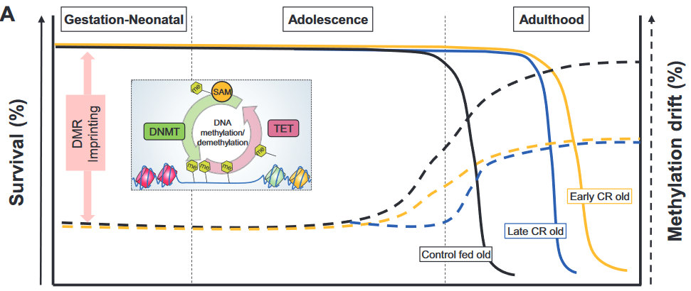
“Cellular adaptation that occurs in response to dietary patterns can be explained by alterations in epigenetic mechanisms such as DNA methylation, histone modifications, and microRNA. Epigenetic reprogramming of the underlying chronic low-grade inflammation by CR can lead to immuno-metabolic adaptations that enhance quality of life, extend lifespan, and delay chronic disease onset.

Short- and long-term CRs produce significant changes in different tissues and across species, in some animal models even with sex-specific effects. Early CR onset may cause a different and even an opposite effect on physiological outcomes in animal models such as body weight.”

https://academic.oup.com/advances/article-abstract/10/3/520/5420411 “Epigenetic Regulation of Metabolism and Inflammation by Calorie Restriction” (not freely available)


1. The review didn’t present evidence to equate survival (left axis) with methylation drift (right axis) per the above graphic. Methylation drift should point in the opposite direction of survival, if anything.

2. No mention was made of the epigenetic clock method of measuring age acceleration, although it’s been available since 2013 and recent diet studies have used it. The sole citation of an age acceleration study was from 2001, which was unacceptable for a review published in 2019.

3. The review provided many cellular-level details about the subject. However, organism-level areas weren’t sufficiently evidenced:

A. Arguments for an effect usually include explanations for no effect as well as for opposite effects. The reviewers didn’t provide direct evidence for why, if caloric restriction extended lifespan, caloric overabundance produced shorter lifespans.

B. Caloric restriction evidence was presented as if only it was responsible for organism-level effects. Other mechanisms may have been involved.

An example of such a mechanism was demonstrated in a 2007 rodent study Reduced Oxidant Stress and Extended Lifespan in Mice Exposed to a Low Glycotoxin Diet which compared two 40%-calorie-restricted diets.

The calories and composition of both diets were identical. However, advanced glycation end product (AGE) levels were doubled in standard chow because heating temperatures were “sufficiently high to inadvertently cause standard mouse chow to be rich in oxidant AGEs.”

The study found that a diet with lower chow heating temperatures increased lifespan and health span irrespective of caloric restriction!

  • The low-AGE calorie-restricted diet group lived an average of 15% longer (>20 human equivalent years) than the CR group.
  • 40% of the low-AGE calorie-restricted diet group were still alive when the last CR group member died.
  • The CR group also had significantly more: 1) oxidative stress damage; 2) glucose and insulin metabolism problems; and 3) kidney, spleen, and liver injuries.

A drug that countered effects of a traumatizing mother

This 2019 US rodent study concerned transmitting poor maternal care to the next generation:

“The quality of parental care received during development profoundly influences an individual’s phenotype, including that of maternal behavior. Infant experiences with a caregiver have lifelong behavioral consequences.

Maternal behavior is a complex behavior requiring the recruitment of multiple brain regions including the nucleus accumbens, bed nucleus of the stria terminalis, ventral tegmental area, prefrontal cortex, amygdala, and medial preoptic area. Dysregulation within this circuitry can lead to altered or impaired maternal responsiveness.

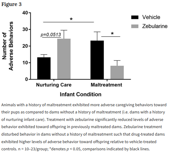
We administered zebularine, a drug known to alter DNA methylation, to dams exposed during infancy to the scarcity-adversity model of low nesting resources, and then characterized the quality of their care towards their offspring.

  1. We replicate that dams with a history of maltreatment mistreat their own offspring.
  2. We show that maltreated-dams treated with zebularine exhibit lower levels of adverse care toward their offspring.
  3. We show that administration of zebularine in control dams (history of nurturing care) enhances levels of adverse care.
  4. We show altered methylation and gene expression in maltreated dams normalized by zebularine.

These findings lend support to the hypothesis that epigenetic alterations resulting from maltreatment causally relate to behavioral outcomes.

Maternal behavior is an intergenerational behavior. It is important to establish the neurobiological underpinnings of aberrant maternal behavior and explore treatments that can improve maternal behavior to prevent the perpetuation of poor maternal care across generations.”


The study authors demonstrated intergenerational epigenetic effects, and missed an opportunity to also investigate transgenerational epigenetically inherited effects. They cited reference 60 for the first part of the above quotation, but the cited reviewer misused the transgenerational term by applying it to grand-offspring instead of the great-grand-offspring.

There were resources available to replicate the study authors’ previous findings, which didn’t show anything new. Why not use such resources to uncover evidence even more applicable to humans by extending experiments to great-grand-offspring that would have no potential germline exposure to the initial damaging cause?

Could a study design similar to A limited study of parental transmission of anxiety/stress-reactive traits have been integrated? That study’s thorough removal of parental behavior would be an outstanding methodology to confirm by falsifiability whether parental behavior is both an intergenerational and a transgenerational epigenetic inheritance mechanism.

Rodent great-grand-offspring can be studied in < 9 months. It takes > 50 years for human studies to reach the great-grand-offspring transgenerational generation.

  • Why not attempt to “prevent the perpetuation of poor maternal care across generations?”
  • Isn’t it a plausible hypothesis that humans “with a history of maltreatment mistreat their own offspring?”
  • Isn’t it worth the extra effort to extend animal research to investigate this unfortunate chain?

https://www.nature.com/articles/s41598-019-46539-4 “Pharmacological manipulation of DNA methylation normalizes maternal behavior, DNA methylation, and gene expression in dams with a history of maltreatment”

Wikipedia is a poor source of information on advanced glycation end products (AGEs)

A link to Wikipedia is usually on the first page of search results. The Wikipedia post on AGEs lacks the evidence that a reader may infer from its text.

For example, the second paragraph of the AGEs post, Dietary Sources, contained the following text and references:

  1. “However, only low molecular weight AGEs are absorbed through diet, and vegetarians have been found to have higher concentrations of overall AGEs compared to non-vegetarians. [4]
  2. Therefore it is unclear whether dietary AGEs contribute to disease and aging, or whether only endogenous AGEs (those produced in the body) matter. [5]
  3. This does not free diet from potentially negatively influencing AGE, but implicates dietary AGE may be less important than other aspects of diet that lead to elevated blood sugar levels and formation of AGEs. [4] [5]”

[4] https://www.sciencedirect.com/science/article/pii/S0278691513004444 “Advanced glycation end products in food and their effects on health” (not freely available) 2013 Denmark.

Please note on this linked page that a German researcher took the time to correct one bias of the Danish reviewers, citing evidence from his studies that:

“The deleterious effects of food-derived AGEs in subjects with type 2 diabetes mellitus are proven.”

[5] https://www.ncbi.nlm.nih.gov/pmc/articles/PMC3257625 “Dietary Advanced Glycation End Products and Aging” 2010 US.


Both of these references were reviews.

Unlike study researchers, reviewers aren’t bound to demonstrate evidence from tested hypotheses. Reviewers are free to:

  • Express their beliefs as facts;
  • Over/under emphasize study limitations; and
  • Disregard and misrepresent evidence as they see fit.

Reviewers also aren’t obligated to make post-publication corrections for their errors and distortions. For example, the Danes didn’t correct their review with any findings the German researcher presented.

As such, reviews can’t be cited for reliable evidence.


A sample of other problems with each of the Wikipedia sentences:

1. “However, only low molecular weight AGEs are absorbed through diet, and vegetarians have been found to have higher concentrations of overall AGEs compared to non-vegetarians. [4]”

The first part of sentence 1 came from the review’s abstract:

“Only LMW AGEs..may be absorbed from the gut and contribute to the body burden of AGEs.”

But the reviewers didn’t support their abstract’s statement with direct evidence from any study!

2. “Therefore it is unclear whether dietary AGEs contribute to disease and aging, or whether only endogenous AGEs (those produced in the body) matter. [5]”

The “therefore” of sentence 2 was misplaced. Sentence 1 didn’t attempt to explain whether “dietary AGEs contribute to disease and aging” or “only endogenous AGEs matter.”

Since sentence 2 wasn’t a consequence of sentence 1, the Wikipedia contributor(s) needed to support sentence 2 with evidence. Citing an “unclear” 2010 reference [5] ignored dozens of studies that provided better clarity.

3. “This does not free diet from potentially negatively influencing AGE, but implicates dietary AGE may be less important than other aspects of diet that lead to elevated blood sugar levels and formation of AGEs. [4] [5]”

Wikipedia contributors tend to cite irrelevant references rather than get flagged with “citation needed.” The value judgment of sentence 3 was an example of this intentionally misleading masquerade.

“Dietary AGE may be less important..” wasn’t unequivocally supported by studies referenced in either review, and didn’t represent an authoritative body of evidence. Contrast those weasel words with:

“The deleterious effects of food-derived AGEs in subjects with type 2 diabetes mellitus are proven.”

Good job, Wikipedia contributors! You used lower-quality reviews to promote misunderstandings that DETRACTED from science.


Wikipedia’s premise is that since the group knows more about any subject than does any individual, everyone is entitled to contribute. The results are usually incoherent narratives that often substitute opinions for evidence.

The second paragraph of the Exogenous section of the Wikipedia glycation post provided an example:

  • Assertions of the first and third sentences needed citations. Did the contributor(s) think these would be unexamined?
  • Someone contributed a cancer reference as the fourth sentence, although it had little to do with the preceding sentences.
  • The fifth sentence was informative on exogenous glycations and AGEs. An editor would have removed “recently” and “recent” though, because the cited source was dated 2005.

Disease and advanced glycation end products (AGEs)

This 2015 French/US review focused on chronic kidney disease, appropriate for its publication in the Journal of the American Society of Nephrology:

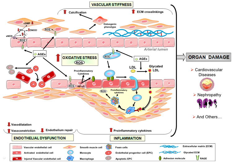
“Advanced glycation end products (AGEs) are formed not only in the presence of hyperglycemia, but also in diseases associated with high levels of oxidative stress, such as CKD. Humans are exposed to exogenous sources of AGE (diet and cigarette smoke) and endogenous sources of AGE when the organism is exposed to high levels of glucose, such as in diabetes.

Accumulation of AGEs in patients with CKD has been shown to result from inflammation, oxidative stress, and diet. AGEs are proinflammatory and pro-oxidative compounds that play a role in the high prevalence of endothelial dysfunction and subsequent cardiovascular disease in patients with CKD.

In view of the many harmful effects of AGEs on cell function, it is essential to develop strategies designed to counteract their effects. AGEs are generated during the thermal processing and storage of foods. Dietary restriction is an effective, feasible, and economic method to reduce levels of toxic AGEs and possibly, the associated cardiovascular mortality.”

https://jasn.asnjournals.org/content/27/2/354 “Uremic Toxicity of Advanced Glycation End Products in CKD”


I came across the AGE subject in the usual Internet way. 🙂 While reading comments on Josh Mitteldorf’s blog post Money in Aging Research, Part I, Dr. Alan Green mentioned Dr. Helen Vlassara’s work. A DuckDuckGo search led to her 44,256 citations, which increase every day.

Another read on the subject is her 2016 book Dr. Vlassara’s AGE-Less Diet: How a Chemical in the Foods We Eat Promotes Disease, Obesity, and Aging and the Steps We Can Take to Stop It. A practical guide is her 2017 book The AGE Food Guide: A Quick Reference to Foods and the AGEs They Contain.

Linking adult neurogenesis to Alzheimer’s disease

This 2019 Spanish human study compared DNA methylation, chromatin and histone modifications in the hippocampus of deceased Alzheimer’s disease patients with controls:

“A significant percentage of the differentially methylated genes were related to neural development and neurogenesis. It was astounding that other biological, cellular, and molecular processes generally associated with neurodegeneration such as apoptosis, autophagy, inflammation, oxidative stress, and mitochondrial or lysosomal dysfunction were not overrepresented.

The results of the present study point to neurogenesis-related genes as targets of epigenetic changes in the hippocampus affected by AD. These methylation changes might be built throughout life due to external and internal cues and would represent an example of epigenetic interaction between environmental and genetic factors in developing AD.

As an alternative explanation, these epigenetic marks might also represent the trace of DNA methylation alterations induced during early developmental stages of the hippocampus, which would remain as a fingerprint in the larger proportion of hippocampal neurons that are not exchanged. This second hypothesis would link AD to early life stages, in concordance with recent studies that revealed abnormal p-tau deposits (pre-tangles) in brains of young individuals under 30, suggesting AD pathology would start earlier in life than it was previously thought. The influence of the genetic risk for AD has also been postulated to begin in early life, and other AD risk factors may be influenced by in utero environment.”


The study cited references to adult neurogenesis:

“Though strongly related to brain development, neurogenesis is also maintained in the adult human brain, mainly in two distinct areas, i.e., the subventricular zone and the subgranular zone of the dentate gyrus in the hippocampus. There is substantial neurogenesis throughout life in the human hippocampus as it is estimated that up to one third of human hippocampal neurons are subject to constant turnover.

Adult neurogenesis is linked to hippocampal-dependent learning and memory tasks and is reduced during aging. Recent evidence suggests that adult neurogenesis is altered in the neurodegenerative process of AD, but it is still controversial with some authors reporting increased neurogenesis, whereas others show reduced neurogenesis. In the human hippocampus, a sharp drop in adult neurogenesis has been observed in subjects with AD.”

One of the study’s limitations was its control group:

“There was a significant difference in age between controls [12, ages 50.7 ± 21.5] and AD patients [26, ages 81.2 ± 12.1], being the latter group older than the former group. Although we adjusted for age in the statistical differential methylation analysis, the accuracy of this correction may be limited as there is little overlap in the age ranges of both groups.”

https://clinicalepigeneticsjournal.biomedcentral.com/track/pdf/10.1186/s13148-019-0672-7 “DNA methylation signature of human hippocampus in Alzheimer’s disease is linked to neurogenesis”

OCD and neural plasticity

Update: this was retracted on February 23, 2021. The retraction note is at https://www.nature.com/articles/s41598-021-84474-5.

This 2019 New York rodent study investigated multiple avenues to uncover mechanisms of obsessive-compulsive disorder:

“Psychophysical models of OCD propose that anxiety (amygdala) and habits (dorsolateral striatum) may be causally linked. Numerous genetic and environmental factors may reduce striatum sensitivity and lead to maladaptive overcompensation, potentially accounting for a significant proportion of cases of pathological OCD-like behaviors.

Our results indicate that both the development and reversal of OCD-like behaviors involve neuroplasticity resulting in circuitry changes in BLA-DLS and possibly elsewhere.”

https://www.nature.com/articles/s41598-019-45325-6.pdf “Amelioration of obsessive-compulsive disorder in three mouse models treated with one epigenetic drug: unraveling the underlying mechanism”


The researchers explored two genetic models of OCD, showed why these insufficiently explained observed phenomena, then followed up with epigenetic investigations. They demonstrated how and the degree to which histone modifications and DNA methylation regulated both the development and reversal of OCD symptoms.

However, the researchers also carelessly cited thirteen papers outside the specific areas of the study to support one statement in the lead paragraph:

“Novel studies propose that modulations in gene expression influenced by environmental factors, are connected to mental health disorders.”

Only one of the thirteen citations was more recent than 2011, and none of them were high-quality studies.

Infant DNA methylation and caregiving

This 2019 US human study attempted to replicate findings of animal studies that associated caregiver behavior with infant DNA methylation of the glucocorticoid receptor gene:

“Greater levels of maternal responsiveness and appropriate touch were related to less DNA methylation of specific regions in NR3c1 exon 1F, but only for females. There was no association with maternal responsiveness and appropriate touch or DNA methylation of NR3c1 exon 1F on prestress cortisol or cortisol reactivity. Our results are discussed in relation to programming models that implicate maternal care as an important factor in programing infant stress reactivity.”


The study had many undisclosed and a few disclosed limitations, one of which was:

“Our free-play session, while consistent with the length of free-play sessions in other studies, was short (5 min). It is unclear whether a longer length of time would have yielded significant different maternal responsiveness and appropriate touch data.”

The final sentence showed the study’s purpose was other than discovering factual evidence:

“Following replication of this work, it could ultimately be used in conjunction with early intervention, or home-visiting programs, to measure the strength of the intervention effect at the epigenetic level.”

https://onlinelibrary.wiley.com/doi/full/10.1002/imhj.21789 “DNA methylation of NR3c1 in infancy: Associations between maternal caregiving and infant sex” (not freely available)

Do delusions have therapeutic value?

This 2019 UK review discussed delusions, aka false beliefs about reality:

“Delusions are characterized by their behavioral manifestations and defined as irrational beliefs that compromise good functioning. In this overview paper, we ask whether delusions can be adaptive notwithstanding their negative features.

We consider different types of delusions and different ways in which they can be considered as adaptive: psychologically (e.g., by increasing wellbeing, purpose in life, intrapsychic coherence, or good functioning) and biologically (e.g., by enhancing genetic fitness).”

https://onlinelibrary.wiley.com/doi/full/10.1002/wcs.1502 “Are clinical delusions adaptive?”


A. Although section 4’s heading was Biological Adaptiveness of Delusions, the reviewers never got around to discussing evolved roles of brain areas and beliefs (delusions). One mention of evolutionary biology was:

“Delusions are biologically adaptive if, as a response to a crisis of some sort (anomalous perception or overwhelming distress), they enhance a person’s chances of reproductive success and survival by conferring systematic biological benefits.”

B. Although section 5’s heading was Psychological Adaptiveness of Delusions, the reviewers didn’t connect feelings and survival sensations as origins of beliefs (delusions) and behaviors. They had a few examples of feelings:

“Delusions of reference and delusions of grandeur can make the person feel important and worthy of admiration.”

and occasionally sniffed a clue:

“Some delusions (especially so‐called motivated delusions) play a defensive function, representing the world as the person would like it to be.”

where “motivated delusions” were later deemed in the Conclusion section to be a:

“Response to negative emotions that could otherwise become overwhelming.”

C. Feelings weren’t extensively discussed until section 6 Delusions in OCD and MDD, which gave readers an impression that feelings were best associated with those diseases.

D. In the Introduction, sections 4, 5, and 7 How Do We Establish and Measure Adaptiveness, the reviewers discussed feeling meaning in life, but without understanding:

  1. Feelings = meaning in life, as I quoted Dr. Arthur Janov in The pain societies instill into children:

    “Without feeling, life becomes empty and sterile. It, above all, loses its meaning.

  2. Beliefs (delusions) defend against feelings.
  3. Consequentially, the stronger and / or more numerous beliefs (delusions) a person has, the less they feel meaning in life.

E. Where, when, why, and how do beliefs (delusions) arise? Where, when, why, and how does a person sense and feel, and what are the connections with beliefs (delusions)?

F. The word “sense” was used 29 times in contexts such as “make sense” and “sense of [anxiety, coherence, control, meaning, purpose, rational agency, reality, self, uncertainty]” but no framework connected biological sensing to delusions. Papers from other fields have detailed cause-and-effect explanations and predecessor-successor diagrams for every step of a process. Not this one.


Regarding any therapeutic value of someone else’s opinion of a patient’s delusions:

I’ll reuse this quotation from the Scientific evidence page of Dr. Janov’s 2011 book “Life Before Birth: The Hidden Script that Rules Our Lives” p.166:

“Primal Therapy differs from other forms of treatment in that the patient is himself a therapist of sorts. Equipped with the insights of his history, he learns how to access himself and how to feel.

The therapist does not heal him; the therapist is only the catalyst allowing the healing forces to take place. The patient has the power to heal himself.

Another way Dr. Janov wrote this was on p.58 of his 2016 book Beyond Belief as quoted in Beyond Belief: The impact of merciless beatings on beliefs:

NO ONE HAS THE ANSWER TO LIFE’S QUESTIONS BUT YOU. How you should lead your life depends on you, not outside counsel.

We do not direct patients, nor dispense wisdom upon them. We have only to put them in touch with themselves; the rest is up to them.

Everything the patient has to learn already resides inside. The patient can make herself conscious. No one else can.”

Another important transgenerational epigenetic inheritance study

This 2019 Washington State University rodent study from Dr. Michael Skinner’s lab found:

“A cascade of epigenetic alterations initiated in PGCs [primordial germ cells of F3 males] appears to be required to alter epigenetic programming during spermatogenesis to modify the sperm epigenome involved in transgenerational epigenetic inheritance phenomenon.

Following fertilization there is a DNA methylation erasure to generate stem cells in the early embryo, which then remethylate in a cell type-specific manner. DNA methylation erasure is thought to, in part, reset deleterious epigenetics in the germline. However, imprinted gene DNA methylation sites and induced transgenerational epimutations appear to be protected from this DNA methylation erasure.

A germline with an altered epigenome has the capacity to alter the early embryo’s stem cell’s epigenome and transcriptome that can subsequently impact epigenomes and transcriptomes of all derived somatic cells. Therefore, an altered sperm epigenome has the capacity to transmit phenotypes transgenerationally. Experiments have demonstrated that epigenetic inheritance can also be transmitted through the female germline.

Previously, agricultural fungicide vinclozolin was found to promote transgenerational inheritance of sperm differential DNA methylation regions (DMRs) termed epimutations that help mediate this epigenetic inheritance. The current study was designed to investigate developmental origins of transgenerational DMRs during gametogenesis.

The current study with vinclozolin-induced transgenerational inheritance demonstrates that sperm DMRs also originate during both spermatogenesis and earlier stages of germline development, but at distinct developmental stages. Fetal exposure initiates a developmental cascade (i.e., distinct developmental origins) of aberrant epigenetic programming, and does not simply induce a specific number of DMRs that are maintained throughout development.”

https://www.tandfonline.com/doi/pdf/10.1080/15592294.2019.1614417?needAccess=true “Transgenerational sperm DNA methylation epimutation developmental origins following ancestral vinclozolin exposure”


The study’s main hypotheses were:

“Following fertilization, the hypothesis is that transgenerational epimutations modify early embryonic transcriptomes and epigenomes to re-establish the cascade for the next generation.

As the individual develops, all somatic cells have altered epigenomes and transcriptomes to promote disease susceptibility later in life.”

Researchers: adopt these hypotheses, and apply them to human studies.

1. Don’t get off track by requiring that the same phenotype must be observed in each generation for there to be transgenerational epigenetic inheritance, because:

“Fetal exposure..does not simply induce a specific number of DMRs that are maintained throughout development.”

Animal transgenerational studies have shown that epigenetic inheritance mechanisms may both express different phenotypes for each generation, and entirely skip a phenotype in one or more generations!

2. Don’t limit your study designs to F1 children as did:

3. Don’t stop at F2 grandchildren as did:

4. Continue studies on to F3 great-grandchildren who had no direct exposure to altering stimulus. Keep in the forefront of your research proposals that there are probably more than 10,000,000 F3 descendants of DES-exposed women just in the US!

The transgenerational impact of Roundup exposure

This 2019 Washington rodent study from Dr. Michael Skinner’s lab found adverse effects in the grand-offspring and great-grand-offspring following their ancestor’s exposure during pregnancy to the world’s most commonly used herbicide:

“Using a transient exposure of gestating F0 generation female rats found negligible impacts of glyphosate on the directly exposed F0 generation, or F1 generation offspring pathology. In contrast, dramatic increases in pathologies in the F2 generation grand-offspring, and F3 transgenerational great-grand-offspring were observed.

The transgenerational pathologies observed include prostate disease, obesity, kidney disease, ovarian disease, and parturition (birth) abnormalities:

  1. Prostate disease in approximately 30% of F3 generation glyphosate lineage males, a three-fold increase in disease rate over controls.
  2. A transgenerational (F3 generation) obese phenotype was observed in approximately 40% of the glyphosate lineage females and 42% of the glyphosate lineage males.
  3. An increased incidence of kidney disease observed in the F3 generation glyphosate lineage females affecting nearly 40% of females.
  4. A significant increase in ovarian disease observed in the F2 [48% vs. 21% for controls] and F3 [36% vs. 15% for controls] generation glyphosate lineage females.
  5. During the gestation of F2 generation mothers with the F3 generation fetuses, dramatic parturition abnormalities were observed in the glyphosate lineage. The frequency of unsuccessful parturition was 35%. To further investigate the parturition abnormalities an outcross of F3 generation glyphosate lineage males with a wildtype female was performed. There were parturition abnormalities observed with a frequency of 30%.

Classic and current toxicology studies only involve direct exposure of the individual, while impacts on future generations are not assessed. The ability of glyphosate and other environmental toxicants to impact our future generations needs to be considered, and is potentially as important as the direct exposure toxicology done today for risk assessment.”


Why isn’t coverage of this study the top story of world news organizations? Is what’s reported more important than reliable evidence of generational consequences to environmental experiences?

Current toxicology practices are a scientific disgrace:

  • What are hypotheses of practices that test only effects on somatic cells, and don’t look for generational effects on germ cells?
  • Are tests selected for their relative convenience instead of chosen for their efficacy?

Why don’t sponsors fund and researchers perform human studies of transgenerational epigenetic inheritance? For example, from Burying human transgenerational epigenetic evidence:

“From the late 1930s through the early 1970s, DES was given to nearly two million pregnant women in the US alone.

Fourth [F3] generation effects of prenatal exposures in humans have not been reported.

Zero studies of probably more than 10,000,000 F3 great-grandchildren of DES-exposed women just here in the US!

There will be abundant human evidence to discover if sponsors and researchers will take their fields seriously.

https://www.nature.com/articles/s41598-019-42860-0.pdf “Assessment of Glyphosate Induced Epigenetic Transgenerational Inheritance of Pathologies and Sperm Epimutations: Generational Toxicology”

Non-emotional memories

This 2019 US review covered memory mechanisms:

“With memory encoding reliant on persistent changes in the properties of synapses, a key question is how can memories be maintained from days to months or a lifetime given molecular turnover? It is likely that positive feedback loops are necessary to persistently maintain the strength of synapses that participate in encoding.

These levels are not isolated, but linked by shared components of feedback loops.”


Despite the review’s exhaustive discussion, the reviewers never came to the point. The word cloud I made of the review’s most frequent thirty words had little to do with why memory occurs:

  • Why do some stimuli evoke a memory in response?
  • Why are almost all of the stimuli an organism receives not remembered?

Much of the discussion was baseless because it excluded emotion. Many of the citations’ memory findings relied on emotion, though.

For example, in the subsection Roles of persistent epigenetic modifications for maintaining LTF [long-term facilitation], LTP [long-term potentiation], and LTM [long-term memory]:

  • Histone acetylation is increased after fear conditioning in the hippocampus and amygdala.
  • Correspondingly, inhibition of histone deacetylase enhances fear conditioning and LTP.
  • Following fear conditioning, histone phosphorylation is also increased.
  • DNA methylation is also up-regulated in the hippocampus and amygdala after fear conditioning, and inhibition of DNA methylation blocks fear LTM.”

http://learnmem.cshlp.org/content/26/5/133.full “How can memories last for days, years, or a lifetime? Proposed mechanisms for maintaining synaptic potentiation and memory”

Statistical inferences vs. biological realities

A 2019 UCLA study introduced a derivative of the epigenetic clock named GrimAge:

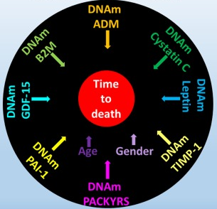
“DNAm GrimAge, a linear combination of chronological age, sex, and DNAm-based surrogate biomarkers for seven plasma proteins and smoking pack-years, outperforms all other DNAm-based biomarkers, on a variety of health-related metrics.

An age-adjusted version of DNAm GrimAge, which can be regarded as a new measure of epigenetic age acceleration (AgeAccelGrim), is associated with a host of age-related conditions, lifestyle factors, and clinical biomarkers. Using large scale validation data from three ethnic groups, we demonstrate that AgeAccelGrim stands out among pre-existing epigenetic clocks in terms of its predictive ability for time-to-death, time-to-coronary heart disease, time-to-cancer, its association with computed tomography data for fatty liver/excess fat, and early age at menopause.”

https://www.ncbi.nlm.nih.gov/pmc/articles/PMC6366976/ “DNA methylation GrimAge strongly predicts lifespan and healthspan”


A miserable attempt at reporting the study’s findings included angles of superstition, fear-of-the-future, and suspicion-by-spurious-association:

“The research has already captured the attention of the life insurance industry. After all, a solid death date could mean real savings when it comes to pricing policies.

The hope is that if and when legitimate anti-aging drugs are developed, GrimAge could be used to test their effectiveness. In a world with functional anti-aging drugs, “doctors could test [your GrimAge number] and say, ‘You know what, you’re aging too quickly. Take this,'” Horvath said.”

https://onezero.medium.com/a-new-test-predicts-when-youll-die-give-or-take-a-few-years-2d08147c8ea6 “A New Test Predicts When You’ll Die (Give or Take a Few Years)”


A detailed blog post from Josh Mitteldorf provided scientific coverage of the study:

“Methylation sites associated with smoking history predicted how long the person would live more accurately than the smoking history itself. Even stranger, the methylation marks most closely associated with smoking were found to be a powerful indication of future health even when the sample was confined to non-smokers.

The DNAm GrimAge clock was developed in two stages, a correlation of a correlation. Curiously, the indirect computation yields the better result.

Horvath’s finding that secondary methylation indicators are more accurate than the underlying primary indicator from which they were derived is provocative, and calls out for a new understanding.”

https://joshmitteldorf.scienceblog.com/2019/03/05/dnam-grimage-the-newest-methylation-clock “DNAm GrimAge—the Newest Methylation Clock”


When there are logical disconnects in findings like the above, it’s time to examine underlying premises. As noted in Group statistics don’t necessarily describe an individual, an assumption required by statistical analyses is that each measured item in the sample is interchangeable with the next.

This presumption is often false, producing individually inapplicable results. For example, Immune memory vs. immune adaptation included this description of the adaptive immune system:

“To be effective, highly specific immune response requires huge diversity of receptors and antibodies, which is achieved by somatic rearrangement of gene segments. Recombination results in millions of TCR [T cell receptor] and antibody variants able to recognize and neutralize millions of various antigens.”

Standard statistics of millions of T cell receptor and antibody variants won’t represent their individually unique properties. But individual differences are both their purpose and benefit to us.

The GrimAge study’s overreach was most apparent in stratifying educational attainment to develop correlations. As mentioned in Does a societal mandate cause DNA methylation? such statistics are poor evidence of each individual’s biological realities.

Neither derivatives of group statistics, nor correlations of correlations, seem to be the techniques needed to understand biological causes of effects. Another commentary on the GrimAge study mentioned but glossed over this point:

“It remains a mystery why exactly the epigenetic clocks work, and whether age-related changes in DNA methylation contribute to the cause of aging or are a result of it.”

Our brains are shaped by our early environments

This 2019 McGill paper reviewed human and animal studies on brain-shaping influences from the fetal period through childhood:

“In neonates, regions of the methylome that are highly variable across individuals are explained by the genotype alone in 25 percent of cases. The best explanation for 75 percent of variably methylated regions is the interaction of genotype with different in utero environments.

A meta-analysis including 45,821 individuals with attention-deficit/hyperactivity disorder and 9,207,363 controls suggests that conditions such as preeclampsia, Apgar score lower than 7 at 5 minutes, breech/transverse presentations, and prolapsed/nuchal cord – all of which involve some sort of poor oxygenation during delivery – are significantly associated with attention-deficit/hyperactivity disorder. The dopaminergic system seems to be one of the brain systems most affected by perinatal hypoxia-ischemia.

Exposure to childhood trauma activates the stress response systems and dysregulates serotonin transmission that can adversely impact brain development. Smaller cerebral, cerebellar, prefrontal cortex, and corpus callosum volumes were reported in maltreated young people as well as reduced hippocampal activity.

Environmental enrichment has a series of beneficial effects associated with neuroplasticity mechanisms, increasing hippocampal volume, and enhancing dorsal dentate gyrus-specific differences in gene expression. Environmental enrichment after prenatal stress decreases depressive-like behaviors and fear, and improves cognitive deficits.”


The reviewers presented strong evidence until the Possible Factors for Reversibility section, which ended with the assertion:

“All these positive environmental experiences mentioned in this section could counterbalance the detrimental effects of early life adversities, making individuals resilient to brain alterations and development of later psychopathology.”

The review’s penultimate sentence recognized that research is seldom done on direct treatments of causes:

“The cross-sectional nature of most epigenetic studies and the tissue specificity of the epigenetic changes are still challenges.”

Cross-sectional studies won’t provide definitive data on cause-and-effect relationships.

The question yet to be examined is: How can humans best address these early-life causes to ameliorate their lifelong effects?

https://onlinelibrary.wiley.com/doi/full/10.1111/dmcn.14182 “Early environmental influences on the development of children’s brain structure and function” (not freely available)

A therapy to reverse cognitive decline

This 2018 human study presented the results of 100 patients’ personalized therapies for cognitive decline:

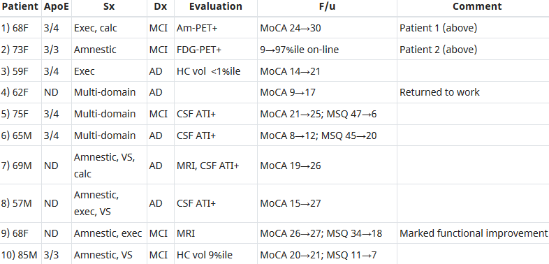
“The first examples of reversal of cognitive decline in Alzheimer’s disease and the pre-Alzheimer’s disease conditions MCI (Mild Cognitive Impairment) and SCI (Subjective Cognitive Impairment) have recently been published..showing sustained subjective and objective improvement in cognition, using a comprehensive, precision medicine approach that involves determining the potential contributors to the cognitive decline (e.g., activation of the innate immune system by pathogens or intestinal permeability, reduction in trophic or hormonal support, specific toxin exposure, or other contributors), using a computer-based algorithm to determine subtype and then addressing each contributor using a personalized, targeted, multi-factorial approach dubbed ReCODE for reversal of cognitive decline.

An obvious criticism of the initial studies is the small number of patients reported. Therefore, we report here 100 patients, treated by several different physicians, with documented improvement in cognition, in some cases with documentation of improvement in electrophysiology or imaging, as well.”

https://www.omicsonline.org/open-access/reversal-of-cognitive-decline-100-patients-2161-0460-1000450-105387.html “Reversal of Cognitive Decline: 100 Patients”


The lead author commented on Josh Mitteldorf’s informative post A cure for Alzheimer’s? Yes, a cure for Alzheimer’s!:

  1. “We have a paper in press, due to appear 10.22.18 (open access, JADP, I’ll send a copy as soon as available), showing 100 patients with documented improvement – some with MRI volumetrics improved, others with quantitative EEG improvements, others with evoked response improvements, and all with quantitative cognitive assessment improvement. Some are very striking – 12 point improvements in MoCA [Montreal Cognitive Assessment], for example – others less so, but all also have subjective improvement. Hopefully this will address some of the criticisms that we haven’t documented improvement in enough people.
  2. We were just turned down again for a randomized, controlled clinical trial, so on the one hand, we are told repeatedly that no one will believe that this approach works until we publish a randomized, controlled study, and on the other hand, we’ve been turned down (first in 2011/12, and now in 2018), with the complaint that we are trying to address more than one variable in the trial (as if AD is a single-variable disease!). Something of a catch-22. We are now resubmitting (unfortunately, the IRBs are not populated by functional medicine physicians, so they are used to seeing old-fashioned drug studies), and we’ll see what happens.
  3. I’ve been extending the studies to other neurodegenerative diseases, and it has been impressive how much of a programmatic response there seems to be in these ‘diseases.’
  4. I agree with you that there are many features in common with aging itself.
  5. You made a good point that APP [amyloid precursor protein] is a dependence receptor, and in fact it functions as an integrating dependence receptor, responding to numerous inputs (Kurakin and Bredesen, 2015).
  6. In the book and the publications, we don’t claim it is a “cure” since we don’t have pathological evidence that the disease process is gone. What we claim is ‘reversal of cognitive decline’ since that is what we document.
  7. As I mentioned in the book, AD is turning out to be a protective response to multiple insults, and this fits well with the finding that Abeta has an antimicrobial effect (Moir and Tanzi’s work). It is a network-downsizing, protective response, which is quite effective – some people live with the ongoing degenerative process for decades.
  8. We have seen several cases now in which a clinical trial of an anti-amyloid antibody made the person much worse in a time-dependent manner (each time there was an injection, the person would get much worse for 5-10 days, then begin to improve back toward where he/she was, but over time, marked decline occurred), and this makes sense for the idea that the amyloid is actually protecting against pathogens or toxins or some other insult.
  9. It is important to note that we’ve never claimed that all people get better – this is not what we’ve seen. People very late in the process, or who don’t follow the protocol, or who don’t address the various insults, do not improve. It is also turning out to be practitioner dependent – some are getting the vast majority of people to improve, others very few, so this is more like surgery than old-fashioned prescriptive medicine – you have to do a somewhat complicated therapeutic algorithm and get it right for best results.
  10. I’m very interested in what is needed to take the next step in people who have shown improvement but who started late in the course. For example, we have people now who have increased MoCA from 0 to 9 (or 0 to 3, etc.), with marked subjective improvement but plateauing at less than normal. These people had extensive synaptic and cellular loss prior to the program. So what do we need to raise the plateau? Stem cells? Intranasal trophic support? Something else?
  11. I haven’t yet seen a mono-etiologic theory of AD or a mono-therapeutic approach that has repeatedly positive results, so although I understand that there are many theories and treatments, there doesn’t seem to be one etiology to the disease, nor does there seem to be one simple treatment that works for most. It is much more like a network failure.”

At a specific level:

  • “There doesn’t seem to be one etiology to the disease,
  • Nor does there seem to be one simple treatment that works for most.
  • We don’t have pathological evidence that the disease process is gone.”

For general concepts, however:

  • “AD is turning out to be a protective response to multiple insults.
  • It is a network-downsizing, protective response, which is quite effective.
  • The amyloid is actually protecting against pathogens or toxins or some other insult.”

For a framework of an AD cure to be valid, each source of each insult that evoked each “protective response” should be traced.

Longitudinal studies would be preferred inside this framework. These study designs would investigate evidence of each insult’s potential modifying effect on each “protective response” that could affect the cumulative disease trajectory of each individual.

In many cases, existing study designs would be adequate if they extended their periods to the end of the subjects’ natural lifetimes. One AD-relevant example would be extending the prenatally-restraint-stressed model used in:

The framework would also encourage extending studies to at least three generations to investigate evidence for transgenerational effects, as were found in: