How much sulforaphane is suitable for healthy people?

This post compares and contrasts two perspectives on how much sulforaphane is suitable for healthy people. One perspective was an October 2019 review from John Hopkins researchers who specialize in sulforaphane clinical trials:

Broccoli or Sulforaphane: Is It the Source or Dose That Matters?

These researchers didn’t give a consumer-practical answer, so I’ve presented a concurrent commercial perspective to the same body of evidence via an October 2019 review from the Australian founder of a company that offers sulforaphane products:

Sulforaphane: Its “Coming of Age” as a Clinically Relevant Nutraceutical in the Prevention and Treatment of Chronic Disease


1. Taste from a clinical trial perspective:

“Harsh taste (a.k.a. back-of-the-throat burning sensation) that is noticed by most people who consume higher doses of sulforaphane, must be acknowledged and anticipated by investigators. This is particularly so at higher limits of dosing with sulforaphane, and not so much of a concern when dosing with glucoraphanin, or even with glucoraphanin-plus-myrosinase.

Presence and/or enzymatic production of levels of sulforaphane in oral doses ranging above about 100 µmol, creates a burning taste that most consumers notice in the back of their throats rather than on the tongue. Higher doses of sulforaphane lead to an increased number of adverse event reports, primarily nausea, heartburn, or other gastrointestinal discomfort.”

Taste wasn’t mentioned in the commercial review. Adverse effects were mentioned in this context:

“Because SFN is derived from a commonly consumed vegetable, it is generally considered to lack adverse effects; safety of broccoli sprouts has been confirmed. However, use of a phytochemical in chemoprevention engages very different biochemical processes when using the same molecule in chemotherapy; biochemical behaviour of cancer cells and normal cells is very different.”

2. Commercial products from a clinical trial perspective:

“Using a dietary supplement formulation of glucoraphanin plus myrosinase (Avmacol®) in tablet form, we observed a median 20% bioavailability with greatly dampened inter-individual variability. Fahey et al. have observed approximately 35% bioavailability with this supplement in a different population.”

Avmacol appeared to be the John Hopkins product of choice, as it was mentioned 15 times in its clinical trials table. A further investigation of Avmacol showed that its supplier for broccoli extract, TrueBroc, was cofounded by a John Hopkins coauthor! Yet the review stated:

“The authors declare no conflict of interest.”

Please disclose easily discoverable ethical and commercial conflicts without prevarication. Other products were downgraded with statements such as:

“5 or 10 g/d of BroccoPhane powder (BSP), reported to be rich in SF, daily x 4 wks (we have assayed previously and found this not to be the case).”

They also disclaimed:

“We have indicated clinical studies in which label results have been used rather than making dose measurements prior to or during intervention.”

No commercial products – not even the author’s own company’s – were directly mentioned in a commercial perspective.

3. Dosage from a clinical trial perspective:

“Reporting of administered dose of glucoraphanin and/or sulforaphane is a poor measure of the bioavailable / bioactive dose of sulforaphane. As a consequence, we propose that the excreted amount of sulforaphane metabolites (sulforaphane + sulforaphane cysteine-glycine + sulforaphane cysteine + sulforaphane N-acetylcysteine) in urine over 24 h (2–3 half-lives), which is a measure of “internal dose”, provides a more revealing and likely consistent view of delivery of sulforaphane to study participants.

Only recently have there been attempts to define minimally effective doses in humans – an outcome made possible by development of consistently formulated, stable, bioavailable broccoli-derived preparations.”

Dosage from a commercial perspective:

“Of available SFN clinical trials associated with genes induced via Nrf2 activation, many demonstrate a linear dose-response. More recently, it has become apparent that SFN can behave hormetically with different effects responsive to different doses. This is in addition to its varying effects on different cell types and consequent to widely varying intracellular concentrations.

A 2017 clinical pilot study examined the effect of an oral dose of 100 μmol (17.3 mg) encapsulated SFN on GSH [reduced glutathione] induction in humans over 7 days. Pre- and postmeasurement of GSH in blood cells that included T cells, B cells, and NK cells showed an increase of 32%. Researchers found that in the pilot group of nine participants, age, sex, and race did not influence the outcome.

Clinical outcomes are achievable in conditions such as asthma with daily SFN doses of around 18 mg daily and from 27 to 40 mg in type 2 diabetes. The daily SFN dose found to achieve beneficial outcomes in most of the available clinical trials is around 20-40 mg.”

The author’s sulforaphane products are available in 100, 250, and 700 mg capsules of enzyme-active broccoli sprout powder.

4. Let’s see how these perspectives treated a 2018 Spanish clinical trial published as Effects of long-term consumption of broccoli sprouts on inflammatory markers in overweight subjects.

From a commercial perspective:

“In a recent study using 30 grams of fresh broccoli sprouts incorporated daily into diet, two key inflammatory cytokines were measured at four time points in forty healthy overweight [BMI 24.9 – 29.9] people. Levels of both interleukin-6 (Il-6) and C-reactive protein (CRP) declined over the 70 days during which sprouts were ingested.

These biomarkers were measured again at day 90, wherein it was found that Il-6 continued to decline, whereas CRP climbed again. When the final measurement was taken at day 160, CRP, although climbing, had not returned to its baseline value. Il-6 remained significantly below baseline level at day 160.

Sprouts contained approximately 51 mg (117 μmol) GRN [glucoraphanin], and plasma and urinary SFN metabolites were measured to confirm that SFN had been produced when sprouts were ingested.”


From a clinical trial perspective, glucoraphanin dosage was “1.67 (GR) μmol/kg BW.” This wasn’t accurate, however. It was assumed into existence by:

“In cases where authors did not indicate dosage in μmol/kg body weight (BW), we have made those calculations using a priori assumption of a 70 kg BW.”

117 μmol / 1.67 μmol/kg = 70 kg.

This study provided overweight subjects’ mean weight in its Table 1 as “85.8 ± 16.7 kg.” So its actual average glucoraphanin dosage per kg body weight was 117 μmol / 85.8 kg = 1.36 μmol/kg. Was making an accurate calculation too difficult?

A clinical trial perspective included this study in Section “3.2. Clinical Studies with Broccoli-Based Preparations: Efficacy” subsection “3.2.8. Diabetes, Metabolic Syndrome, and Related Disorders.” This was somewhat misleading, as it was grouped with studies such as a 2012 Iranian Effects of broccoli sprout with high sulforaphane concentration on inflammatory markers in type 2 diabetic patients: A randomized double-blind placebo-controlled clinical trial (not freely available).

A commercial perspective pointed out substantial differences between these two studies:

“Where the study described above by Lopez-Chillon et al. investigated healthy overweight people to assess effects of SFN-yielding broccoli sprout homogenate on biomarkers of inflammation, Mirmiran et al. in 2012 had used a SFN-yielding supplement in T2DM patients. Although the data are not directly comparable, the latter study using the powdered supplement resulted in significant lowering of Il-6, hs-CRP, and TNF-α over just 4 weeks.

It is not possible to further compare the two studies due to vastly different time periods over which each was conducted.”


The commercial perspective impressed as more balanced than the clinical trial perspective. The clinical trial perspective also had an undisclosed conflict of interest!

A. The clinical trial perspective:

  • Effectively promoted one commercial product whose supplier was associated with a coauthor;
  • Downgraded several other commercial products; and
  • Tried to shift responsibility for the lack of “minimally effective doses in humans” to commercial products with:

    “Only recently have there been attempts to define minimally effective doses in humans – an outcome made possible by the development of consistently formulated, stable, bioavailable broccoli-derived preparations.”

But unless four years previous is “recently,” using commercial products to excuse slow research progress can be dismissed. A coauthor of the clinical trial perspective was John Hopkins’ lead researcher for a November 2015 Sulforaphane Bioavailability from Glucoraphanin-Rich Broccoli: Control by Active Endogenous Myrosinase, which commended “high quality, commercially available broccoli supplements” per:

“We have now discontinued making BSE [broccoli sprout extract], because there are several high quality, commercially available broccoli supplements on the market.”

The commercial perspective didn’t specifically mention any commercial products.

B. The commercial perspective didn’t address taste, which may be a consumer acceptance problem.

C. The commercial perspective provided practical dosage recommendations, reflecting their consumer orientation. These recommendations didn’t address how much sulforaphane is suitable for healthy people, though.

The clinical trial perspective will eventually have to make practical dosage recommendations after they stop dodging their audience – which includes clinicians trying to apply clinical trial data – with unhelpful statements such as:

“Reporting of administered dose of glucoraphanin and/or sulforaphane is a poor measure of the bioavailable / bioactive dose of sulforaphane.”

How practical was their “internal dose” recommendation for non-researcher readers?


Here’s what I’m doing to answer how much sulforaphane is suitable for healthy people.

I’d like to posthumously credit my high school literature teachers Dorothy Jasiecki and Martin Obrentz for this post’s compare-and-contrast approach. They both required their students to read at least two books monthly, then minimally handwrite a 3-page (single-spaced) paper comparing and contrasting those books.

Each monthly assignment was individualized so that students couldn’t undo the assignment’s purpose – to think for yourself – with parasitical collaboration. This former practice remains a good measure of intentional dumbing-down of young people, the intent of which has become clearer.

You can see from these linked testimonials that their approach was in a bygone era, back when some teachers considered a desired outcome of public education to be that each individual learned to think for themself. My younger brother contributed:

“I can still remember everything Mr. Obrentz ever assigned for me to read. He was the epitome of what a teacher should be.”

Microwave broccoli to increase sulforaphane levels

This 2020 Chinese/USDA study investigated effects on sulforaphane amount from heating broccoli in water and microwaving at different power settings to different temperatures:

“Microwave treatment causes a sudden collapse of cell structure due to the increase in osmotic pressure difference over vacuole membrane. Mild heating could increase SFR [sulforaphane] level, possibly explained by the increased activity of MYR [the enzyme myrosinase] which can hydrolyze GLR [glucoraphanin] into SFR at high temperature (up to 60°C).

Microwave‐cooked broccoli had higher levels of these two compounds compared to broccoli heated in water. The broccoli sample without cooking as a control showed the least amount of GLR, indicating that microwave heating did help to release more GLR from the cell.

In the temperature range of 50–60°C, a positive correlation was observed between GLR or SFR contents and temperature. However, these two physiochemical contents were negatively correlated with temperature when it increased to 70°C.

The glucoraphanin (GLR) and sulforaphane (SFR) contents (μmol/g DW) in florets of broccoli during microwaving at 40, 50, 60, and 70°C using low power level (LL) or high power level (HL). Data are reported as the mean ± SD (n = 3). Values with different letters are significantly (p < .05) different.

[For example, sulforaphane levels of the control (raw), LL40, LL70, and HL40 conditions weren’t significantly different, and the HL70 level was significantly lower than those levels]. The microwave using high level at 60°C showed the greatest SFR level (2.45 µmol/g DW).”

Table S1 from the supporting material:

Temperature

(°C)

Time

(S)

Power level

(W)

Heating in water 40 185 NA
50 230
60 262
70 290
Microwave (HL) 40 65 950
50 90
60 108
70 120
Microwave (LL) 40 115 475
50 148
60 178
70 200

https://onlinelibrary.wiley.com/doi/10.1002/fsn3.1493 “Microwave cooking increases sulforaphane level in broccoli”


The researchers demonstrated a more effective method of increasing sulforaphane than did the cited and widely discussed 2004 Heating decreases epithiospecifier protein activity and increases sulforaphane formation in broccoli (not freely available). The older study methods were difficult to implement in kitchens, and evaluated heating temperature as the only factor.

The present study added microwave power level irradiation effects as a factor, and simplified heating temperature implementation. People can use Table S1 to maximize broccoli floret sulforaphane content in their kitchens. See Week 2 of Changing an inflammatory phenotype with broccoli sprouts for changes.

The study provided an optimal sulforaphane end result of “(2.45 µmol/g DW)”. I asked a study author for additional data, and they replied:

“The control GLR and SLR amount was 2.18 and 0.22 µmol/g DW, respectively, while the HL60 GLR amount was 2.78 µmol/g DW.”

Microwaving 10 grams of broccoli florets to 60°C (140°F) increased the sulforaphane amount by 1,114% (2.45 / .22)! That also increased the glucoraphanin amount by 27% (2.78 / 2.18) for further processing into sulforaphane after eating.

I replied: That’s an exciting result, increasing sulforaphane more than 11 times, while also increasing glucoraphanin! I haven’t found similar experiments with broccoli sprouts. Would you expect similar results?

The study author responded:

“We didn’t expect this result, and think microwave irradiation might help to release more conjugated forms of glucosinolates and then get hydrolyzed by released myrosinase. Further studies are being carried out.”


The study also measured broccoli stems:

“GLR and SFR were hardly detected in stems. Less than 52% of GLR was detected in the [50/50] mixture of florets and stems compared to florets.

Microwaved at 60°C, the florets had a concentration of GLR and SFR at 2.78 and 2.45 µmol/g DW, respectively, which was significantly higher than the levels detected in mixture of florets and stems (1.21 and 0.82 µmol/g DW, respectively).”

The 50% florets / 50% stems mixture’s glucoraphanin amount of 1.21 µmol was roughly comparable with the 1.08 µmol glucoraphanin amount of mature broccoli extract in item 2 below.

Reminders from Eat broccoli sprouts today:

  1. A 1 mg sulforaphane weight equals a 5.64 μmol sulforaphane amount.
  2. “Content of glucoraphanin in extract from broccoli sprouts was 16.6 μmol per gram of fresh weight. In contrast, mature broccoli extract contained 1.08 μmol per gram of fresh weight.”
  3. The bioavailability of sulforaphane in a broccoli sprout extract with the myrosinase enzyme 100 μmol gelcap was 36.1% which weighed 6.4 mg.
  4. The question of how much sulforaphane is suitable for healthy people remains unanswered.

Eat broccoli sprouts today!

This 2020 Korean letter to a journal editor cited 23 recent papers in support of sulforaphane’s positive effects, mainly in anti-cancer treatments:

“Gene expression is mediated by chromatin epigenetic changes, including DNA methylation, histone modifications, promoter-enhancer interactions, and non-coding RNA (microRNA and long non-coding RNA)-mediated regulation. Approximately 50% of all tumor suppressor genes are inactivated through epigenetic modifications, rather than by genetic mechanisms, in sporadic cancers. Accumulating evidence suggests that epigenetic modulators are important tools to improve the efficacy of disease prevention strategies.

Because sulforaphane (SFN) induces the nuclear factor erythroid 2-related factor 2 (Nrf2)-antioxidant response element pathway that induces the cellular defense against oxidative stress, SFN has received increased attention because it acts as an antioxidant, antimicrobial, anti-inflammatory, and anticancer agent.”

https://www.ncbi.nlm.nih.gov/pmc/articles/PMC7068201/ “A recent overview on sulforaphane as a dietary epigenetic modulator”


Letters to the editor aren’t peer-reviewed, though. One of the cited papers was a 2018 Czech mini-review that included metabolism, preparation and processing evidence:

“Sulforaphane is a phytochemical that occurs in plants in the form of biological inactive precursor glucoraphanin. This precursor belongs to the group of phytochemicals – glucosinolates – that are rapidly converted to isothiocyanate by the enzyme myrosinase.

The process of transformation takes place after a disruption of plant tissues by biting, chewing, slicing, and other destruction of tissues, when myrosinase is released from plant tissues. When myrosinase is destroyed during meal preparation (during cooking, steam cooking, or microwave treatment), a likely source of isothiocyanates is microbial degradation of glucosinolates by intestinal microflora. However, hydrolysis by microflora has been reported to be not very efficient, and in humans it is very diverse and variable.

Content of glucoraphanin in extract from broccoli sprouts was 16.6 μmol per gram of fresh weight. In contrast, mature broccoli extract contained 1.08 μmol per gram of fresh weight. Total amount of glucosinolates in young broccoli sprouts is 22.7 μmol per gram of fresh weight and 3.37 μmol per gram of fresh weight for mature broccoli.

Percentage amount of sulforaphane formed from its precursor glucoraphanin in broccoli which had not been heat treated and had been lyophilized [freeze-dried] was 22.8%. Broccoli steaming (5 min) and its lyophilization decrease the amount of sulforaphane formed to 4.2%.”

https://www.liebertpub.com/doi/full/10.1089/jmf.2018.0024 “Isothiocyanate from Broccoli, Sulforaphane, and Its Properties (not freely available)


Information about 43 completed sulforaphane clinical trials is here. Among them, the 2014 Effect of Broccoli Sprouts on Nasal Response to Live Attenuated Influenza Virus in Smokers: A Randomized, Double-Blind Study was of particular interest, stating:

“Nutritional interventions aimed at boosting antioxidants may be most effective in individuals who are relatively antioxidant-deficient at baseline, a condition likely to be more prevalent in smokers.”

I didn’t notice regular supplement dosage studies. Maybe I didn’t read control group information carefully enough?


https://pubchem.ncbi.nlm.nih.gov/compound/sulforaphane lists sulforaphane’s molecular weight as 177.3 g/mol. A 1 mg sulforaphane capsule weight equals a 5.64 μmol sulforaphane amount (.001 / 177.3).

From the 2015 Sulforaphane Bioavailability from Glucoraphanin-Rich Broccoli: Control by Active Endogenous Myrosinase:

  • Figure 4 showed bioavailability of sulforaphane in a broccoli sprout extract with myrosinase 100 μmol gelcap was 36.1% which weighed 6.4 mg (36.1 / 5.64).
  • Figure 3 showed bioavailability of sulforaphane in freeze-dried broccoli sprouts in pineapple-lime juice was 40.5% in 50, 100, and 200 μmol amounts and 33.8% with 100 μmol gel caps. You do the weight math.
  • Figure 2 showed that if broccoli sprout extract didn’t have the enzyme, bioavailability of sulforaphane was 10.4% whether the amount was 69 or 230 μmol, weighing 1.27 mg (69 x .104) / 5.64 and 4.24 mg (230 x .104) / 5.64.

Bioavailability ranged from Figure 2’s 10.4% to Figure 4’s 36.1%. The question of how much sulforaphane is suitable for healthy people remains unanswered.


Aging as a disease

This 2020 interview was with UC Berkeley researchers:

“Lack of cure goes hand in hand with inability to accept that this [aging] is disease. For example, there was some resistance to accept tuberculosis as the actual disease. When there was no antibiotics or cure against it, people tended to discard it and said, oh, it’s just nerves, you need to go to a sanatorium and relax.

It used to be that, please do not diagnose that there’s bacterial meningitis, because there is no cure. Whatever else you can come up with, do it first. Now, diagnose it as fast as possible, so we can put patients on antibiotics immediately. My prediction is that the same will happen to aging.

We and others have demonstrated that you can, from the outside, either by some signal or blood therapy, parabiosis, something like that, some intervention, jump-start aged resident stem cells in tissue and get them to behave as, by whatever means you’re measuring it, young or a lot closer to young than they would normally be. Intrinsic capacity of them to act that way is there.

As we grow old, the environment of differentiated niche stem cells does not provide productive instruction. It provides counterproductive instruction, which, overall, tells them just to remain quiescent and do nothing.

It’s not a program to kill you. It’s the lack of a program to keep you young and healthy for longer than 90 years.

If your program was that whenever you’re a damaged, differentiated cell, you simply trigger apoptosis and activate stem cells to make new cells, we would live much longer and healthy. The program right now is to resist being dead and replaced as much as you can for as long as you can.

So cells produce too much TGF beta [transforming growth factor-β] because it helps them to keep functioning even when they’re damaged. That too much TGF beta, ironically, inhibits resident stem cells, so they are not replacing old cells with new ones. It’s almost like you have old bureaucrats that are running an organization and do not want to be replaced.

Our thoughts are probably different from most people, because we go to the data and the data show that they’re not really fully what authors wrote in the abstract or conclusion. When you look at that, my thought is that much more work needs to be done before it [partial cellular reprogramming] could be even thought to be commercialized.”

https://www.lifespan.io/news/apheresis-with-profs-irina-michael-conboy/ “Irina & Michael Conboy – Resetting Aged Blood to Restore Youth”


Keep in mind that although the interviewers’ organization had changed, their advocacy position as displayed in A blood plasma aging clock persisted. One of the interviewees is on the interviewers’ organization scientific advisory board, and they also have an interest in downgrading competing approaches.

Despite caveats, this interview was these researchers’ perspective in their decades-long investigations of aging. I included a graphic and below quote from Organismal aging and cellular senescence to note how their paradigm compared with other aging researchers:

“In our view, recent evidence that

  • Senescence is based on an unterminated developmental growth program and finding that
  • The concept of post-mitotic senescence requires activation of expansion, or ‘growth’ factors as a second hit,

favor the assumption that aging underlies a grating of genetic determination similarly to what is summarized above under the pseudo-programmed causative approach.”

Recover your sanity

Men, it has been well said:

  • Think in herds; it will be seen that
  • They go mad in herds, while
  • They only recover their senses slowly, one by one.

During the great plague, which ravaged all Europe between the years 1345 and 1350, it was generally considered that the end of the world was at hand.

‘But the facts, my dear fellow,’ said his friend, ‘the facts do not agree with your theory.’ ‘Don’t they?’ replied the philosopher, shrugging his shoulders, ‘then, tant pis pour les faits’ – so much the worse for the facts!”

Charles Mackay, 1841

https://www.gutenberg.org/ebooks/24518 “Memoirs of Extraordinary Popular Delusions and the Madness of Crowds”

Alfred Jacob Miller “Hunting buffalo” 1837
“Establishing the hidden communication networks in large self-organized groups facilitates a quantitative understanding of behavioral contagion.

An individual will be more likely to respond (is more susceptible) if it:

  • Is strongly connected to the initiator (short path length), and if it
  • Has neighbors which are strongly connected to each other.”

Why is it so difficult to live your own life?

The WHO has a financial incentive to declare COVID-19 a pandemic

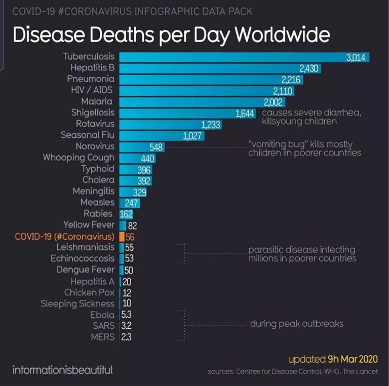
“The percent of the population being affected has not even reached 1/10th of one percent. When we look at this Coronavirus scare, it does not even compare to all the deaths taking place every single day from a host of other diseases nobody bothers to ever mention. This really makes me wonder just who is orchestrating this panic and is the purpose really to expand government powers?”

The Cyclical Nature of Disease

“The WHO has a financial incentive in declaring this a pandemic. It issued Pandemic Bonds paying 7% interest which now the WHO does not have to repay.”

Half-Billion $ Pandemic Derivatives

See Well done, WHO, carefully played to follow up.

Train your immune system every day!

This 2019 US review subject was β-glucan:

“β-1,3-Glucans (hereafter referred to as glucan) are natural molecules able to significantly improve our health. In human studies, the tested (and suggested) daily dose remains in the range of 100–500 mg for stimulation of the immune system, whereas for a decrease in cholesterol levels a daily dose of 3 g is recommended.

Glucan does not represent essential nutrients, but it might be successfully used not only for:

  • Improvement of immune functions, but also to improve the general quality of life via
  • Improvements of immune status,
  • Lowering cholesterol,
  • Improving blood glucose levels and
  • Reduction of stress.

ClinicalTrials.gov summarizes 177 [now 207 with 110 completed] β-glucan clinical trials, mostly in cancer, gastrointestinal tract therapy, lowering cholesterol and improvements of immune reactions.

The question is not if glucans will move from food supplement to widely accepted drug, but how soon.

Reactions known to be influenced by glucan are represented in white, reactions where glucan has no confirmed effects are shown in black. The first defensive body response to infection results from formation of the anorexia cytokines (IL-1, IL-6, IL-8, and TNF-α).”

https://www.mdpi.com/1420-3049/24/7/1251/htm “Beta Glucan: Supplement or Drug? From Laboratory to Clinical Trials”

The review is also indexed at https://www.betaglucan.org/i-p/ under “Immunomodulator”


I’m curating this review on Day 12 of a self-quarantine after coming back from Milano, Italy, Monday, February 24, 2020. The previous Friday into Saturday I flew to Milano sitting with a group of elderly Italians who were returning from vacation.

On Saturday my traveling companion and I used the Milano rail and crowded subway system to go downtown. On Sunday we used their crowded rail, crowded bus, and crowded ferry systems to visit Como, Bellagio, and Menaggio.

I don’t think we could have mixed in more with people during transits, touristing, and Carnevale celebrations.

IMG_9539

After returning to our hotel Sunday evening, we heard about the coronavirus outbreak south of Milano and the closing of ten towns. We changed flights and departed for the US early Monday morning.

Neither of us have had any signature symptoms of COVID-19 (fever, shortness of breath, dry cough). Our daily diet the past few years included β-glucan from steel-cut oats (~3 g) and from a 1/3, 1/6 yeast cell wall supplement.

Coincidence?

Each of our futures will depend on what we do Now to prepare.

Take responsibility for your one precious life.

Moonrise at sunrise with Venus

Masters of manipulating their host

This 2020 French review subject was parasitical influences on host epigenetic processes:

“Parasites have become masters of manipulating their host cells, exploiting signaling, and metabolic pathways to hijack host gene expression to their own advantage. These intracellular parasites have developed a wide range of strategies that affect transcriptional machineries and epigenetic events in the host cell nucleus.

Parasite effectors regulate host transcription. Secretion of numerous parasite effector proteins are key processes during parasite infection. Parasite effectors deregulate host expression profile which lead to host cell transformation, or escape from the host immune system to allow parasite persistence and survival.”

parasites
The first two of the six strategies discussed are shown above:

  1. “Induction of a host epigenetic enzyme. Parasite infection leads to upregulation of SMYD3, a methyltransferase that activates genes involved in host transformation, through H3K4 trimethylation.
  2. Secreting effector proteins that drive epigenetic repression of host genes. TEEGR activates a host chromatin modifier able to repress transcription of immune system genes through H3K27 trimethylation.”

https://link.springer.com/article/10.1007/s00281-020-00779-z “The clever strategies used by intracellular parasites to hijack host gene expression” (not freely available)


I used a “parasites” paradigm while living in the Washington DC area for three decades to help understand what goes on there. Moved away several years ago, but haven’t changed my thinking that all six of this paper’s parasite strategies had analogous human actions.

Other curated papers that explored the review’s topic include:

Drink tea today

This 2020 Chinese paper reviewed this century’s research into tea:

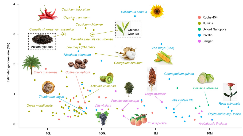
“Tea plants contain rich and unique characteristic secondary metabolites, such as catechins, theanine, and caffeine, which are essential to the formation of tea quality. It is not only the three major types of secondary metabolites but also the volatile terpenoids, saponins, polysaccharides, and other phenolic conjugates that contribute to the beneficial health effects and the enjoyable flavors of various teas.

The contents of these secondary metabolites vary greatly among different varieties and Camellia species. They also differ significantly in several morphological traits (e.g., leaf size) and stress resistance characteristics (e.g., cold tolerance), showing a divergent genetic makeup. The genome sequence of a single individual of a tea plant variety cannot represent the entire gene pool.

Modern transgenic breeding technology has provided us a new solution for the molecular design of breeding strategies. Although great progress has been made in the last two decades, the genomics and molecular biology of tea plants are still not fully understood. Compared to other crops such as rice, there is a long way to go.”

https://www.nature.com/articles/s41438-019-0225-4 “Tea plant genomics: achievements, challenges and perspectives”

Trained immunity responses to bacterial infections

This 2019 Swiss rodent study investigated immune responses to five types of bacterial infections:

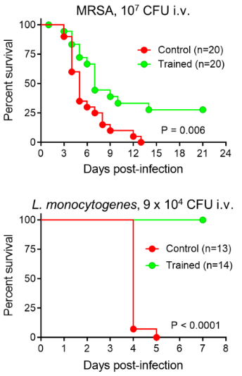
“The innate immune system recalls a challenge to adapt to a secondary challenge, a phenomenon called trained immunity. Trained immunity protected mice from a large panel of clinically relevant bacterial pathogens inoculated systematically and locally to induce peritonitis, enteritis and pneumonia.

Induction of trained immunity remodeled bone marrow and blood cellular compartments, providing efficient barriers against bacterial infections. Protection was remarkably broad when considering the pathogens and sites of infection tested.

We are running experiments to delineate the length of protection conferred by trained immunity. Trained immunity is most typically induced with β-glucan.

Mice were injected with methicillin-resistant Staphylococcus aureus (MRSA). Trained mice survived better than control mice (31% vs. 0% survival) and had 10-fold less bacteria in blood 2 days post-infection.

Mice were challenged with a lethal dose of Listeria monocytogenes. Most strikingly, all trained mice survived infection while all control mice died within 5 days. Bacteria were not detected in blood collected from trained mice 2 and 3 days post-infection.”

https://academic.oup.com/jid/advance-article/doi/10.1093/infdis/jiz692/5691195 “Trained immunity confers broad-spectrum protection against bacterial infections”


One of the coauthors also published:

Epigenetic inheritance and microRNAs

This 2019 Canadian rodent study found:

“Folic acid (FA) supplementation mitigates sperm miRNA profiles transgenerationally following in utero paternal exposure to POPs [persistent organic pollutants]. Across the F1 – F4 generations, sperm miRNA profiles were less perturbed with POPs + FA compared to sperm from descendants of dams treated with POPs alone..and only in F1 sperm.

The POPs mixture represents the pollutant composition found in Ringed seal blubber of Northern Quebec which is a traditional food of Inuit people in that region.

F0 founder dams were gavaged with the POPs mixture corresponding to 500 µg PCBs/kg body weight or corn oil (CTRL) thrice weekly and were fed the AIN-93G diet containing either 2 mg/kg (1X) or 6 mg/kg (3X) of FA ad libitum. Treatments were only administered to F0 founder dams for 9 weeks in total; 5 weeks before mating to untreated males at postnatal day 90 and until parturition. Subsequent lineages, F1 through F4, were neither exposed to POPs nor 3X FA – instead they received 1X FA diet ad libitum.”


Folic acid’s mechanisms weren’t clear:

“The protective role of FA supplementation in the F1 sperm may be partly explained by its antioxidant activity if the miRNA changes are caused by oxidative stress induced by POPs exposure. If, however, the miRNA changes in POPs exposed sperm are due to an altered methylation capacity or dysregulated nucleotide synthesis or mutations, then the increased availability of methyl groups provided by FA supplementation may mitigate the POPs effect by supporting DNA repair through nucleotide synthesis. Additional studies of the interaction between POPs and FA are required.”

Epigenetic inheritance mechanisms were also unclear:

“It remains puzzling how environmentally perturbed paternal miRNAs can persist across multiple generations. To become heritable, parts of the sperm chromatin must escape reprogramming, leading to the possibility that sperm miRNA profiles are subsequently modified by environmental factors. There are clear examples of sperm DNA methylation that escape reprogramming and histones can be involved.”

The study may have produced more clarity had its design investigated DNA methylation as Epigenetic transgenerational inheritance extends to the great-great-grand offspring did. That study also had an intercross breeding scheme with the populations for the F1 – F3 generations before an outcross for the F4 generation because:

“An intercross within the exposure lineage population (with no sibling or cousin breeding to avoid inbreeding artifacts) provides the optimal phenotypes (i.e. pathology) and germline epigenetic alterations.”

Which breeding scheme do you think would more fairly represent the humans of this study? I’d guess that intercross would – if all Inuits eat Ringed seal blubber and have children with other Inuits.

https://academic.oup.com/eep/article/5/4/dvz024/5677505 “Folic acid supplementation reduces multigenerational sperm miRNA perturbation induced by in utero environmental contaminant exposure”

Prenatal stress heightened adult chronic pain

This 2019 McGill rodent study found:

Prenatal stress exacerbates pain after injury. Analysis of mRNA expression of genes related to epigenetic regulation and stress responses in the frontal cortex and hippocampus, brain structures implicated in chronic pain, showed distinct sex and region-specific patterns of dysregulation.

In general, mRNA expression was most frequently altered in the male hippocampus and effects of prenatal stress were more prevalent than effects of nerve injury. Recent studies investigating chronic pain-related pathology in the hippocampus in humans and in rodent models demonstrate functional abnormalities in the hippocampus, changes in associated behavior, and decreases in adult hippocampal neurogenesis.

The change in expression of epigenetic- and stress-related genes is not a consequence of nerve injury but rather precedes nerve injury, consistent with the hypothesis that it might play a causal role in modulating the phenotypic response to nerve injury. These findings demonstrate the impact of prenatal stress on behavioral sensitivity to a painful injury.

Decreased frontal mRNA expression of BDNF and BDNF IV in male offspring following neuropathic pain or prenatal stress respectively. Relative mRNA expression of other stress-related genes (GR17, FKBP5) and epigenetic-related genes (DNMTs, TETs, HDACs, MBDs, MeCP2) in male offspring.

A drastic decrease in expression of HDAC1 was observed in all groups compared to sham-control animals. CCI: chronic constriction injury.”


The study’s design was similar to the PRS (prenatal restraint stress) model, except that the PRS procedure covered gestational days 11 to 21 (birth):

“Prenatal stress was induced on Embryonic days 13 to 17 by restraining the pregnant dams in transparent cylinder with 5 mm water, under bright light exposure, 3 times per day for 45 min.”

None of the French, Italian, and Swiss PRS studies were cited.

The limitation section included:

  1. “Although our study shows significant changes in expression of epigenetic enzymes, it didn’t examine the impact of these changes on genes that are epigenetically regulated by this machinery or their involvement in intensifying pain responses.
  2. The current study is limited by the focus on changes in gene expression which do not necessarily correlate with changes in protein expression.
  3. Another limitation of this study is the inability to distinguish the direct effects of stress in utero vs. changes in the dam’s maternal behavior due to stress during pregnancy; cross-fostering studies are needed to address this issue.
  4. Functional experiments that involve up and down regulation of epigenetic enzymes in specific brain regions are required to establish a causal role for these processes in chronic pain.”

What do you think about possible human applicability of this study’s “effects of prenatal stress were more prevalent than effects of nerve injury” finding?

Are there any professional therapeutic frameworks that instruct trainees to recognize that if a person’s mother was stressed while pregnant, their prenatal experiences could cause more prevalent biological and behavioral effects than a recent injury?

https://www.sciencedirect.com/science/article/pii/S0166432819315219 “Prenatal maternal stress is associated with increased sensitivity to neuropathic pain and sex-specific changes in supraspinal mRNA expression of epigenetic- and stress-related genes in adulthood” (not freely available)

A transgenerational view of the rise in obesity

This 2019 Washington State University rodent study found epigenetically inherited transgenerational effects in great-grand offspring due to their great-grandmothers’ toxicant exposures during pregnancy:

“Previous studies found an increased susceptibility to obesity in F3 generation rats ancestrally exposed to the pesticide DDT, and an increase in a lean phenotype in the F3 generation rats ancestrally exposed to the herbicide atrazine. The present study investigated whether there were common DMR [differential DNA methylated region] and associated genes between the control, DDT, and atrazine lineage male and female adipocytes in order to identify potential novel gene pathways modulated by DNA methylation.

Comparison of epigenetic alterations indicated that there were substantial overlaps between the different treatment lineage groups for both the lean and obese phenotypes. Novel correlated genes and gene pathways associated with DNA methylation were identified, and may aid in the discovery of potential therapeutic targets for metabolic diseases such as obesity.

Given that the first widespread [DDT] exposures to gestating human females started in the 1950s, the majority of the subsequent F3 generation are adults today. Ancestral exposures to environmental toxicants like DDT may have had a role in the dramatic rise in obesity rates worldwide.”


This same research group noted in Transgenerational diseases caused by great-grandmother DDT exposure:

“DDT was banned in the USA in 1973, but it is still recommended by the World Health Organization for indoor residual spray. India is by far the largest consumer of DDT worldwide.

India has experienced a 5-fold increase of type II diabetes over the last three decades with a predisposition to obesity already present at birth in much of the population. Although a large number of factors may contribute to this increased incidence of obesity, the potential contribution of ancestral toxicant exposures in the induction of obesity susceptibility requires further investigation.”

https://www.tandfonline.com/doi/full/10.1080/21623945.2019.1693747 “Adipocyte epigenetic alterations and potential therapeutic targets in transgenerationally inherited lean and obese phenotypes following ancestral exposures”

Maternal obesity causes heart disease in every offspring generation

This 2019 St. Louis rodent study found:

“We hypothesized that maternal obesity induces cardiac mitochondrial dysfunction in the offspring via transgenerational inheritance of abnormal oocyte mitochondria. All F1 to F3 descendants bred via the female in each generation were nonobese and demonstrated cardiac mitochondrial abnormalities.

Contrary to our hypothesis, male F1 also transmitted these effects to their offspring, ruling out maternal mitochondria as the primary mode of transmission. We conclude that transmission of obesity-induced effects in the oocyte nucleus rather than abnormal mitochondria underlie transgenerational inheritance of cardiac mitochondrial defects in descendants of obese females.”


For some reason, the researchers didn’t cite any of Dr. Michael Skinner’s research on epigenetic transgenerational inheritance. Their time, efforts, and resources would have been more productive had they used Dr. Skinner’s studies – such as the 2018 Epigenetic transgenerational inheritance of ovarian disease – as guides.

A podcast with the researchers is available here.

https://www.physiology.org/doi/abs/10.1152/ajpheart.00013.2019 “Maternal High-Fat, High-Sucrose Diet Induces Transgenerational Cardiac Mitochondrial Dysfunction Independent of Maternal Mitochondrial Inheritance” (not freely available)

Emotional responses and BDNF methylation

This 2019 German human study found:

“A critical role of BDNF [brain-derived neurotrophic factor] methylation in human amygdala response to negative emotional stimuli, whereby:

  • High BDNF methylation rates were for the first time shown to be associated with a high reactivity in the amygdala; and
  • High BDNF methylation and high amygdala reactivity were associated with low novelty seeking.

There was no interaction or main effect of the Val66Met polymorphism on amygdala reactivity.

Our data adds evidence to the hypothesis that epigenetic modifications of BDNF can result in an endophenotype associated with anxiety and mood disorders. However, since correlations do not prove causality:

  • A direct link between human BDNF mRNA/protein levels, methylation, amygdala reactivity and psychiatric disorders is still missing, demanding further research.
  • Determining the underlying directions of the relations between BDNF methylation, amygdala reactivity, and NS [novelty seeking] cannot be accomplished based on our data and must await further research.

The fact that our results mainly involve the right amygdala is in line with previous studies. Recent reviews suggest a general right hemisphere dominance for all kinds of emotions, and, more specifically, a critical role of the right amygdala in the early assessment of emotional stimuli.

The experimental fMRI paradigm utilized a face‐processing task (faces with anger or fear expressions), alternating with a sensorimotor control task. Harm avoidance, novelty seeking, and reward dependence were measured using the Tridimensional Personality Questionnaire.”

https://onlinelibrary.wiley.com/doi/full/10.1002/hbm.24825 “The role of BDNF methylation and Val 66 Met in amygdala reactivity during emotion processing”