Reversing epigenetic T cell exhaustion

This 2019 worldwide discussion among 18 experts concerned T cell exhaustion:

“‘T cell exhaustion’ is a broad term that has been used to describe the response of T cells to chronic antigen stimulation, first in the setting of chronic viral infection but more recently in response to tumours.

Key questions remain about the potential to reverse the epigenetic programme of exhaustion and how this might affect the persistence of T cell populations.”


There were nearly a dozen viewpoints on “What do we mean by T cell exhaustion and/or dysfunction and how would you define this state?” 🙂

Answers to the question “What are the key controversies and outstanding research questions?” included:

  • “What are the cellular signalling and transcriptional pathways that drive the conversion to an exhausted T cell phenotype, and how can the chromatin and transcriptional changes of exhaustion be reversed in individual exhausted cells?
  • Whether and how we can manipulate signalling pathways to both activate and maintain T cell responses remain open questions, as does the question of whether pharmacological manipulations can reverse the epigenetic changes associated with exhaustion versus expand less-exhausted populations.
  • We need to define better the effects of the microenvironment on the induction of T cell exhaustion, the developmental trajectories of exhaustion and the point at which and extent to which exhaustion can be reversed. Understanding the consequences of unleashing T cells from exhaustion will also be crucial to designing the most effective therapeutic interventions.
  • When and how exhausted T cell populations are formed. The original view that they are terminally differentiated descendants of formerly ‘normal’ effector T cells has been challenged.
  • Whether the predysfunctional T cells themselves, or their more differentiated (and phenotypically dysfunctional) progeny, form the ultimate effector pool for control of human tumours.
  • How do the functions and states (subpopulations) of exhausted T cells change over time? Can the epigenetic state of exhaustion be reversed to form true effector or memory T cells, and is this required for improved cancer immunotherapy?
  • There is no definitive marker for exhausted T cells, although TOX may prove to be useful. Transcriptional profiles are informative, but epigenetic changes are more specific and robust. A major clinical question is whether exhausted T cells can be, or indeed need to be, reprogrammed to achieve therapeutic benefit.”

https://www.nature.com/articles/s41577-019-0221-9 “Defining ‘T cell exhaustion'” (not freely available)

Get outside today

This 2019 Finnish review focused on vitamin D’s immune system effects:

“The epigenome of human monocytes is at multiple levels sensitive to vitamin D. These data served as the basis for the chromatin model of vitamin D signaling, which mechanistically explains the activation of a few hundred primary vitamin D target genes.

Vitamin D and its receptor are able to antagonize the pro-inflammatory actions of the transcription factors nuclear factor activated T cells (NF-AT) and nuclear factor κ-light-chain-enhancer of activated B cells (NF-κB) in T cells. In this way, vitamin D reduces autoimmunity, such as the onset and progression of multiple sclerosis, as well as chronic inflammation.

Population-wide recommendations do not take inter-individual variations into account, such as a different molecular response to vitamin D, which are expressed by the vitamin D response index. Instead of population-based recommendations for vitamin D3 supplementation there should be personalized recommendations in order to reach a vitamin D status that is optimized for an individual’s health protection.

Trained immunity implies that immune cells memorize challenges, to which they are exposed in their rather short lifespan, in form of changes of their epigenome leading to subtype specification. The stabilization of the epigenomes of the subtypes of monocytes, macrophages and dendritic cells by vitamin D can prevent or delay the onset of common age-related diseases.”

https://www.ncbi.nlm.nih.gov/pmc/articles/PMC6753645/ “Vitamin D Signaling in the Context of Innate Immunity: Focus on Human Monocytes”


One of the five elements of the clinical trial Reversal of aging and immunosenescent trends was daily 3,000 IU vitamin D3 supplementation for nine months. That study’s monocyte findings included:

“Analysis of CyTOF‐defined immune cell populations revealed the most robust changes to be decreases in total and CD38‐positive monocytes and resulting increases in the lymphocyte‐to‐monocyte ratio (LMR). The changes in mean monocyte populations persisted 6 months after discontinuation of treatment, and the increase in LMR remained highly significant at 18 months as well.”

Too cheap for clinical trials

Let’s compare and contrast a 2019 meta-analysis and a 2017 review of using acetyl-L-carnitine to treat diabetic neuropathy.

A 2019 Brazilian meta-analysis Acetyl‐L‐carnitine for the treatment of diabetic peripheral neuropathy of four previous trials stated:

  • “The risk of bias was high in both trials of different ALC doses and low in the other two trials.
  • No included trial measured the proportion of participants with at least moderate (30%) or substantial (50%) pain relief.
  • At doses greater than 1500 mg/day, ALC reduced pain more than placebo. This subgroup analysis should be viewed with caution as the evidence was even less certain than the overall analysis, which was already of very low certainty.
  • The placebo-controlled studies did not measure functional impairment and disability scores.
  • No study used validated symptom scales.
  • Two studies were funded by the manufacturer of ALC and the other two studies had at least one co-author who was a consultant for an ALC manufacturer.

Authors’ conclusions:

  • We are very uncertain whether ALC causes a reduction in pain after 6 to 12 months treatment in people with DPN, when compared with placebo, as the evidence is sparse and of low certainty.
  • Data on functional and sensory impairment and symptoms are lacking, or of very low certainty.
  • The evidence on adverse events is too uncertain to make any judgements on safety.”

A 2017 Italian review Effects of acetyl-L-carnitine in diabetic neuropathy and other geriatric disorders stated:

“A long history of diabetes mellitus and increasing age are associated with the onset of diabetic neuropathy, a painful and highly disabling complication with a prevalence peaking at 50% among elderly diabetic patients. The management of diabetic neuropathy is extremely difficult: in addition to the standard analgesics used for pain control, common treatments include opioids, anticonvulsants, antidepressants, and local anesthetics, alone or in combination. Such therapies still show a variable, often limited efficacy, however.

Many patients do not spontaneously report their symptoms to physicians, but, if asked, they often describe having experienced a persistent and non-abating pain for many years. The prevalence of painful symptoms is just as high in patients with mild neuropathy as in those with more advanced DPN.

Through the donation of acetyl groups, ALC exerts a positive action on mitochondrial energy metabolism. ALC has cytoprotective, antioxidant, and antiapoptotic effects in the nervous system.

ALC has also been proposed for the treatment of other neurological and psychiatric diseases, such as mood disorders and depression, dementia, Alzheimer’s disease, and Parkinson’s disease, given that synaptic energy states and mitochondrial dysfunctions are core factors in their pathogenesis. Compared to other treatments, ALC is safe and extremely well tolerated.

In nerve injury, the mGlu2 receptor overexpressed by ALC binds the glutamate, reducing its concentration in the synapses with an analgesic effect. ALC may improve nerve regeneration and damage repair after primary nerve trauma.”


Where will the money come from to realize what the 2017 review promised, as well as provide what the 2019 meta-analysis required?

Do we prefer the current “limited efficacy” treatments of “opioids, anticonvulsants, antidepressants, and local anesthetics?”

Who will initiate clinical trials of a multiple of the normal dietary supplement dose (500 mg at $.25 a day, retail)? How profitable is a product whose hypothetical effective dosage for diabetic neuropathy (3000 mg) sells for only $1.50 a day?

Online dating cuts out the middlemen

This information is from a 2019 prepublication Stanford study:

“We present new data from a nationally representative 2017 survey showing that meeting online has continued to grow for heterosexual couples, and meeting through friends has continued its sharp decline. As a result of the continued rise of meeting online and the decline of meeting through friends, online has become the most popular way heterosexual couples in the U.S.

Meeting through friends and family provided guarantees that any potential partner had been personally vetted and vouched for by trusted alters. We would expect any rise in Internet dating to reinforce rather than to displace the traditional roles of friends and family as introducers and intermediaries. [Hypothesis 2]

Results reflect support of Hypothesis 1, as the percentage of heterosexual couples meeting online has surged in the post‐2009 smart phone era. Because the results show that meeting online has displaced meeting through friends and meeting through family, we find evidence to reject Hypothesis 2, which led us to expect that online dating would reinforce existing face‐to‐face social networks.

Figure 1’s apparent post‐2010 rise in meeting through bars and restaurants for heterosexual couples is due entirely to couples who met online and subsequently had a first in‐person meeting at a bar or restaurant or other establishment where people gather and socialize. If we exclude the couples who first met online from the bar/restaurant category, the bar/restaurant category was significantly declining after 1995 as a venue for heterosexual couples to meet.”


Are there examples where it wouldn’t potentially improve a person’s life to choose their information sources? Friends, family, and other social groups – and religious, educational, and other institutions – have had their middlemen / guarantor time, and have been found lacking.

Make your own choices for your one precious life. Similar themes are explored in:

https://web.stanford.edu/~mrosenfe/Rosenfeld_et_al_Disintermediating_Friends.pdf “Disintermediating your friends”

Perinatal stress and sex differences in circadian activity

This 2019 French/Italian rodent study used the PRS model to investigate its effects on circadian activity:

“The aim of this study was to explore the influence of PRS on the circadian oscillations of gene expression in the SCN [suprachiasmatic nucleus of the hypothalamus] and on circadian locomotor behavior, in a sex-dependent manner.

Research on transcriptional rhythms has shown that more than half of all genes in the human and rodent genome follow a circadian pattern. We focused on genes belonging to four functional classes, namely the circadian clock, HPA axis stress response regulation, signaling and glucose metabolism in male and female adult PRS rats.

Our findings provide evidence for a specific profile of dysmasculinization induced by PRS at the behavioral and molecular level, thus advocating the necessity to include sex as a biological variable to study the set-up of circadian system in animal models.”

“There was a clear-cut effect of sex on the effect of PRS on the levels of activity:

  • During the period of lower activity (light phase), both CONT and PRS females were more active than males. During the light phase, PRS increased activity in males, which reached levels of CONT females.
  • More interestingly, during the period of activity (dark phase), male PRS rats were more active than male CONT rats. In contrast, female PRS rats were less active than CONT females.
  • During the dark phase, CONT female rats were less active than CONT male rats.

The study presented evidence for sex differences in circadian activity of first generation offspring that was caused by stress experienced by the pregnant mother:

“Exposure to gestational stress and altered maternal behavior programs a life-long disruption in the reactive adaptation such as:

  •  A hyperactive response to stress and
  • A defective feedback of the hypothalamus-pituitary-adrenal (HPA) axis together with
  • Long-lasting modifications in stress/anti-stress gene expression balance in the hippocampus.”

It would advance science if these researchers carried out experiments to two more generations to investigate possible transgenerational epigenetic inheritance of effects caused by PRS. What intergenerational and transgenerational effects would they possibly find by taking a few more months and extending research efforts to F2 and F3 generations? Wouldn’t these findings likely help humans?


One aspect of the study was troubling. One of the marginally-involved coauthors was funded by the person described in How one person’s paradigms regarding stress and epigenetics impedes relevant research. Although no part of the current study was sponsored by that person, there were three gratuitous citations of their work.

All three citations were reviews. Unlike study researchers, reviewers aren’t bound to demonstrate evidence from tested hypotheses. Reviewers are free to:

  • Express their beliefs as facts;
  • Over/under emphasize study limitations; and
  • Disregard and misrepresent evidence as they see fit.

Fair or not, comparisons of reviews with Cochrane meta-analyses of the same subjects consistently show the extent of reviewers’ biases. Reviewers also aren’t obligated to make post-publication corrections for their errors and distortions.

As such, reviews can’t be cited for reliable evidence. Higher-quality studies that were more relevant and recent than a 1993 review could have elucidated points.

Sucking up to the boss and endorsing their paradigm was predictable. Since that coauthor couldn’t constrain themself to funder citations only in funder studies, it was the other coauthors’ responsibilities to edit out unnecessary citations.

https://www.frontiersin.org/articles/10.3389/fnmol.2019.00089/full “Perinatal Stress Programs Sex Differences in the Behavioral and Molecular Chronobiological Profile of Rats Maintained Under a 12-h Light-Dark Cycle”

Disease and advanced glycation end products (AGEs)

This 2015 French/US review focused on chronic kidney disease, appropriate for its publication in the Journal of the American Society of Nephrology:

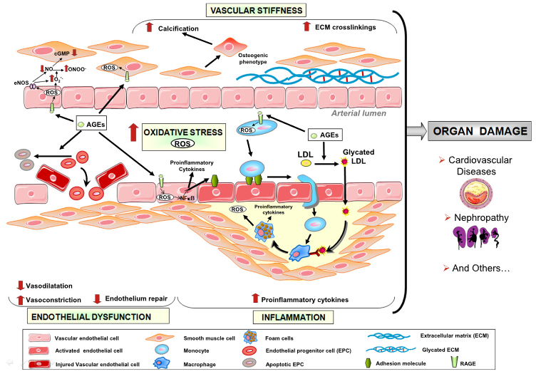
“Advanced glycation end products (AGEs) are formed not only in the presence of hyperglycemia, but also in diseases associated with high levels of oxidative stress, such as CKD. Humans are exposed to exogenous sources of AGE (diet and cigarette smoke) and endogenous sources of AGE when the organism is exposed to high levels of glucose, such as in diabetes.

Accumulation of AGEs in patients with CKD has been shown to result from inflammation, oxidative stress, and diet. AGEs are proinflammatory and pro-oxidative compounds that play a role in the high prevalence of endothelial dysfunction and subsequent cardiovascular disease in patients with CKD.

In view of the many harmful effects of AGEs on cell function, it is essential to develop strategies designed to counteract their effects. AGEs are generated during the thermal processing and storage of foods. Dietary restriction is an effective, feasible, and economic method to reduce levels of toxic AGEs and possibly, the associated cardiovascular mortality.”

https://jasn.asnjournals.org/content/27/2/354 “Uremic Toxicity of Advanced Glycation End Products in CKD”


I came across the AGE subject in the usual Internet way. 🙂 While reading comments on Josh Mitteldorf’s blog post Money in Aging Research, Part I, Dr. Alan Green mentioned Dr. Helen Vlassara’s work. A DuckDuckGo search led to her 44,256 citations, which increase every day.

Another read on the subject is her 2016 book Dr. Vlassara’s AGE-Less Diet: How a Chemical in the Foods We Eat Promotes Disease, Obesity, and Aging and the Steps We Can Take to Stop It. A practical guide is her 2017 book The AGE Food Guide: A Quick Reference to Foods and the AGEs They Contain.

A therapy to reverse cognitive decline

This 2018 human study presented the results of 100 patients’ personalized therapies for cognitive decline:

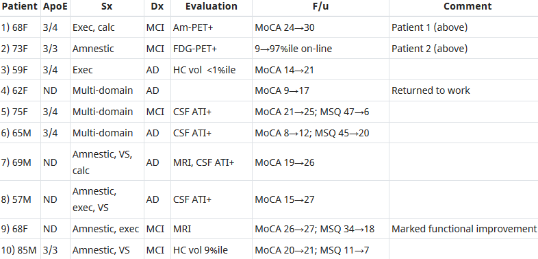
“The first examples of reversal of cognitive decline in Alzheimer’s disease and the pre-Alzheimer’s disease conditions MCI (Mild Cognitive Impairment) and SCI (Subjective Cognitive Impairment) have recently been published..showing sustained subjective and objective improvement in cognition, using a comprehensive, precision medicine approach that involves determining the potential contributors to the cognitive decline (e.g., activation of the innate immune system by pathogens or intestinal permeability, reduction in trophic or hormonal support, specific toxin exposure, or other contributors), using a computer-based algorithm to determine subtype and then addressing each contributor using a personalized, targeted, multi-factorial approach dubbed ReCODE for reversal of cognitive decline.

An obvious criticism of the initial studies is the small number of patients reported. Therefore, we report here 100 patients, treated by several different physicians, with documented improvement in cognition, in some cases with documentation of improvement in electrophysiology or imaging, as well.”

https://www.omicsonline.org/open-access/reversal-of-cognitive-decline-100-patients-2161-0460-1000450-105387.html “Reversal of Cognitive Decline: 100 Patients”


The lead author commented on Josh Mitteldorf’s informative post A cure for Alzheimer’s? Yes, a cure for Alzheimer’s!:

  1. “We have a paper in press, due to appear 10.22.18 (open access, JADP, I’ll send a copy as soon as available), showing 100 patients with documented improvement – some with MRI volumetrics improved, others with quantitative EEG improvements, others with evoked response improvements, and all with quantitative cognitive assessment improvement. Some are very striking – 12 point improvements in MoCA [Montreal Cognitive Assessment], for example – others less so, but all also have subjective improvement. Hopefully this will address some of the criticisms that we haven’t documented improvement in enough people.
  2. We were just turned down again for a randomized, controlled clinical trial, so on the one hand, we are told repeatedly that no one will believe that this approach works until we publish a randomized, controlled study, and on the other hand, we’ve been turned down (first in 2011/12, and now in 2018), with the complaint that we are trying to address more than one variable in the trial (as if AD is a single-variable disease!). Something of a catch-22. We are now resubmitting (unfortunately, the IRBs are not populated by functional medicine physicians, so they are used to seeing old-fashioned drug studies), and we’ll see what happens.
  3. I’ve been extending the studies to other neurodegenerative diseases, and it has been impressive how much of a programmatic response there seems to be in these ‘diseases.’
  4. I agree with you that there are many features in common with aging itself.
  5. You made a good point that APP [amyloid precursor protein] is a dependence receptor, and in fact it functions as an integrating dependence receptor, responding to numerous inputs (Kurakin and Bredesen, 2015).
  6. In the book and the publications, we don’t claim it is a “cure” since we don’t have pathological evidence that the disease process is gone. What we claim is ‘reversal of cognitive decline’ since that is what we document.
  7. As I mentioned in the book, AD is turning out to be a protective response to multiple insults, and this fits well with the finding that Abeta has an antimicrobial effect (Moir and Tanzi’s work). It is a network-downsizing, protective response, which is quite effective – some people live with the ongoing degenerative process for decades.
  8. We have seen several cases now in which a clinical trial of an anti-amyloid antibody made the person much worse in a time-dependent manner (each time there was an injection, the person would get much worse for 5-10 days, then begin to improve back toward where he/she was, but over time, marked decline occurred), and this makes sense for the idea that the amyloid is actually protecting against pathogens or toxins or some other insult.
  9. It is important to note that we’ve never claimed that all people get better – this is not what we’ve seen. People very late in the process, or who don’t follow the protocol, or who don’t address the various insults, do not improve. It is also turning out to be practitioner dependent – some are getting the vast majority of people to improve, others very few, so this is more like surgery than old-fashioned prescriptive medicine – you have to do a somewhat complicated therapeutic algorithm and get it right for best results.
  10. I’m very interested in what is needed to take the next step in people who have shown improvement but who started late in the course. For example, we have people now who have increased MoCA from 0 to 9 (or 0 to 3, etc.), with marked subjective improvement but plateauing at less than normal. These people had extensive synaptic and cellular loss prior to the program. So what do we need to raise the plateau? Stem cells? Intranasal trophic support? Something else?
  11. I haven’t yet seen a mono-etiologic theory of AD or a mono-therapeutic approach that has repeatedly positive results, so although I understand that there are many theories and treatments, there doesn’t seem to be one etiology to the disease, nor does there seem to be one simple treatment that works for most. It is much more like a network failure.”

At a specific level:

  • “There doesn’t seem to be one etiology to the disease,
  • Nor does there seem to be one simple treatment that works for most.
  • We don’t have pathological evidence that the disease process is gone.”

For general concepts, however:

  • “AD is turning out to be a protective response to multiple insults.
  • It is a network-downsizing, protective response, which is quite effective.
  • The amyloid is actually protecting against pathogens or toxins or some other insult.”

For a framework of an AD cure to be valid, each source of each insult that evoked each “protective response” should be traced.

Longitudinal studies would be preferred inside this framework. These study designs would investigate evidence of each insult’s potential modifying effect on each “protective response” that could affect the cumulative disease trajectory of each individual.

In many cases, existing study designs would be adequate if they extended their periods to the end of the subjects’ natural lifetimes. One AD-relevant example would be extending the prenatally-restraint-stressed model used in:

The framework would also encourage extending studies to at least three generations to investigate evidence for transgenerational effects, as were found in:

Fitting data

Let’s start out the new year with a repost of a cautionary reminder:

“Both “predict and “explain” imply that investigators have uncovered a reliable structure to phenomena, the latter involving hypotheses describing unseen mechanisms, leading to a new ability to control events and produce formerly unpredicted/unpredictable outcomes. This is clearly not a fair description of post hoc correlation-fishing.

The current publication system almost forces authors to make causal statements using filler verbs (e.g. to drive, alter, promote) as a form of storytelling (Gomez-Marin, 2017); without such a statement they are often accused of just collecting meaningless facts.”

https://mythsofvisionscience.wordpress.com/2018/12/30/neuroscience-newspeak-or-how-to-publish-meaningless-facts/ “Neuroscience Newspeak, Or How to Publish Meaningless Facts”

Eat your oats!

Here’s some motivation to replenish your oats supply.

From a 2013 Canadian human review:

“Review of human studies investigating the post-prandial blood-glucose lowering ability of oat and barley food products” https://www.nature.com/articles/ejcn201325

“Change in glycaemic response (expressed as incremental area under the post-prandial blood-glucose curve) was greater for intact grains than for processed foods. For processed foods, glycaemic response was more strongly related to the β-glucan dose alone than to the ratio of β-glucan to the available carbohydrate.”

The review found that people don’t have to eat a lot of carbohydrates to get the glycemic-response benefits of β-glucan. Also, eating ~3 grams of β-glucan in whole oats and barley will deliver the same glycemic-response benefits as eating ~4 grams of β-glucan in processed oats and barley.

However, the glycemic index used in the review is a very flawed measure. What’s the point of indexing healthy choices like whole grains to unhealthy choices that healthy people aren’t going to make anyway?


The reviewer somewhat redeemed herself by participating in a 2018 review:

“Processing of oat: the impact on oat’s cholesterol lowering effect” https://www.ncbi.nlm.nih.gov/pmc/articles/PMC5885279/

“For a similar dose of β-glucan:

  1. Liquid oat-based foods seem to give more consistent, but moderate reductions in cholesterol than semi-solid or solid foods where the results are more variable;
  2. The quantity of β-glucan and the molecular weight at expected consumption levels (∼3 g day) play a role in cholesterol reduction; and
  3. Unrefined β-glucan-rich oat-based foods (where some of the plant tissue remains intact) often appear more efficient at lowering cholesterol than purified β-glucan added as an ingredient.”

The review’s sections 3. Degree of processing and functionality and 4. Synergistic action of oat constituents were informative:

“Both in vitro and in vivo studies clearly demonstrated the beneficial effect of oat on cholesterolemia, which is unlikely to be due exclusively to β-glucan, but rather to a combined and synergetic action of several oat compounds acting together to reduce blood cholesterol levels.”


Another use of β-glucan is to improve immune response. Here’s a 2016 Netherlands study where the researchers used β-glucan to get a dozen people well after making them sick with lipopolysaccharide as is often done in animal studies:

β-Glucan Reverses the Epigenetic State of LPS-Induced Immunological Tolerance” https://www.ncbi.nlm.nih.gov/pmc/articles/PMC5927328/

“The innate immune ‘training stimulus’ β-glucan can reverse macrophage tolerance ex vivo.”

I’ve curated other research on β-glucan’s immune-response benefits in:

Adverse epigenetic effects of prenatal and perinatal anesthesia

This 2018 Chinese animal review subject was prenatal and perinatal anesthesia’s adverse epigenetic effects on a fetus/neonate:

“Accumulating evidence from rodent and primate studies has demonstrated that in utero or neonatal exposure to commonly used inhaled and intravenous general anesthetics is associated with neural degeneration and subsequent neurocognitive impairments, manifested in learning and memory disabilities.

So far, conflicting data exist about the effect of anesthetic agents on neurodevelopment in humans and no definite conclusion has been given yet.”

The inhibitors in the above graphic counter anesthesia’s effects on the fetus/neonate, summarized as:

“Epigenetic targeting of DNA methyltransferases and/or histone deacetylases may have some therapeutic value.”


Do physicians consider possible epigenetic alterations of a newborn’s chromatin structure and gene expression when they administer anesthesia to mothers during childbirth?

https://www.ncbi.nlm.nih.gov/pmc/articles/PMC6079265/ “Epigenetic Alterations in Anesthesia-Induced Neurotoxicity in the Developing Brain”

Epigenetic clock statistics and methods

This 2018 Chinese study was a series of statistical and methodological counter-arguments to a previous epigenetic clock study finding that:

“Only [CpG] sites mapping to the ELOVL2 promoter constitute cell and tissue-type independent aDMPs [age-associated differentially methylated positions].”

The study used external data sets and the newer epigenetic clock’s fibroblast data in its analyses to find:

“While we agree that specific sites mapping to ELOVL2 are special aDMPs in the sense that their effect sizes are particularly large across a number of different tissue-types, our analysis suggests that most aDMPs are valid across multiple different tissue types, suggesting that shared aDMPs are common.”

The details of each of the study’s counter-arguments were compelling. For example:

“We analyzed Illumina 850k data from an EWAS profiling blood, buccal and cervical samples from a common set of 263 women. Because blood is a complex mixture of many immune-cell subtypes, and buccal and cervical samples are highly contaminated by immune cells, we identified aDMPs in each tissue after adjustment for batch effects and cell-type heterogeneity.

Using either an FDR [false discovery rate] < 0.05 or Bonferroni adjusted P-value < 0.05 thresholds, the overlap of aDMPs between the 3 tissues was highly significant, mimicking the result obtained on blood cell subtypes. We observed a total of 2200 aDMPs in common between blood, buccal and cervix, an overlap which cannot be explained by random chance.”

The study’s Discussion section provided qualifications and limitations such as:

“It is important to point out that even if age-associated DNAm changes are widespread across the genome, downstream functional effects may be rare. While specific aDMPs may be shared between tissue-types, it is only in specific tissues or cell-types that any associated functional deregulation may be of biological and clinical significance.

https://www.aging-us.com/article/101666/text “Cell and tissue type independent age-associated DNA methylation changes are not rare but common”


The November 2018 issue of Aging also contained other articles of interest:

https://www.aging-us.com/article/101626/text “Accelerated DNA methylation age and the use of antihypertensive medication among older adults”

“DNAmAge and AA [age acceleration] may not be able to capture the preventive effects of AHMs [antihypertensive medications] that reduce cardiovascular risks and mortality.”

https://www.aging-us.com/article/101633/text “Azithromycin and Roxithromycin define a new family of senolytic drugs that target senescent human fibroblasts”

“Azithromycin preferentially targets senescent cells, removing approximately 97% of them with great efficiency. This represents a near 25-fold reduction in senescent cells.”

https://www.aging-us.com/article/101647/text “Disease or not, aging is easily treatable”

“Aging consists of progression from (pre)-pre-diseases (early aging) to diseases (late aging associated with functional decline). Aging is NOT a risk factor for these diseases, as aging consists of these diseases: aging and diseases are inseparable.”

Reductionism vs. reductionism

This 2004 essay by an evolutionary biologist reviewed his field’s direction in the current century:

“Science is impelled by two main factors, technological advance and a guiding vision (overview). A properly balanced relationship between the two is key to the successful development of a science.

Without the proper technological advances the road ahead is blocked. Without a guiding vision there is no road ahead; the science becomes an engineering discipline, concerned with temporal practical problems.

Empirical reductionism is in essence methodological; it is simply a mode of analysis, the dissection of a biological entity or system into its constituent parts in order better to understand it. Empirical reductionism makes no assumptions about the fundamental nature, an ultimate understanding, of living things.

Fundamentalist reductionism (the reductionism of 19th century classical physics), on the other hand, is in essence metaphysical. It is ipso facto a statement about the nature of the world: living systems (like all else) can be completely understood in terms of the properties of their constituent parts.

This is a view that flies in the face of what classically trained biologists tended to take for granted, the notion of emergent properties. Whereas emergence seems to be required to explain numerous biological phenomena, fundamentalist reductionism flatly denies its existence: in all cases the whole is no more than the sum of its parts.”

Regarding cellular evolution:

“Modern concepts of cellular evolution are effectively petrified versions of 19th century speculations. Try to imagine a biology released from the intellectual shackles of mechanism, reductionism, and determinism.

Evolution, as a complex dynamic process, will encounter critical points in its course, junctures that result in phase transitions (drastic changes in the character of the system as a whole). Human language is a development that has set Homo sapiens worlds apart from its otherwise very close primate relatives, adding new dimensions to the phase space within which human evolution occurs. Another good critical-point candidate is the advent of (eucaryotic) multicellularity.

Nowhere in thinking about a symbiotic origin of the eucaryotic cell has consideration been given to the fact that the process as envisioned would involve radical change in the designs of the cells involved. You can’t just tear cell designs apart and willy-nilly construct a new type of design from the parts.

The organization of the mitochondrial endosymbiont is radically changed during its evolution, but that change is a degeneration to a far simpler “cell-like” design. The mitochondrial design could never evolve back to the level of complexity that its free-living [bacterial] ancestor had.

A common thread that links language and multicellularity is communication (interaction at a distance). In each case a complex, sophisticated network of interactions forms the medium within which the new level of organization (entities) comes into existence.

Our experience with variation and selection in the modern context does not begin to prepare us for understanding what happened when cellular evolution was in its very early, rough-and-tumble phase(s) of spewing forth novelty. Cellular evolution began in a highly multiplex fashion, from many initial independent ancestral starting points, not just a single one.”

https://mmbr.asm.org/content/68/2/173 “A New Biology for a New Century”


I came across this review by it being referenced in this researcher’s blog post:

Chinese Longevity Herb
I often don’t agree with him, but I subscribe to his blog because it’s interesting.

The arrogance of a paradigm exceeding its evidence

This 2018 commentary from the American College of Emergency Physicians by 7 physicians discussed the harm that will result from imposing a mandatory paradigm of sepsis treatment. I’ll quote sections that mention evidence:

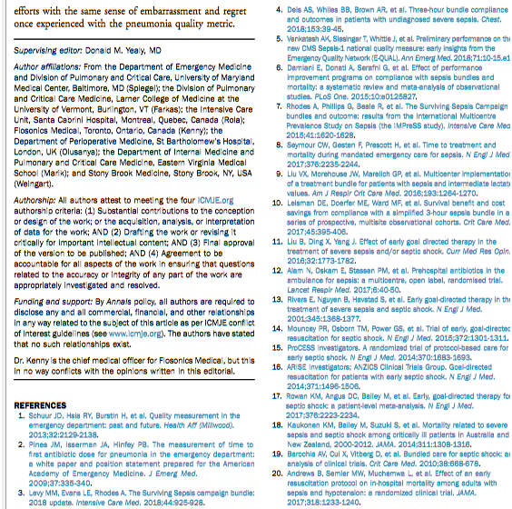
“These metrics [for pneumonia treatment] had little evidentiary basis but led to an institutional-fostered culture of overdiagnosis and overtreatment. Have we learned from this folly or does a new sepsis guideline promote similar time-based treatment strategies with little direct supporting evidence?

Like the pneumonia quality measure, this resource-heavy care flows from an overreaching interpretation of evidence. Despite that evidence consistently fails to find a benefit of a single treatment strategy, the Surviving Sepsis Campaign continues to promote recommendations that bypass the individual clinician’s judgment.

Although well intentioned, the current sepsis bundles and the potential penalties associated with noncompliance lay a heavy weight on ED [emergency department] care absent evidence that a net benefit will follow. The proposed Surviving Sepsis Campaign abbreviated bundle heightens the burden by further restricting the time allotted for the identification and treatment of patients with suspected sepsis, all without any evidence of benefit or knowledge of the logistic consequences or cost.”

The paradigm’s promoters didn’t learn the appropriate lessons in the above page regarding “the sense of embarrassment and regret once experienced with the pneumonia quality metric.”


What do you think are the root causes of the Surviving Sepsis Campaign’s agenda?

  • Did it start with lawyers? Lawsuits can force hospitals into actions for which the primary reason is to avoid “the potential penalties associated with noncompliance.”
  • Is it due to governments? Governments can force hospitals into actions “without any evidence of benefit or knowledge of the logistic consequences or cost” when the hospitals accept government reimbursement.
  • Did it start with other groups of unaccountable people who think they know better than everyone else about how others should act?

https://www.sciencedirect.com/science/article/pii/S0196064418306073 “The 2018 Surviving Sepsis Campaign’s Treatment Bundle: When Guidelines Outpace the Evidence Supporting Their Use” (not freely available)

Epigenetic transgenerational inheritance of ovarian disease

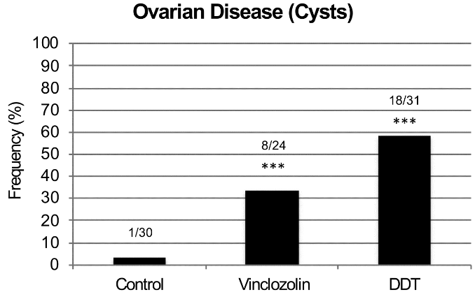
This 2018 Washington rodent study investigated ovarian disease in F3 great-granddaughters caused by their F0 great-grandmothers’ exposures to DDT or vinclozolin while pregnant:

“Two of the most prevalent ovarian diseases affecting women’s fertility and health are Primary Ovarian Insufficiency (POI) and Polycystic Ovarian Syndrome (PCOS). POI is characterized by a marked reduction in the primordial follicle pool of oocytes and the induction of menopause prior to age 40. POI currently affects approximately 1% of female population. While genetic causes can be ascribed to a minority of patients, around 90% of POI cases are considered idiopathic, with no apparent genetic link nor known cause.

PCOS is a multi-faceted disease that affects 6-18% of women. It is characterized by infrequent ovulation or anovulation, high androgen levels in the blood, and the presence of multiple persistent ovarian cysts.

For both PCOS and POI other underlying causes such as epigenetic transgenerational inheritance of disease susceptibility have seldom been considered. Epigenetic transgenerational inheritance is defined as “the germline transmission of epigenetic information and phenotypic change across generations in the absence of any continued direct environmental exposure or genetic manipulation.” Epigenetic factors include:

    • DNA methylation,
    • Histone modifications,
    • Expression of noncoding RNA,
    • RNA methylation, and
    • Alterations in chromatin structure.

The majority of transgenerational studies have examined sperm transmission of epigenetic changes due to limitations in oocyte numbers for efficient analysis.

There was no increase in ovarian disease in direct fetal exposed F1 [grandmothers] or germline exposed F2 [mothers] generation vinclozolin or DDT lineage rats compared to controls.

F3 generation ovarian disease

The transgenerational molecular mechanism is distinct and involves the germline (sperm or egg) having an altered epigenome that following fertilization may modify the embryonic stem cells epigenome and transcriptome. This subsequently impacts the epigenetics and transcriptome of all somatic cell types derived from these stem cells.

Therefore, all somatic cells in the transgenerational [F3] animal have altered epigenomes and transcriptomes and those sensitive to this alteration will be susceptible to develop disease. The F3 generation can have disease while the F1 and F2 generations do not, due to this difference in the molecular mechanisms involved.

The epimutations and gene expression differences observed are present in granulosa cells in the late pubertal female rats at 22-24 days of age, which is long before any visible signs of ovarian disease are detectable. This indicates that the underlying factors that can contribute to adult-onset diseases like PCOS and POI appear to be present early in life.

Ancestral exposure to toxicants is a risk factor that must be considered in the molecular etiology of ovarian disease.”


1. The study highlighted a great opportunity for researchers of any disease that frequently has an “idiopathic” diagnosis. It said a lot about research priorities that “around 90% of POI cases are considered idiopathic, with no apparent genetic link nor known cause.”

It isn’t sufficiently explanatory for physicians to continue using categorization terminology from thousands of years ago. Science has progressed enough with measured evidence to discard the “idiopathic” category and express probabilistic understanding of causes.

2. One of this study’s coauthors made a point worth repeating in The imperative of human transgenerational studies: What’s keeping researchers from making a significant difference in their fields with human epigenetic transgenerational inheritance studies?

3. Parts of the study’s Discussion section weren’t supported by its evidence. The study didn’t demonstrate:

  • That “all somatic cells in the transgenerational animal have altered epigenomes and transcriptomes”; and
  • The precise “molecular mechanisms involved” that exactly explain why “the F3 generation can have disease while the F1 and F2 generations do not.”

https://www.tandfonline.com/doi/abs/10.1080/15592294.2018.1521223 “Environmental Toxicant Induced Epigenetic Transgenerational Inheritance of Ovarian Pathology and Granulosa Cell Epigenome and Transcriptome Alterations: Ancestral Origins of Polycystic Ovarian Syndrome and Primary Ovarian Insuf[f]iency” (not freely available)

The imperative of human transgenerational studies

The coauthor of:

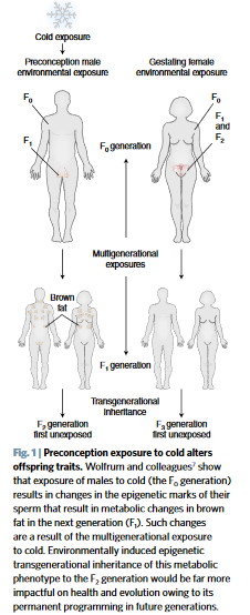
pointed out the opportunity for the researchers of A seasonal epigenetic effect of conception on BMI to have their work make a difference in their field:

“The ability of environmental epigenetics to promote an adaptive phenotype to cold has impacts on evolution. However, the impacts would be far greater if the phenomenon was transgenerational.

Future studies are now needed to determine whether the cold-induced thrifty metabolic phenotype is transmitted to subsequent generations. If exposure not only impacts the health of offspring, but also of all subsequent generations, the impact is significant.”


Every human alive today has observable lasting epigenetic effects caused by environmental factors:

  • During the earliest parts of our lives;
  • From our parents’ exposures and experiences before we’re conceived – many of which are inadequately researched; and
  • Potentially from some of our earlier ancestors’ exposures and experiences.

Aren’t animal studies’ evidence for epigenetic transgenerational inheritance sufficient to compel serious human follow-on research efforts by research sponsors and study designers?

The same comments about epigenetic effects caused by temperature potentially inherited by multiple human generations can also be made about other environmental factors, such as:

  • Nutrition,
  • Toxins – the commentator’s usual area of study, and
  • Stress.

I hope that these researchers value their professions enough to make a difference with this or other areas of their expertise. And that sponsors won’t thwart researchers’ desires for difference-making science by putting them into endless funding queues.

https://www.nature.com/articles/s41591-018-0187-3 “Preconception cold-induced epigenetic inheritance” (not freely available)