A strawman argument against epigenetic clocks

This 2019 review of epigenetic clocks by Washington cancer researchers ignored the elephant in the room: Their epigenetic drift paradigm is generally inapplicable to humans because the vast majority of our cells don’t divide/proliferate. They repeatedly returned to an argument for randomness as a cause for aging and disease:

“A time-dependent stochastic event process, like epigenetic drift, could lead to cancer formation through the accumulation of random epigenetic alterations that, through chance, eventually alter epigenetic driver gene expression leading to a clone of cells destined to become cancer.

It is plausible that the stochastic process inherent in epigenetic drift can induce aberrant methylation events that accumulate in normal cells and eventually induce cancer formation.

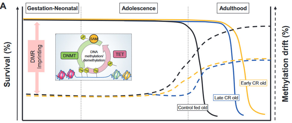
Epigenetic drift relates to a biological process that changes the DNA methylome with age via stochastic gains or losses of DNA methylation. Epigenetic drift can be understood in terms of errors in DNA methylation maintenance during DNA-replication.

The phenomenon of (epi)genetic drift is generally associated with phenotypic neutrality.

For patients who develop cancer around age 80, the most likely initiation time for the founder adenoma cell is predicted to be very early in life, roughly between the ages 15 to 20 years. This unexpected and provocative finding suggests that the optimal age-range for prevention of colorectal cancer may be in adolescence and early adulthood (and ideally through lifelong) dietary and lifestyle interventions.”


The reviewers’ strawman arguments intentionally mischaracterized aspects of the epigenetic clock:

1. The epigenetic clock founder’s actual view on aging was in The epigenetic clock theory of aging:

“The proposed epigenetic clock theory of ageing views biological ageing as an unintended consequence of both developmental programmes and maintenance programmes, the molecular footprints of which give rise to DNAm age estimators.”

The reviewers omitted this intrinsic view of aging, which didn’t fit into the above graphic.

2. Another misrepresentation was:

“In contrast to epigenetic clocks, epigenetic drift refers to a stochastic process that involves both gains and losses of the methylation state of CpG dinucleotides over time.”

A reader of the original 2013 epigenetic clock study would understand that epigenetic clocks measure “both gains and losses of methylation” as in:

“The 193 positively and 160 negatively correlated CpGs get hypermethylated and hypomethylated with age, respectively.”

3. These reviewers omitted recent epigenetic clock significant developments. For example, there was no mention of the GrimAge study, although it was published before this review was submitted.

4. Epigenetic drift as the cause of aging and disease has abundant contrary evidence. These reviewers tossed in a little toward the end of their directed narrative:

“We found only a small number of drift-related CpG island-gene pairs for which drift correlated positively and significantly with gene expression.

The functional consequences of epigenetic drift need to be further elucidated.”

However, they never acknowledged the elephant in the room!

https://cancerres.aacrjournals.org/content/early/2019/11/06/0008-5472.CAN-19-0924 “Epigenetic aging: more than just a clock when it comes to cancer” (not freely available)

Do genes or maternal environments shape fetal brains?

This 2019 Singapore human study used Diffusion Tensor Imaging on 5-to-17-day old infants to find:

“Our findings showed evidence for region-specific effects of genotype and GxE on individual differences in human fetal development of the hippocampus and amygdala. Gene x Environment models outcompeted models containing genotype or environment only, to best explain the majority of measures but some, especially of the amygdaloid microstructure, were best explained by genotype only.

Models including DNA methylation measured in the neonate umbilical cords outcompeted the Gene and Gene x Environment models for the majority of amygdaloid measures and minority of hippocampal measures. The fact that methylation models outcompeted gene x environment models in many instances is compatible with the idea that DNA methylation is a product of GxE.

A genome-wide association study of SNP [single nucleotide polymorphism] interactions with the prenatal environments (GxE) yielded genome wide significance for 13 gene x environment models. The majority (10) explained hippocampal measures in interaction with prenatal maternal mental health and SES [socioeconomic status]. The three genome-wide significant models predicting amygdaloid measures, explained right amygdala volume in interaction with maternal depression.

The transcription factor CUX1 was implicated in the genotypic variation interaction with prenatal maternal health to shape the amygdala. It was also a central node in the subnetworks formed by genes mapping to the CpGs in neonatal umbilical cord DNA methylation data associating with both amygdala and hippocampus structure and substructure.

Our results implicated the glucocorticoid receptor (NR3C1) in population variance of neonatal amygdala structure and microstructure.

Estrogen in the hippocampus affects learning, memory, neurogenesis, synapse density and plasticity. In the brain testosterone is commonly aromatized to estradiol and thus the estrogen receptor mediates not only the effects of estrogen, but also that of testosterone.”

https://onlinelibrary.wiley.com/doi/full/10.1111/gbb.12576 “Neonatal amygdalae and hippocampi are influenced by genotype and prenatal environment, and reflected in the neonatal DNA methylome” (not freely available)

Emotional responses and BDNF methylation

This 2019 German human study found:

“A critical role of BDNF [brain-derived neurotrophic factor] methylation in human amygdala response to negative emotional stimuli, whereby:

  • High BDNF methylation rates were for the first time shown to be associated with a high reactivity in the amygdala; and
  • High BDNF methylation and high amygdala reactivity were associated with low novelty seeking.

There was no interaction or main effect of the Val66Met polymorphism on amygdala reactivity.

Our data adds evidence to the hypothesis that epigenetic modifications of BDNF can result in an endophenotype associated with anxiety and mood disorders. However, since correlations do not prove causality:

  • A direct link between human BDNF mRNA/protein levels, methylation, amygdala reactivity and psychiatric disorders is still missing, demanding further research.
  • Determining the underlying directions of the relations between BDNF methylation, amygdala reactivity, and NS [novelty seeking] cannot be accomplished based on our data and must await further research.

The fact that our results mainly involve the right amygdala is in line with previous studies. Recent reviews suggest a general right hemisphere dominance for all kinds of emotions, and, more specifically, a critical role of the right amygdala in the early assessment of emotional stimuli.

The experimental fMRI paradigm utilized a face‐processing task (faces with anger or fear expressions), alternating with a sensorimotor control task. Harm avoidance, novelty seeking, and reward dependence were measured using the Tridimensional Personality Questionnaire.”

https://onlinelibrary.wiley.com/doi/full/10.1002/hbm.24825 “The role of BDNF methylation and Val 66 Met in amygdala reactivity during emotion processing”

Transgenerational epigenetic inheritance of thyroid hormone sensitivity

My 500th curation is a 2019 Portuguese human study of Azorean islanders:

“This study demonstrates a transgenerational epigenetic inheritance in humans produced by exposure to high TH [thyroid hormone] in fetal life, in the absence of maternal influences secondary to thyrotoxicosis. The inheritance is along the male line.

The present work took advantage of the relatively frequent occurrence of fetal exposure to high TH levels in the Azorean island of São Miguel. This is the consequence of a missense mutation in the THRB gene causing the amino-acid replacement R243Q, resulting in reduced affinity of the TH receptor beta (TRβ) for TH and thus RTHβ.

Its origin has been traced to a couple who lived at the end of the 19th century. F0 represented the third generation and F3 the sixth and seventh generation descendant.”


These researchers provided the first adequately evidenced human transgenerational epigenetic inheritance study! However, the lead sentence in its Abstract wasn’t correct:

“Evidence for transgenerational epigenetic inheritance in humans is still controversial, given the requirement to demonstrate persistence of the phenotype across three generations.”

Although found in this study, there is no “requirement to demonstrate persistence of the phenotype.” Observing the same phenotype in each generation is NOT required for human transgenerational epigenetic inheritance to exist!

Animal transgenerational studies have shown that epigenetic inheritance mechanisms may both express different phenotypes for each generation:

and entirely skip a phenotype in one or more generations!

  • Transgenerational pathological traits induced by prenatal immune activation found a F2 and F3 generation phenotype of impaired sociability, abnormal fear expression and behavioral despair – effects that weren’t present in the F1 offspring;
  • The transgenerational impact of Roundup exposure “Found negligible impacts of glyphosate on the directly exposed F0 generation, or F1 generation offspring pathology. In contrast, dramatic increases in pathologies in the F2 generation grand-offspring, and F3 transgenerational great-grand-offspring were observed.” (a disease phenotype similarly skipped the first offspring generation);
  • Epigenetic transgenerational inheritance mechanisms that lead to prostate disease “There was also no increase in prostate histopathology in the directly exposed F1 or F2 generation.” (a prostate disease phenotype skipped the first two male offspring generations before it was observed in the F3 male offspring); and
  • Epigenetic transgenerational inheritance of ovarian disease “There was no increase in ovarian disease in direct fetal exposed F1 or germline exposed F2 generation. The F3 generation can have disease while the F1 and F2 generations do not, due to this difference in the molecular mechanisms involved.” (an ovarian disease phenotype similarly skipped the first two female offspring generations before it was observed in the F3 female offspring).

Details of epigenetic inheritance mechanisms were provided in Another important transgenerational epigenetic inheritance study. Mechanisms from fetal exposure to the fungicide vinclozolin were compared with mechanisms from fetal DDT exposure, and summarized as:

The fetal exposure initiates a developmental cascade of aberrant epigenetic programming, and does NOT simply induce a specific number of DMRs [DNA methylation regions] that are maintained throughout development.

I emailed references to the studies in the first five above curations to the current study’s corresponding coauthor. They replied “What is the mechanism for the transgenerational inheritance you describe?” and my reply included a link to the sixth curation’s study.

Are there still other transgenerational epigenetically inherited effects due to fetal exposure to high thyroid hormone levels?

https://www.liebertpub.com/doi/full/10.1089/thy.2019.0080 “Reduced Sensitivity to Thyroid Hormone as a Transgenerational Epigenetic Marker Transmitted Along the Human Male Line”

Preliminary findings from a senolytics clinical trial

This 2019 US human clinical trial reported preliminary results. See Reanalysis of findings from a senolytics clinical trial for strikeout changes.

Senescent cells, which can release factors that cause inflammation and dysfunction, the senescence-associated secretory phenotype (SASP), accumulate with ageing and at etiological sites in multiple chronic diseases. Senolytics, including the combination of Dasatinib and Quercetin (D + Q), selectively eliminate senescent cells by transiently disabling pro-survival networks that defend them against their own apoptotic environment.

Since the target of senolytics is senescent cells, these drugs do not need to be continuously present in the circulation in the same way as drugs whose mechanism of action is to occupy a receptor, modulate an enzyme, or act on a particular biochemical pathway, at least in mice. Intermittently administering D + Q effectively circumvents any potential off-target effects due to continuous receptor occupancy or modulation of an enzyme or biochemical pathway.

To test whether intermittent D + Q is effective in targeting senescent cells in humans, we administered a single 3 day course of oral D + Q and assayed senescent cell abundance 11 days after the last dose in subjects with DKD [diabetic kidney disease], the most common cause of end-stage kidney failure and which is characterized by increased senescent cell burden.

In this interim report of findings, we found the single brief course of D + Q:

  • Attenuated adipose tissue and skin senescent cell burden,
  • Decreased resulting adipose tissue macrophage accumulation,
  • Enhanced adipocyte progenitor replicative potential, and
  • Reduced key circulating SASP factors.”

gr2_lrg.jpg

“In adipose tissue D + Q significantly reduced raw numbers of:

  • p16INK4A+ cells by 35%;
  • p21CIP1+ cells by 17%;
  • SAβgal+ cells by 62%;
  • CD68+ macrophages by 28%; and
  • Crown-like structures by 86%.”

https://www.ebiomedicine.com/article/S2352-3964(19)30591-2/fulltext “Senolytics decrease senescent cells in humans: Preliminary report from a clinical trial of Dasatinib plus Quercetin in individuals with diabetic kidney disease”


In a referenced 2019 rodent study by many of the same researchers:

“We also found that even Q alone can prevent high fat diet-induced increases in markers of senescence, renal fibrosis, decreases in renal oxygenation, and increased creatinine in mice, although Q alone did not prevent insulin resistance.”

The rodent study’s 50 mg/kg quercetin dose scaled human-equivalent dose would be (0.081 x 50 mg) = 13.3 mg/kg. This was 375% higher than a 1,000 mg/75 kg quercetin dose (clinical trial participants’ weights weren’t disclosed.)

https://onlinelibrary.wiley.com/doi/full/10.1111/acel.12950 “Targeting senescent cells alleviates obesity‐induced metabolic dysfunction”

PNAS politics in the name of science

This 2019 Germany/Canada human fetal cell study was a Proceedings of the National Academy of Sciences of the United States of America direct submission:

“In a human hippocampal progenitor cell line, we assessed the short- and long-term effects of GC [glucocorticoid] exposure during neurogenesis on messenger RNA expression and DNA methylation profiles. Our data suggest that early exposure to GCs can change the set point of future transcriptional responses to stress by inducing lasting DNAm changes.”


The study’s basic finding was that cells had initial responses to stressors that primed them for subsequent stressors. Since this finding wasn’t new, the researchers tried to make it exciting by applying it to novel contexts that were yet circumscribed by official paradigms.

Hypothesis-seeking associations of human fetal hippocampal cell behaviors with human behaviors were flimsy stretches, as were correlations to placental measurements. These appeared to have been efforts to find headline-making effects.

There wasn’t even a hint of the principle described in Epigenetic variations in metabolism:

“Because of the extreme interconnectivity of cell regulatory networks, even at the cellular level, predicting the impact of a sequence variant is difficult as the resultant variation acts:

  • In the context of all other variants and
  • Their potential additive, synergistic and antagonistic interactions.

This phenomenon is known as epistasis.”

It would have condemned pet models of reality to admit that a cell exists in multiple contexts of other cells with potential additive, synergistic, and antagonistic interactions.

A research proposal to trace a specific cell type’s behaviors – while isolated from their extremely interconnected networks – to trillion-celled human behaviors would be rejected in less-politicized organizations.

Sanctioned speculations manifested in this paper with phrases such as “although not significant..” and “although not directly tested..” The study’s title was probably a disappointment in that it conformed to the study’s evidence.

Involvements of psychiatry departments at the pictured Kings College, Harvard, etc., as part of PNAS entrenched politics, retard advancements of science past approved paradigms.

This is my final curation of PNAS papers.

https://www.pnas.org/content/pnas/early/2019/08/08/1820842116.full.pdf “Glucocorticoid exposure during hippocampal neurogenesis primes future stress response by inducing changes in DNA methylation”

Too cheap for clinical trials

Let’s compare and contrast a 2019 meta-analysis and a 2017 review of using acetyl-L-carnitine to treat diabetic neuropathy.

A 2019 Brazilian meta-analysis Acetyl‐L‐carnitine for the treatment of diabetic peripheral neuropathy of four previous trials stated:

  • “The risk of bias was high in both trials of different ALC doses and low in the other two trials.
  • No included trial measured the proportion of participants with at least moderate (30%) or substantial (50%) pain relief.
  • At doses greater than 1500 mg/day, ALC reduced pain more than placebo. This subgroup analysis should be viewed with caution as the evidence was even less certain than the overall analysis, which was already of very low certainty.
  • The placebo-controlled studies did not measure functional impairment and disability scores.
  • No study used validated symptom scales.
  • Two studies were funded by the manufacturer of ALC and the other two studies had at least one co-author who was a consultant for an ALC manufacturer.

Authors’ conclusions:

  • We are very uncertain whether ALC causes a reduction in pain after 6 to 12 months treatment in people with DPN, when compared with placebo, as the evidence is sparse and of low certainty.
  • Data on functional and sensory impairment and symptoms are lacking, or of very low certainty.
  • The evidence on adverse events is too uncertain to make any judgements on safety.”

A 2017 Italian review Effects of acetyl-L-carnitine in diabetic neuropathy and other geriatric disorders stated:

“A long history of diabetes mellitus and increasing age are associated with the onset of diabetic neuropathy, a painful and highly disabling complication with a prevalence peaking at 50% among elderly diabetic patients. The management of diabetic neuropathy is extremely difficult: in addition to the standard analgesics used for pain control, common treatments include opioids, anticonvulsants, antidepressants, and local anesthetics, alone or in combination. Such therapies still show a variable, often limited efficacy, however.

Many patients do not spontaneously report their symptoms to physicians, but, if asked, they often describe having experienced a persistent and non-abating pain for many years. The prevalence of painful symptoms is just as high in patients with mild neuropathy as in those with more advanced DPN.

Through the donation of acetyl groups, ALC exerts a positive action on mitochondrial energy metabolism. ALC has cytoprotective, antioxidant, and antiapoptotic effects in the nervous system.

ALC has also been proposed for the treatment of other neurological and psychiatric diseases, such as mood disorders and depression, dementia, Alzheimer’s disease, and Parkinson’s disease, given that synaptic energy states and mitochondrial dysfunctions are core factors in their pathogenesis. Compared to other treatments, ALC is safe and extremely well tolerated.

In nerve injury, the mGlu2 receptor overexpressed by ALC binds the glutamate, reducing its concentration in the synapses with an analgesic effect. ALC may improve nerve regeneration and damage repair after primary nerve trauma.”


Where will the money come from to realize what the 2017 review promised, as well as provide what the 2019 meta-analysis required?

Do we prefer the current “limited efficacy” treatments of “opioids, anticonvulsants, antidepressants, and local anesthetics?”

Who will initiate clinical trials of a multiple of the normal dietary supplement dose (500 mg at $.25 a day, retail)? How profitable is a product whose hypothetical effective dosage for diabetic neuropathy (3000 mg) sells for only $1.50 a day?

Effects of advanced glycation end products on quality of life and lifespan

This 2018 Chinese review concerned advanced glycation end products (AGE) mobility interventions:

“Only a limited number of studies have focused on measuring the effects of low AGEs levels or AGEs inhibitors on mobility, although many observational human studies and in vitro studies have reported the correlation of AGEs with and the contribution of AGEs to mobility, particular in diseases such as:

  • osteoporosis,
  • cartilage degradation,
  • osteoarthritis and
  • sarcopenia.

There is insufficient information from previous animal and human studies for use as a reference to determine the intervention period. Although serum AGEs levels can be easily affected by a lower AGEs diet or AGEs inhibitors, it may take longer to see the changes in certain organs or tissues, as a result of a reduction in AGEs accumulation.”

“Effect of AGEs on apoptosis signalling. AP-1, activator protein 1; ERK, extracellular signal-regulated protein kinases; IGF-I, insulin-like growth factor I; IL-6, interleukin-6; JAK, Janus kinase; JNK, c-Jun N-terminal kinases; MEK, mitogen-activated protein kinase; NF-κB, nuclear factor kappa B; p38 MAPK, p38 mitogen-activated protein kinase; RAGE, receptor for AGEs; STAT3, signal transducers and activators of transcription 3; TGF-β, transforming growth factor-β”

https://www.ncbi.nlm.nih.gov/pmc/articles/PMC6180645/ “Role of advanced glycation end products in mobility and considerations in possible dietary and nutritional intervention strategies”


Citations aren’t validations of the reference’s quality and strength of evidence. This review would have benefited from not citing reviews that contained misrepresentations, such as one mentioned in Wikipedia is a poor source of information on advanced glycation end products (AGEs).

I came across this review as a result of it citing the excellent 2008 rodent study Oral Glycotoxins Determine the Effects of Calorie Restriction on Oxidant Stress, Age-Related Diseases, and Lifespan which found:

“Higher levels of oxidant AGEs in offspring of Reg-F0 dams may be attributable to placental transmission from mothers with high AGE levels. These high intrauterine AGE levels may predispose the offspring to the development of chronic inflammation and diseases in adulthood, such as insulin resistance and diabetes.

Increasing the intake of AGEs in the diet erases the benefits of CR [calorie restriction]. OS [oxidant stress] can be reduced, and healthspan increased, in mice fed a diet that is restricted in the content of AGEs.

The beneficial effects of a CR diet may be partly related to reduced oxidant intake rather than decreased energy intake.”

Perinatal stress and sex differences in circadian activity

This 2019 French/Italian rodent study used the PRS model to investigate its effects on circadian activity:

“The aim of this study was to explore the influence of PRS on the circadian oscillations of gene expression in the SCN [suprachiasmatic nucleus of the hypothalamus] and on circadian locomotor behavior, in a sex-dependent manner.

Research on transcriptional rhythms has shown that more than half of all genes in the human and rodent genome follow a circadian pattern. We focused on genes belonging to four functional classes, namely the circadian clock, HPA axis stress response regulation, signaling and glucose metabolism in male and female adult PRS rats.

Our findings provide evidence for a specific profile of dysmasculinization induced by PRS at the behavioral and molecular level, thus advocating the necessity to include sex as a biological variable to study the set-up of circadian system in animal models.”

“There was a clear-cut effect of sex on the effect of PRS on the levels of activity:

  • During the period of lower activity (light phase), both CONT and PRS females were more active than males. During the light phase, PRS increased activity in males, which reached levels of CONT females.
  • More interestingly, during the period of activity (dark phase), male PRS rats were more active than male CONT rats. In contrast, female PRS rats were less active than CONT females.
  • During the dark phase, CONT female rats were less active than CONT male rats.

The study presented evidence for sex differences in circadian activity of first generation offspring that was caused by stress experienced by the pregnant mother:

“Exposure to gestational stress and altered maternal behavior programs a life-long disruption in the reactive adaptation such as:

  •  A hyperactive response to stress and
  • A defective feedback of the hypothalamus-pituitary-adrenal (HPA) axis together with
  • Long-lasting modifications in stress/anti-stress gene expression balance in the hippocampus.”

It would advance science if these researchers carried out experiments to two more generations to investigate possible transgenerational epigenetic inheritance of effects caused by PRS. What intergenerational and transgenerational effects would they possibly find by taking a few more months and extending research efforts to F2 and F3 generations? Wouldn’t these findings likely help humans?


One aspect of the study was troubling. One of the marginally-involved coauthors was funded by the person described in How one person’s paradigms regarding stress and epigenetics impedes relevant research. Although no part of the current study was sponsored by that person, there were three gratuitous citations of their work.

All three citations were reviews. Unlike study researchers, reviewers aren’t bound to demonstrate evidence from tested hypotheses. Reviewers are free to:

  • Express their beliefs as facts;
  • Over/under emphasize study limitations; and
  • Disregard and misrepresent evidence as they see fit.

Fair or not, comparisons of reviews with Cochrane meta-analyses of the same subjects consistently show the extent of reviewers’ biases. Reviewers also aren’t obligated to make post-publication corrections for their errors and distortions.

As such, reviews can’t be cited for reliable evidence. Higher-quality studies that were more relevant and recent than a 1993 review could have elucidated points.

Sucking up to the boss and endorsing their paradigm was predictable. Since that coauthor couldn’t constrain themself to funder citations only in funder studies, it was the other coauthors’ responsibilities to edit out unnecessary citations.

https://www.frontiersin.org/articles/10.3389/fnmol.2019.00089/full “Perinatal Stress Programs Sex Differences in the Behavioral and Molecular Chronobiological Profile of Rats Maintained Under a 12-h Light-Dark Cycle”

Caloric restriction’s epigenetic effects

This 2019 US review subject was caloric restriction (CR) without malnutrition:

“Cellular adaptation that occurs in response to dietary patterns can be explained by alterations in epigenetic mechanisms such as DNA methylation, histone modifications, and microRNA. Epigenetic reprogramming of the underlying chronic low-grade inflammation by CR can lead to immuno-metabolic adaptations that enhance quality of life, extend lifespan, and delay chronic disease onset.

Short- and long-term CRs produce significant changes in different tissues and across species, in some animal models even with sex-specific effects. Early CR onset may cause a different and even an opposite effect on physiological outcomes in animal models such as body weight.”

https://academic.oup.com/advances/article-abstract/10/3/520/5420411 “Epigenetic Regulation of Metabolism and Inflammation by Calorie Restriction” (not freely available)


1. The review didn’t present evidence to equate survival (left axis) with methylation drift (right axis) per the above graphic. Methylation drift should point in the opposite direction of survival, if anything.

2. No mention was made of the epigenetic clock method of measuring age acceleration, although it’s been available since 2013 and recent diet studies have used it. The sole citation of an age acceleration study was from 2001, which was unacceptable for a review published in 2019.

3. The review provided many cellular-level details about the subject. However, organism-level areas weren’t sufficiently evidenced:

A. Arguments for an effect usually include explanations for no effect as well as for opposite effects. The reviewers didn’t provide direct evidence for why, if caloric restriction extended lifespan, caloric overabundance produced shorter lifespans.

B. Caloric restriction evidence was presented as if only it was responsible for organism-level effects. Other mechanisms may have been involved.

An example of such a mechanism was demonstrated in a 2007 rodent study Reduced Oxidant Stress and Extended Lifespan in Mice Exposed to a Low Glycotoxin Diet which compared two 40%-calorie-restricted diets.

The calories and composition of both diets were identical. However, advanced glycation end product (AGE) levels were doubled in standard chow because heating temperatures were “sufficiently high to inadvertently cause standard mouse chow to be rich in oxidant AGEs.”

The study found that a diet with lower chow heating temperatures increased lifespan and health span irrespective of caloric restriction!

  • The low-AGE calorie-restricted diet group lived an average of 15% longer (>20 human equivalent years) than the CR group.
  • 40% of the low-AGE calorie-restricted diet group were still alive when the last CR group member died.
  • The CR group also had significantly more: 1) oxidative stress damage; 2) glucose and insulin metabolism problems; and 3) kidney, spleen, and liver injuries.

A drug that countered effects of a traumatizing mother

This 2019 US rodent study concerned transmitting poor maternal care to the next generation:

“The quality of parental care received during development profoundly influences an individual’s phenotype, including that of maternal behavior. Infant experiences with a caregiver have lifelong behavioral consequences.

Maternal behavior is a complex behavior requiring the recruitment of multiple brain regions including the nucleus accumbens, bed nucleus of the stria terminalis, ventral tegmental area, prefrontal cortex, amygdala, and medial preoptic area. Dysregulation within this circuitry can lead to altered or impaired maternal responsiveness.

We administered zebularine, a drug known to alter DNA methylation, to dams exposed during infancy to the scarcity-adversity model of low nesting resources, and then characterized the quality of their care towards their offspring.

  1. We replicate that dams with a history of maltreatment mistreat their own offspring.
  2. We show that maltreated-dams treated with zebularine exhibit lower levels of adverse care toward their offspring.
  3. We show that administration of zebularine in control dams (history of nurturing care) enhances levels of adverse care.
  4. We show altered methylation and gene expression in maltreated dams normalized by zebularine.

These findings lend support to the hypothesis that epigenetic alterations resulting from maltreatment causally relate to behavioral outcomes.

Maternal behavior is an intergenerational behavior. It is important to establish the neurobiological underpinnings of aberrant maternal behavior and explore treatments that can improve maternal behavior to prevent the perpetuation of poor maternal care across generations.”


The study authors demonstrated intergenerational epigenetic effects, and missed an opportunity to also investigate transgenerational epigenetically inherited effects. They cited reference 60 for the first part of the above quotation, but the cited reviewer misused the transgenerational term by applying it to grand-offspring instead of the great-grand-offspring.

There were resources available to replicate the study authors’ previous findings, which didn’t show anything new. Why not use such resources to uncover evidence even more applicable to humans by extending experiments to great-grand-offspring that would have no potential germline exposure to the initial damaging cause?

Could a study design similar to A limited study of parental transmission of anxiety/stress-reactive traits have been integrated? That study’s thorough removal of parental behavior would be an outstanding methodology to confirm by falsifiability whether parental behavior is both an intergenerational and a transgenerational epigenetic inheritance mechanism.

Rodent great-grand-offspring can be studied in < 9 months. It takes > 50 years for human studies to reach the great-grand-offspring transgenerational generation.

  • Why not attempt to “prevent the perpetuation of poor maternal care across generations?”
  • Isn’t it a plausible hypothesis that humans “with a history of maltreatment mistreat their own offspring?”
  • Isn’t it worth the extra effort to extend animal research to investigate this unfortunate chain?

https://www.nature.com/articles/s41598-019-46539-4 “Pharmacological manipulation of DNA methylation normalizes maternal behavior, DNA methylation, and gene expression in dams with a history of maltreatment”

Wikipedia is a poor source of information on advanced glycation end products (AGEs)

A link to Wikipedia is usually on the first page of search results. The Wikipedia post on AGEs lacks the evidence that a reader may infer from its text.

For example, the second paragraph of the AGEs post, Dietary Sources, contained the following text and references:

  1. “However, only low molecular weight AGEs are absorbed through diet, and vegetarians have been found to have higher concentrations of overall AGEs compared to non-vegetarians. [4]
  2. Therefore it is unclear whether dietary AGEs contribute to disease and aging, or whether only endogenous AGEs (those produced in the body) matter. [5]
  3. This does not free diet from potentially negatively influencing AGE, but implicates dietary AGE may be less important than other aspects of diet that lead to elevated blood sugar levels and formation of AGEs. [4] [5]”

[4] https://www.sciencedirect.com/science/article/pii/S0278691513004444 “Advanced glycation end products in food and their effects on health” (not freely available) 2013 Denmark.

Please note on this linked page that a German researcher took the time to correct one bias of the Danish reviewers, citing evidence from his studies that:

“The deleterious effects of food-derived AGEs in subjects with type 2 diabetes mellitus are proven.”

[5] https://www.ncbi.nlm.nih.gov/pmc/articles/PMC3257625 “Dietary Advanced Glycation End Products and Aging” 2010 US.


Both of these references were reviews.

Unlike study researchers, reviewers aren’t bound to demonstrate evidence from tested hypotheses. Reviewers are free to:

  • Express their beliefs as facts;
  • Over/under emphasize study limitations; and
  • Disregard and misrepresent evidence as they see fit.

Reviewers also aren’t obligated to make post-publication corrections for their errors and distortions. For example, the Danes didn’t correct their review with any findings the German researcher presented.

As such, reviews can’t be cited for reliable evidence.


A sample of other problems with each of the Wikipedia sentences:

1. “However, only low molecular weight AGEs are absorbed through diet, and vegetarians have been found to have higher concentrations of overall AGEs compared to non-vegetarians. [4]”

The first part of sentence 1 came from the review’s abstract:

“Only LMW AGEs..may be absorbed from the gut and contribute to the body burden of AGEs.”

But the reviewers didn’t support their abstract’s statement with direct evidence from any study!

2. “Therefore it is unclear whether dietary AGEs contribute to disease and aging, or whether only endogenous AGEs (those produced in the body) matter. [5]”

The “therefore” of sentence 2 was misplaced. Sentence 1 didn’t attempt to explain whether “dietary AGEs contribute to disease and aging” or “only endogenous AGEs matter.”

Since sentence 2 wasn’t a consequence of sentence 1, the Wikipedia contributor(s) needed to support sentence 2 with evidence. Citing an “unclear” 2010 reference [5] ignored dozens of studies that provided better clarity.

3. “This does not free diet from potentially negatively influencing AGE, but implicates dietary AGE may be less important than other aspects of diet that lead to elevated blood sugar levels and formation of AGEs. [4] [5]”

Wikipedia contributors tend to cite irrelevant references rather than get flagged with “citation needed.” The value judgment of sentence 3 was an example of this intentionally misleading masquerade.

“Dietary AGE may be less important..” wasn’t unequivocally supported by studies referenced in either review, and didn’t represent an authoritative body of evidence. Contrast those weasel words with:

“The deleterious effects of food-derived AGEs in subjects with type 2 diabetes mellitus are proven.”

Good job, Wikipedia contributors! You used lower-quality reviews to promote misunderstandings that DETRACTED from science.


Wikipedia’s premise is that since the group knows more about any subject than does any individual, everyone is entitled to contribute. The results are usually incoherent narratives that often substitute opinions for evidence.

The second paragraph of the Exogenous section of the Wikipedia glycation post provided an example:

  • Assertions of the first and third sentences needed citations. Did the contributor(s) think these would be unexamined?
  • Someone contributed a cancer reference as the fourth sentence, although it had little to do with the preceding sentences.
  • The fifth sentence was informative on exogenous glycations and AGEs. An editor would have removed “recently” and “recent” though, because the cited source was dated 2005.

Disease and advanced glycation end products (AGEs)

This 2015 French/US review focused on chronic kidney disease, appropriate for its publication in the Journal of the American Society of Nephrology:

“Advanced glycation end products (AGEs) are formed not only in the presence of hyperglycemia, but also in diseases associated with high levels of oxidative stress, such as CKD. Humans are exposed to exogenous sources of AGE (diet and cigarette smoke) and endogenous sources of AGE when the organism is exposed to high levels of glucose, such as in diabetes.

Accumulation of AGEs in patients with CKD has been shown to result from inflammation, oxidative stress, and diet. AGEs are proinflammatory and pro-oxidative compounds that play a role in the high prevalence of endothelial dysfunction and subsequent cardiovascular disease in patients with CKD.

In view of the many harmful effects of AGEs on cell function, it is essential to develop strategies designed to counteract their effects. AGEs are generated during the thermal processing and storage of foods. Dietary restriction is an effective, feasible, and economic method to reduce levels of toxic AGEs and possibly, the associated cardiovascular mortality.”

https://jasn.asnjournals.org/content/27/2/354 “Uremic Toxicity of Advanced Glycation End Products in CKD”


I came across the AGE subject in the usual Internet way. 🙂 While reading comments on Josh Mitteldorf’s blog post Money in Aging Research, Part I, Dr. Alan Green mentioned Dr. Helen Vlassara’s work. A DuckDuckGo search led to her 44,256 citations, which increase every day.

Another read on the subject is her 2016 book Dr. Vlassara’s AGE-Less Diet: How a Chemical in the Foods We Eat Promotes Disease, Obesity, and Aging and the Steps We Can Take to Stop It. A practical guide is her 2017 book The AGE Food Guide: A Quick Reference to Foods and the AGEs They Contain.

Linking adult neurogenesis to Alzheimer’s disease

This 2019 Spanish human study compared DNA methylation, chromatin and histone modifications in the hippocampus of deceased Alzheimer’s disease patients with controls:

“A significant percentage of the differentially methylated genes were related to neural development and neurogenesis. It was astounding that other biological, cellular, and molecular processes generally associated with neurodegeneration such as apoptosis, autophagy, inflammation, oxidative stress, and mitochondrial or lysosomal dysfunction were not overrepresented.

The results of the present study point to neurogenesis-related genes as targets of epigenetic changes in the hippocampus affected by AD. These methylation changes might be built throughout life due to external and internal cues and would represent an example of epigenetic interaction between environmental and genetic factors in developing AD.

As an alternative explanation, these epigenetic marks might also represent the trace of DNA methylation alterations induced during early developmental stages of the hippocampus, which would remain as a fingerprint in the larger proportion of hippocampal neurons that are not exchanged. This second hypothesis would link AD to early life stages, in concordance with recent studies that revealed abnormal p-tau deposits (pre-tangles) in brains of young individuals under 30, suggesting AD pathology would start earlier in life than it was previously thought. The influence of the genetic risk for AD has also been postulated to begin in early life, and other AD risk factors may be influenced by in utero environment.”


The study cited references to adult neurogenesis:

“Though strongly related to brain development, neurogenesis is also maintained in the adult human brain, mainly in two distinct areas, i.e., the subventricular zone and the subgranular zone of the dentate gyrus in the hippocampus. There is substantial neurogenesis throughout life in the human hippocampus as it is estimated that up to one third of human hippocampal neurons are subject to constant turnover.

Adult neurogenesis is linked to hippocampal-dependent learning and memory tasks and is reduced during aging. Recent evidence suggests that adult neurogenesis is altered in the neurodegenerative process of AD, but it is still controversial with some authors reporting increased neurogenesis, whereas others show reduced neurogenesis. In the human hippocampus, a sharp drop in adult neurogenesis has been observed in subjects with AD.”

One of the study’s limitations was its control group:

“There was a significant difference in age between controls [12, ages 50.7 ± 21.5] and AD patients [26, ages 81.2 ± 12.1], being the latter group older than the former group. Although we adjusted for age in the statistical differential methylation analysis, the accuracy of this correction may be limited as there is little overlap in the age ranges of both groups.”

https://clinicalepigeneticsjournal.biomedcentral.com/track/pdf/10.1186/s13148-019-0672-7 “DNA methylation signature of human hippocampus in Alzheimer’s disease is linked to neurogenesis”

OCD and neural plasticity

Update: this was retracted on February 23, 2021. The retraction note is at https://www.nature.com/articles/s41598-021-84474-5.

This 2019 New York rodent study investigated multiple avenues to uncover mechanisms of obsessive-compulsive disorder:

“Psychophysical models of OCD propose that anxiety (amygdala) and habits (dorsolateral striatum) may be causally linked. Numerous genetic and environmental factors may reduce striatum sensitivity and lead to maladaptive overcompensation, potentially accounting for a significant proportion of cases of pathological OCD-like behaviors.

Our results indicate that both the development and reversal of OCD-like behaviors involve neuroplasticity resulting in circuitry changes in BLA-DLS and possibly elsewhere.”

https://www.nature.com/articles/s41598-019-45325-6.pdf “Amelioration of obsessive-compulsive disorder in three mouse models treated with one epigenetic drug: unraveling the underlying mechanism”


The researchers explored two genetic models of OCD, showed why these insufficiently explained observed phenomena, then followed up with epigenetic investigations. They demonstrated how and the degree to which histone modifications and DNA methylation regulated both the development and reversal of OCD symptoms.

However, the researchers also carelessly cited thirteen papers outside the specific areas of the study to support one statement in the lead paragraph:

“Novel studies propose that modulations in gene expression influenced by environmental factors, are connected to mental health disorders.”

Only one of the thirteen citations was more recent than 2011, and none of them were high-quality studies.