To follow up How much sulforaphane is suitable for healthy people? I’ve started growing broccoli sprouts, and a “30 60 grams of fresh broccoli sprouts incorporated daily into the diet” [1] program. See Week 2 of Changing an inflammatory phenotype with broccoli sprouts for changes.
I loosely follow [2]‘s sprouting guidelines. One preparation difference is microwaving per [3]‘s findings as follows:
My current microwaving time for 60 grams of 3-day-old broccoli sprouts in 100 ml of water with a 1000 W microwave on full power is 35 seconds. The temperature gets up to 57°C. See Enhancing sulforaphane content for changes. I immediately dump the broccoli sprouts into a colander and spray with cold water to stop heating at the desired temperature.
The first batch of broccoli sprouts was a mild, cabbage-tasting side dish to the home-style chicken soup on page 238 of [4].
The a priori hypotheses:
-
- 30 grams of fresh broccoli sprouts will not have “51 mg (117 μmol)” of glucoraphanin [1] because they “Used the elicitor methyl jasmonate (MeJA) by priming the seeds as well as by spraying daily. MeJA at concentrations of 156 μM act as stressor in the plant and enhances the biosynthesis of the phytochemicals glucosinolates. Compared to control plants without MeJA treatment, the content of compounds as the aliphatic glucosinolate glucoraphanin was enhanced up to 70%.” 117 μmol / 1.70 = 69 μmol is the expected glucoraphanin amount in 30 grams weight of fresh broccoli sprouts. 69 x 2 = 138 μmol in 60 grams.
One measurement [5] of how much sulforaphane is present in fresh broccoli sprouts before microwaving is 100 μmol / 111 g = .9 μmol / g. (.9 x 30 g) = 27 μmol is the expected sulforaphane amount in 30 grams of fresh broccoli sprouts. Changed assumption to 0 μmol sulforaphane due to 2013 Sulforaphane: translational research from laboratory bench to clinic “Broccoli sprouts are correctly described as releasing, generating, or yielding but not containing SFN [sulforaphane].”- Last week a [3] coauthor agreed to make the data available to facilitate calculations. While I’m waiting… The study said the Figure 3 HL60 sulforaphane amount was 2.45 μmol / g. Eyeball estimate of the below Figure 3 control (raw broccoli florets) is a glucoraphanin amount of ~2.2 μmol / g. I assume that the broccoli florets and sprouts conversion would be the same at a 2.45 μmol / 2.2 μmol ≈ 1.11 ratio. I expect that microwaving the raw broccoli sprouts to 60°C will convert the 138 μmol of glucoraphanin to a 153 μmol amount of sulforaphane at this assumed 1.11 conversion ratio.
- The estimated sulforaphane weight per [6] would be (153 μmol / 5.64) = 27 mg which is comparable to clinical trial dosages listed in [7] and [8].
- I’ve been sitting around a lot since returning from Milano, Italy, on February 24, 2020, and probably weigh around 75 kg. The estimated dosage represents 153 μmol of sulforaphane / 75 kg = 2.04 μmol of sulforaphane / kg, compared to the 1.36 μmol of glucoraphanin / kg average of [1]. (The study provided the subjects’ mean weight in Table 1 as “85.8 ± 16.7 kg.” The average dosage per kg body weight was 117 μmol of glucoraphanin / 85.8 kg = 1.36 μmol of glucoraphanin / kg.)
- Don’t have a practical estimate of the amount of sulforaphane I metabolize from post-microwave glucoraphanin that would add to the calculated 153 μmol of sulforaphane. Both [7] and [8] cited a 2012 study that found: “Some conversion of GRN [glucoraphanin] to SFN can occur in response to metabolism by the gut microflora; however, the response is inefficient, having been shown to vary ‘from about 1% to more than 40% of the dose.’”
- Don’t have a practical estimate of the “internal dose” [8] that would result from 153+ μmol of sulforaphane.
- 30 grams of fresh broccoli sprouts will not have “51 mg (117 μmol)” of glucoraphanin [1] because they “Used the elicitor methyl jasmonate (MeJA) by priming the seeds as well as by spraying daily. MeJA at concentrations of 156 μM act as stressor in the plant and enhances the biosynthesis of the phytochemicals glucosinolates. Compared to control plants without MeJA treatment, the content of compounds as the aliphatic glucosinolate glucoraphanin was enhanced up to 70%.” 117 μmol / 1.70 = 69 μmol is the expected glucoraphanin amount in 30 grams weight of fresh broccoli sprouts. 69 x 2 = 138 μmol in 60 grams.
I don’t have a laboratory in my kitchen 🙂 and won’t have quantified results. See Grow a broccoli sprouts Victory Garden today! for August 2020 practices.
References in order of citation:
[1] 2018 Effects of long-term consumption of broccoli sprouts on inflammatory markers in overweight subjects
[2] 2017 You Need Sulforaphane – How and Why to Grow Broccoli Sprouts
[3] 2020 Microwave cooking increases sulforaphane level in broccoli curated in Microwave broccoli to increase sulforaphane levels

[4] 2016 Dr. Vlassara’s AGE-Less Diet: How a Chemical in the Foods We Eat Promotes Disease, Obesity, and Aging and the Steps We Can Take to Stop It
[5] 2016 Effect of Broccoli Sprouts and Live Attenuated Influenza Virus on Peripheral Blood Natural Killer Cells: A Randomized, Double-Blind Study
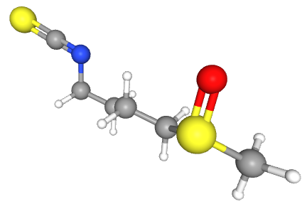
[6] 2020 https://pubchem.ncbi.nlm.nih.gov/compound/sulforaphane lists sulforaphane’s molecular weight as 177.3 g / mol. A 1 mg weight of sulforaphane equals a 5.64 μmol sulforaphane amount (.001 / 177.3).
[7] 2019 Sulforaphane: Its “Coming of Age” as a Clinically Relevant Nutraceutical in the Prevention and Treatment of Chronic Disease
[8] 2019 Broccoli or Sulforaphane: Is It the Source or Dose That Matters? Note that a coauthor didn’t disclose their business’ conflict of interest for an effectively promoted commercial product.


 Moonrise at sunrise with Venus