An epigenetic clock review by committee

This 2019 worldwide review of epigenetic clocks was a semi-anonymous mishmash of opinions, facts, hypotheses, unwarranted extrapolations, and beliefs. Diversity of viewpoints among the 21 coauthors wasn’t evident.

1. Citations of coauthors’ works seemed excessive, and they apologized for omissions. However:

  • Challenge 5 was titled “Single-cell analysis of aging changes and disease” and
  • Table 1 “Major biological and analytic issues with epigenetic DNA methylation clocks” had single-cell analysis as the Proposed solution to five Significant issues.

Yet studies such as High-Resolution Single-Cell DNA Methylation Measurements Reveal Epigenetically Distinct Hematopoietic Stem Cell Subpopulations were unmentioned.

2. Some coauthors semi-anonymously expressed faith that using current flawed methodologies in the future – only more thoroughly, with newer equipment, etc. – would yield better results. If all 21 coauthors were asked their viewpoints of Proposed solutions to the top three Significant issues of epigenetic clocks, what would they emphasize when quoted?

3. Techniques were praised:

“Given the precision with which DNA methylation clock age can be estimated and evolving measures of biological, phenotype-, and disease-related age (e.g., PhenoAge, GrimAge)..”

Exactly why these techniques have at times produced inexplicable results wasn’t examined, though. Two examples:

  • In Reversal of aging and immunosenescent trends, Levine PhenoAge methodology estimated that the 51-65 year old subjects’ biological ages at the beginning of the study averaged 17.5 years less than their chronological age. Comparing that to Horvath average biological age of 3.95 years less raised the question: exactly why did PhenoAge show such a large difference?
  • The paper mentioned GrimAge methodology findings about “smoking-related changes.” But it didn’t explain why GrimAge methylation findings most closely associated with smoking history also accurately predicted future disease risk with non-smokers.

Eluding explanations for these types of findings didn’t help build confidence in methodologies.

4. A more readable approach to review by committee could have coauthors – in at least one section – answer discussion questions, as Reversing epigenetic T cell exhaustion did with 18 experts.

https://genomebiology.biomedcentral.com/articles/10.1186/s13059-019-1824-y “DNA methylation aging clocks: challenges and recommendations”

A review of fetal adverse events

This 2019 Australian review subject was fetal adversities:

“Adversity during the perinatal period is a significant risk factor for the development of neurodevelopmental disorders long after the causative event. Despite stemming from a variety of causes, perinatal compromise appears to have similar effects on the developing brain, thereby resulting in behavioural disorders of a similar nature.

These behavioural disorders occur in a sex‐dependent manner, with males affected more by externalizing behaviours such as attention deficit hyperactivity disorder (ADHD) and females by internalizing behaviours such as anxiety. The term ‘perinatal compromise’ serves as an umbrella term for intrauterine growth restriction, maternal immune activation, prenatal stress, early life stress, premature birth, placental dysfunction, and perinatal hypoxia.

The above conditions are associated with imbalanced excitatory-inhibitory pathways resulting from reduced GABAergic signalling. Methylation of the GAD1/GAD67 gene, which encodes the key glutamate‐to‐GABA synthesizing enzyme Glutamate Decarboxylase 1, resulting in increased levels of glutamate is one epigenetic mechanism that may account for a tendency towards excitation in disorders such as ADHD.

The posterior cerebellum’s role in higher executive functioning is becoming well established due to its connections with the prefrontal cortex, association cortices, and limbic system. It is now suggested that disruptions to cerebellar development, which can occur due to late gestation compromises such as preterm birth, can have a major impact on the region of the brain to which it projects.

Activation of the maternal hypothalamic-pituitary adrenal (HPA) axis and placental protection. Psychological stress is perceived by the maternal HPA axis, which stimulates cortisol release from the maternal adrenal gland.

High levels of maternal cortisol are normally prevented from reaching the fetus by the 11β-hydroxysteroid dehydrogenase 2 (HSD11B2) enzyme, which converts cortisol to the much less active cortisone. Under conditions of high maternal stress, this protective mechanism can be overwhelmed, with the gene encoding the enzyme becoming methylated, which reduces its expression allowing cortisol to cross the placenta and reach the fetus.”


The reviewers extrapolated many animal study findings to humans, although most of their own work was with guinea pigs. The “suggest” and “may” qualifiers were used often – 22 and 37 times, respectively. More frequent use of the “appears,” “hypothesize,” “propose,” and “possible” terms was justified.

As a result, many reviewed items such as the above graphic and caption should be viewed as hypothetical for humans rather than reflecting solid evidence from quality human studies.

The reviewers focused on the prenatal (before birth) period more than the perinatal (last trimester of pregnancy to one month after birth) period. There were fewer mentions of birth and early infancy adversities.

https://onlinelibrary.wiley.com/doi/abs/10.1111/jne.12814 “Perinatal compromise contributes to programming of GABAergic and Glutamatergic systems leading to long-term effects on offspring behaviour” (not freely available)

A transgenerational view of the rise in obesity

This 2019 Washington State University rodent study found epigenetically inherited transgenerational effects in great-grand offspring due to their great-grandmothers’ toxicant exposures during pregnancy:

“Previous studies found an increased susceptibility to obesity in F3 generation rats ancestrally exposed to the pesticide DDT, and an increase in a lean phenotype in the F3 generation rats ancestrally exposed to the herbicide atrazine. The present study investigated whether there were common DMR [differential DNA methylated region] and associated genes between the control, DDT, and atrazine lineage male and female adipocytes in order to identify potential novel gene pathways modulated by DNA methylation.

Comparison of epigenetic alterations indicated that there were substantial overlaps between the different treatment lineage groups for both the lean and obese phenotypes. Novel correlated genes and gene pathways associated with DNA methylation were identified, and may aid in the discovery of potential therapeutic targets for metabolic diseases such as obesity.

Given that the first widespread [DDT] exposures to gestating human females started in the 1950s, the majority of the subsequent F3 generation are adults today. Ancestral exposures to environmental toxicants like DDT may have had a role in the dramatic rise in obesity rates worldwide.”


This same research group noted in Transgenerational diseases caused by great-grandmother DDT exposure:

“DDT was banned in the USA in 1973, but it is still recommended by the World Health Organization for indoor residual spray. India is by far the largest consumer of DDT worldwide.

India has experienced a 5-fold increase of type II diabetes over the last three decades with a predisposition to obesity already present at birth in much of the population. Although a large number of factors may contribute to this increased incidence of obesity, the potential contribution of ancestral toxicant exposures in the induction of obesity susceptibility requires further investigation.”

https://www.tandfonline.com/doi/full/10.1080/21623945.2019.1693747 “Adipocyte epigenetic alterations and potential therapeutic targets in transgenerationally inherited lean and obese phenotypes following ancestral exposures”

A GWAS meta-analysis of two epigenetic clocks

This 2019 UK human study conducted a meta-analysis of genome-wide association studies of two epigenetic clocks using 13,493 European-ancestry individuals aged between ten and 98 years:

“Horvath-EAA, described in previous publications as ‘intrinsic’ epigenetic age acceleration (IEAA), can be interpreted as a measure of cell-intrinsic ageing that exhibits preservation across multiple tissues, appears unrelated to lifestyle factors, and probably indicates a fundamental cell ageing process that is largely conserved across cell types.

In contrast, Hannum-EAA, referred to in previous studies as ‘extrinsic’ epigenetic age acceleration (EEAA), can be considered a biomarker of immune system ageing, explicitly incorporating aspects of immune system decline such as age-related changes in blood cell counts, correlating with lifestyle and health-span related characteristics, and thus yielding a stronger predictor of all-cause mortality.

The meta-analysis of Horvath-EAA identified ten independent associated SNPs [single nucleotide polymorphisms], doubling the number reported to date, and highlighted 21 genes involved in Horvath-based epigenetic ageing. Four of the ten Horvath-EAA-associated SNPs are mQTL [methylation quantitative trait loci] for CpGs used in the Horvath/Hannum epigenetic clocks. A possible interpretation of this is that the functional mechanism by which these SNPs influence the rate of biological ageing is via altering methylation levels.

Father’s age at death, a rough proxy for lifespan, was nominally significantly correlated with both EAA measures, and parents’ age at death was additionally correlated with Hannum-EAA. Aside from these, genetic correlations with age-related traits were surprisingly few: it is possible that this could reflect an overly conservative correction for the multiple tests carried out, or low statistical power, rather than a genuine lack of correlations.

Genetic correlation analysis should be restricted to GWAS with a heritability Z-score of 4 or more, on the grounds of interpretability and power, so the Horvath-based results particularly should be interpreted with caution.”


A non-apologetic way to explain the above graphic is that NONE of these 218 “health and behavioral traits” were any more associated with the studied genetic measurements than would be expected by chance!

Fervent believers in the GWAS methodology’s capability to exactly predict individual phenotypes eventually become victims of the scientific method. These GWAS researchers griped about “overly conservative correction, or low statistical power” and other predictable shortfalls, and ended a long limitations statement with:

“While we have identified a number of SNPs and genes significantly associated with EAA, including genes already known to be related to ageing, the analyses presented here fall short of providing a mechanistic explanation for how these variants and genes act to influence biological age.”

Outside of beliefs, it’s hard to understand why research money keeps pouring into the GWAS dead end. If these researchers and their employing institution and sponsors want to make a difference in human lives, they need to get busy in other areas.

These researchers were employed by the same institution that couldn’t be bothered to scrape together six more weeks of funds to study the transgenerational damaging effects of acetaminophen – an analgesic available to billions of people – in Epigenetics research that was designed to fall one step short of wonderful.

https://journals.plos.org/plosgenetics/article?id=10.1371/journal.pgen.1008104 “A meta-analysis of genome-wide association studies of epigenetic age acceleration”

Organismal aging and cellular senescence

I’ll curate this 2019 German review through its figures:

“With the discovery of beneficial aspects of cellular senescence and evidence of senescence being not limited to replicative cellular states, a redefinition of our comprehension of aging and senescence appears scientifically overdue.

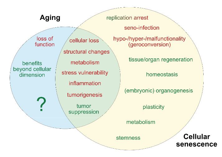
Figure 1. Current determinants and relevant open questions, marking the processes of aging and senescence as discussed in the text. Aspects represented in green are considered as broadly accepted or scientifically consolidated. Novel aspects that are yet unproven, or are under debate, are highlighted in red.

SASP = senescence-associated secretory phenotype. AASP = putative aging-associated secretory phenotype as suggested in the text.

Figure 2. Theories on the causality and purpose of aging. Graphically summarized are four contrasting concepts crystallized from current evidence addressing the inductive driving force of aging. Apart from a stochastic deleteriome, there are arguments for a pseudo-programmed, programmed or at least partially programmed nature of aging.

Figure 3. Comparative representation of the aging and senescence processes highlighting different levels of interaction and putative sites of interventions.

(1) As discussed in the text, causative mechanisms of aging are still not well understood, however, multiple factors including genetic, epigenetic and stress-related effects seem to have an orchestrated role in the progression of aging. Senescence on the other hand, is seen as a programmed response to different kinds of stressors, which proceed in defined stages. Whether, in analogy, aging also follows a defined program or sequential stages is not known.

(2) Senescence involves autocrine and paracrine factors, which are responsible for a ‘seno-infection’ or bystander effect in neighboring cells. There is currently no direct evidence for a similar factor composition propagating the aging process via a kind of ‘gero-infection’.

(3) Accumulation of senescent cells has been described as a hallmark of aging; however, whether they are a causative factor or a consequence of tissue and organismal aging is still unknown. As discussed in the text, it appears possible that aging and senescence mutually influence each other through positive feedback at this level, leading to accelerated tissue damage and aging.

(4,5) Clearance of senescent or aging cells might constitute putative targets for interventional approaches aimed to reduce or reverse the impact of aging and improve cell and tissue homeostasis by inducing a ‘rejuvenation’ process.

Figure 4. Pathological and beneficial functions of aging and senescence, according to current knowledge. In red are represented pathological consequences and in green beneficial functions of aging and senescence.

The impact of aging has mainly been described at the organismal level, since a complete cellular functional profile has not yet been established. Accordingly, whether beneficial consequences of the aging process exist at the cellular level is unclear.”


The assertion of Figure 3 (2) that:

“There is currently no direct evidence for a similar factor composition propagating the aging process via a kind of ‘gero-infection.”

was shown to be false in Reevaluate findings in another paradigm:

“It was demonstrated that increased aging occurred as a result of lack of gonadotropin-releasing hormone and that increased lifespan resulted from its provision during aging.

In this manner:

  1. Aging of hypothalamic microglia leads to
  2. Aging of the hypothalamus, which leads to
  3. Aging elsewhere in the body.

So here we have a multi-level interaction:

  1. Activation of NF-κB leads to
  2. Cellular aging, leading to
  3. A diminished production of GnRH, which then
  4. Acts (through cells with a receptor for it, or indirectly as a result of changes to GnRH-receptor-possessing cells) to decrease lifespan.

So the age state of hypothalamic cells, at least with respect to NF-κB activation, is communicated to other cells via reduced output of GnRH.”


The reviewers’ position on Figure 2 was:

“In our view, recent evidence that

  • Senescence is based on an unterminated developmental growth program and the finding that
  • The concept of post-mitotic senescence requires the activation of expansion, or ‘growth’ factors as a second hit,

favor the assumption that aging underlies a grating of genetic determination similarly to what is summarized above under the pseudo-programmed causative approach.”

Their position on Figure 4’s beneficial effects of aging began with the sentence:

“If we assume that aging already starts before birth, it can be considered simply a developmental stage, required to complete the evolutionary program associated with species-intrinsic biological functions such as reproduction, survival, and selection.”

Cited studies included:

https://www.mdpi.com/2073-4409/8/11/1446 “Dissecting Aging and Senescence-Current Concepts and Open Lessons”

A strawman argument against epigenetic clocks

This 2019 review of epigenetic clocks by Washington cancer researchers ignored the elephant in the room: Their epigenetic drift paradigm is generally inapplicable to humans because the vast majority of our cells don’t divide/proliferate. They repeatedly returned to an argument for randomness as a cause for aging and disease:

“A time-dependent stochastic event process, like epigenetic drift, could lead to cancer formation through the accumulation of random epigenetic alterations that, through chance, eventually alter epigenetic driver gene expression leading to a clone of cells destined to become cancer.

It is plausible that the stochastic process inherent in epigenetic drift can induce aberrant methylation events that accumulate in normal cells and eventually induce cancer formation.

Epigenetic drift relates to a biological process that changes the DNA methylome with age via stochastic gains or losses of DNA methylation. Epigenetic drift can be understood in terms of errors in DNA methylation maintenance during DNA-replication.

The phenomenon of (epi)genetic drift is generally associated with phenotypic neutrality.

For patients who develop cancer around age 80, the most likely initiation time for the founder adenoma cell is predicted to be very early in life, roughly between the ages 15 to 20 years. This unexpected and provocative finding suggests that the optimal age-range for prevention of colorectal cancer may be in adolescence and early adulthood (and ideally through lifelong) dietary and lifestyle interventions.”


The reviewers’ strawman arguments intentionally mischaracterized aspects of the epigenetic clock:

1. The epigenetic clock founder’s actual view on aging was in The epigenetic clock theory of aging:

“The proposed epigenetic clock theory of ageing views biological ageing as an unintended consequence of both developmental programmes and maintenance programmes, the molecular footprints of which give rise to DNAm age estimators.”

The reviewers omitted this intrinsic view of aging, which didn’t fit into the above graphic.

2. Another misrepresentation was:

“In contrast to epigenetic clocks, epigenetic drift refers to a stochastic process that involves both gains and losses of the methylation state of CpG dinucleotides over time.”

A reader of the original 2013 epigenetic clock study would understand that epigenetic clocks measure “both gains and losses of methylation” as in:

“The 193 positively and 160 negatively correlated CpGs get hypermethylated and hypomethylated with age, respectively.”

3. These reviewers omitted recent epigenetic clock significant developments. For example, there was no mention of the GrimAge study, although it was published before this review was submitted.

4. Epigenetic drift as the cause of aging and disease has abundant contrary evidence. These reviewers tossed in a little toward the end of their directed narrative:

“We found only a small number of drift-related CpG island-gene pairs for which drift correlated positively and significantly with gene expression.

The functional consequences of epigenetic drift need to be further elucidated.”

However, they never acknowledged the elephant in the room!

https://cancerres.aacrjournals.org/content/early/2019/11/06/0008-5472.CAN-19-0924 “Epigenetic aging: more than just a clock when it comes to cancer” (not freely available)

Epigenetic transgenerational inheritance extends to the great-great-grand offspring

This 2019 rodent study by the Washington State University labs of Dr. Michael Skinner continued to F4 generation great-great-grand offspring, and demonstrated that epigenetic inheritance mechanisms are similar to imprinted genes:

“Epigenetic transgenerational inheritance potentially impacts disease etiology, phenotypic variation, and evolution. An increasing number of environmental factors from nutrition to toxicants have been shown to promote the epigenetic transgenerational inheritance of disease.

Imprinted genes are a special class of genes since their DNA methylation patterns are unchanged over the generation and are not affected by the methylation erasure occurring early in development. The transgenerational epigenetic alterations in the germline appear to be permanently reprogrammed like imprinted genes, and appear protected from this DNA methylation erasure and reprogramming at fertilization in the subsequent generations. Similar to imprinted genes, the epigenetic transgenerational germline epimutations appear to have a methylation erasure in the primordial germ cells involving an epigenetic molecular memory.

Comparison of the transgenerational F3 generation, with the outcross to the F4 generation through the paternal or maternal lineages, allows an assessment of parent-of-origin transmission of disease or pathology. Observations provided examples of the following:

  1. Pathology that required combined contribution of both paternal and maternal alleles to promote disease [testis and ovarian disease];
  2. Pathology that is derived from the opposite sex allele such as father to daughter [kidney disease] or mother to son [prostate disease];
  3. Pathology that is derived from either parent-of-origin alleles independently [obesity];
  4. Pathology that is transmitted within the same sex, such as maternal to daughter [mammary tumor development]; and
  5. Pathology that is observed only following a specific parent-of-origin outcross [both F4 male obesity and F4 female kidney disease in the vinclozolin lineage].”

https://www.sciencedirect.com/science/article/pii/S0012160619303471 “Epigenetic transgenerational inheritance of parent-of-origin allelic transmission of outcross pathology and sperm epimutations”


This study showed that epigenetically inherited legacies extend to the fifth generation. Do any of us know our ancestors’ medical histories back to our great-great-grandparents?

Will toxicologists take their jobs seriously, catch up to current science, and investigate possible effects in at least the F3 generation that had no direct toxicant exposure?

Do genes or maternal environments shape fetal brains?

This 2019 Singapore human study used Diffusion Tensor Imaging on 5-to-17-day old infants to find:

“Our findings showed evidence for region-specific effects of genotype and GxE on individual differences in human fetal development of the hippocampus and amygdala. Gene x Environment models outcompeted models containing genotype or environment only, to best explain the majority of measures but some, especially of the amygdaloid microstructure, were best explained by genotype only.

Models including DNA methylation measured in the neonate umbilical cords outcompeted the Gene and Gene x Environment models for the majority of amygdaloid measures and minority of hippocampal measures. The fact that methylation models outcompeted gene x environment models in many instances is compatible with the idea that DNA methylation is a product of GxE.

A genome-wide association study of SNP [single nucleotide polymorphism] interactions with the prenatal environments (GxE) yielded genome wide significance for 13 gene x environment models. The majority (10) explained hippocampal measures in interaction with prenatal maternal mental health and SES [socioeconomic status]. The three genome-wide significant models predicting amygdaloid measures, explained right amygdala volume in interaction with maternal depression.

The transcription factor CUX1 was implicated in the genotypic variation interaction with prenatal maternal health to shape the amygdala. It was also a central node in the subnetworks formed by genes mapping to the CpGs in neonatal umbilical cord DNA methylation data associating with both amygdala and hippocampus structure and substructure.

Our results implicated the glucocorticoid receptor (NR3C1) in population variance of neonatal amygdala structure and microstructure.

Estrogen in the hippocampus affects learning, memory, neurogenesis, synapse density and plasticity. In the brain testosterone is commonly aromatized to estradiol and thus the estrogen receptor mediates not only the effects of estrogen, but also that of testosterone.”

https://onlinelibrary.wiley.com/doi/full/10.1111/gbb.12576 “Neonatal amygdalae and hippocampi are influenced by genotype and prenatal environment, and reflected in the neonatal DNA methylome” (not freely available)

Emotional responses and BDNF methylation

This 2019 German human study found:

“A critical role of BDNF [brain-derived neurotrophic factor] methylation in human amygdala response to negative emotional stimuli, whereby:

  • High BDNF methylation rates were for the first time shown to be associated with a high reactivity in the amygdala; and
  • High BDNF methylation and high amygdala reactivity were associated with low novelty seeking.

There was no interaction or main effect of the Val66Met polymorphism on amygdala reactivity.

Our data adds evidence to the hypothesis that epigenetic modifications of BDNF can result in an endophenotype associated with anxiety and mood disorders. However, since correlations do not prove causality:

  • A direct link between human BDNF mRNA/protein levels, methylation, amygdala reactivity and psychiatric disorders is still missing, demanding further research.
  • Determining the underlying directions of the relations between BDNF methylation, amygdala reactivity, and NS [novelty seeking] cannot be accomplished based on our data and must await further research.

The fact that our results mainly involve the right amygdala is in line with previous studies. Recent reviews suggest a general right hemisphere dominance for all kinds of emotions, and, more specifically, a critical role of the right amygdala in the early assessment of emotional stimuli.

The experimental fMRI paradigm utilized a face‐processing task (faces with anger or fear expressions), alternating with a sensorimotor control task. Harm avoidance, novelty seeking, and reward dependence were measured using the Tridimensional Personality Questionnaire.”

https://onlinelibrary.wiley.com/doi/full/10.1002/hbm.24825 “The role of BDNF methylation and Val 66 Met in amygdala reactivity during emotion processing”

Reversing epigenetic T cell exhaustion

This 2019 worldwide discussion among 18 experts concerned T cell exhaustion:

“‘T cell exhaustion’ is a broad term that has been used to describe the response of T cells to chronic antigen stimulation, first in the setting of chronic viral infection but more recently in response to tumours.

Key questions remain about the potential to reverse the epigenetic programme of exhaustion and how this might affect the persistence of T cell populations.”


There were nearly a dozen viewpoints on “What do we mean by T cell exhaustion and/or dysfunction and how would you define this state?” 🙂

Answers to the question “What are the key controversies and outstanding research questions?” included:

  • “What are the cellular signalling and transcriptional pathways that drive the conversion to an exhausted T cell phenotype, and how can the chromatin and transcriptional changes of exhaustion be reversed in individual exhausted cells?
  • Whether and how we can manipulate signalling pathways to both activate and maintain T cell responses remain open questions, as does the question of whether pharmacological manipulations can reverse the epigenetic changes associated with exhaustion versus expand less-exhausted populations.
  • We need to define better the effects of the microenvironment on the induction of T cell exhaustion, the developmental trajectories of exhaustion and the point at which and extent to which exhaustion can be reversed. Understanding the consequences of unleashing T cells from exhaustion will also be crucial to designing the most effective therapeutic interventions.
  • When and how exhausted T cell populations are formed. The original view that they are terminally differentiated descendants of formerly ‘normal’ effector T cells has been challenged.
  • Whether the predysfunctional T cells themselves, or their more differentiated (and phenotypically dysfunctional) progeny, form the ultimate effector pool for control of human tumours.
  • How do the functions and states (subpopulations) of exhausted T cells change over time? Can the epigenetic state of exhaustion be reversed to form true effector or memory T cells, and is this required for improved cancer immunotherapy?
  • There is no definitive marker for exhausted T cells, although TOX may prove to be useful. Transcriptional profiles are informative, but epigenetic changes are more specific and robust. A major clinical question is whether exhausted T cells can be, or indeed need to be, reprogrammed to achieve therapeutic benefit.”

https://www.nature.com/articles/s41577-019-0221-9 “Defining ‘T cell exhaustion'” (not freely available)

Transgenerational epigenetic inheritance of thyroid hormone sensitivity

My 500th curation is a 2019 Portuguese human study of Azorean islanders:

“This study demonstrates a transgenerational epigenetic inheritance in humans produced by exposure to high TH [thyroid hormone] in fetal life, in the absence of maternal influences secondary to thyrotoxicosis. The inheritance is along the male line.

The present work took advantage of the relatively frequent occurrence of fetal exposure to high TH levels in the Azorean island of São Miguel. This is the consequence of a missense mutation in the THRB gene causing the amino-acid replacement R243Q, resulting in reduced affinity of the TH receptor beta (TRβ) for TH and thus RTHβ.

Its origin has been traced to a couple who lived at the end of the 19th century. F0 represented the third generation and F3 the sixth and seventh generation descendant.”


These researchers provided the first adequately evidenced human transgenerational epigenetic inheritance study! However, the lead sentence in its Abstract wasn’t correct:

“Evidence for transgenerational epigenetic inheritance in humans is still controversial, given the requirement to demonstrate persistence of the phenotype across three generations.”

Although found in this study, there is no “requirement to demonstrate persistence of the phenotype.” Observing the same phenotype in each generation is NOT required for human transgenerational epigenetic inheritance to exist!

Animal transgenerational studies have shown that epigenetic inheritance mechanisms may both express different phenotypes for each generation:

and entirely skip a phenotype in one or more generations!

  • Transgenerational pathological traits induced by prenatal immune activation found a F2 and F3 generation phenotype of impaired sociability, abnormal fear expression and behavioral despair – effects that weren’t present in the F1 offspring;
  • The transgenerational impact of Roundup exposure “Found negligible impacts of glyphosate on the directly exposed F0 generation, or F1 generation offspring pathology. In contrast, dramatic increases in pathologies in the F2 generation grand-offspring, and F3 transgenerational great-grand-offspring were observed.” (a disease phenotype similarly skipped the first offspring generation);
  • Epigenetic transgenerational inheritance mechanisms that lead to prostate disease “There was also no increase in prostate histopathology in the directly exposed F1 or F2 generation.” (a prostate disease phenotype skipped the first two male offspring generations before it was observed in the F3 male offspring); and
  • Epigenetic transgenerational inheritance of ovarian disease “There was no increase in ovarian disease in direct fetal exposed F1 or germline exposed F2 generation. The F3 generation can have disease while the F1 and F2 generations do not, due to this difference in the molecular mechanisms involved.” (an ovarian disease phenotype similarly skipped the first two female offspring generations before it was observed in the F3 female offspring).

Details of epigenetic inheritance mechanisms were provided in Another important transgenerational epigenetic inheritance study. Mechanisms from fetal exposure to the fungicide vinclozolin were compared with mechanisms from fetal DDT exposure, and summarized as:

The fetal exposure initiates a developmental cascade of aberrant epigenetic programming, and does NOT simply induce a specific number of DMRs [DNA methylation regions] that are maintained throughout development.

I emailed references to the studies in the first five above curations to the current study’s corresponding coauthor. They replied “What is the mechanism for the transgenerational inheritance you describe?” and my reply included a link to the sixth curation’s study.

Are there still other transgenerational epigenetically inherited effects due to fetal exposure to high thyroid hormone levels?

https://www.liebertpub.com/doi/full/10.1089/thy.2019.0080 “Reduced Sensitivity to Thyroid Hormone as a Transgenerational Epigenetic Marker Transmitted Along the Human Male Line”

Reversal of aging and immunosenescent trends

The title of this post is essentially the same as the 2019 human clinical trial:

“Epigenetic aging can be reversed in humans. Using a protocol intended to regenerate the thymus, we observed protective immunological changes, improved risk indices for many age‐related diseases, and a mean epigenetic age approximately 1.5 years less than baseline after 1 year of treatment.

This is to our knowledge the first report of an increase, based on an epigenetic age estimator, in predicted human lifespan by means of a currently accessible aging intervention.

Analysis of CyTOF‐defined immune cell populations revealed the most robust changes to be decreases in total and CD38‐positive monocytes and resulting increases in the lymphocyte‐to‐monocyte ratio (LMR). The changes in mean monocyte populations persisted 6 months after discontinuation of treatment, and the increase in LMR remained highly significant at 18 months as well.

Example of treatment‐induced change in thymic MRI appearance. Darkening corresponds to replacement of fat with nonadipose tissue. White lines denote the thymic boundary. Volunteer 2 at 0 (a) and 9 (b) months”

https://onlinelibrary.wiley.com/doi/full/10.1111/acel.13028 “Reversal of epigenetic aging and immunosenescent trends in humans”


Here’s a 2017 interview with the clinical trial lead author:

“You might also say that what also happened was to just postpone death from infectious diseases to after 60-65 years of age, which means that the same basic problem still remains.”


The popular press botched the facts as they usually do. I won’t link the UK Independent article because they couldn’t be bothered to even define epigenetic clock correctly.

A science journal article did a better job of explaining the study to readers. However, they often used hyperbole instead of trying to promote understanding.

Josh Mitteldorf’s blog post 1st Age Reversal Results—Is it HGH or Something Else? provided the most informative explanations:

“In 2015, Fahy finally had funding and regulatory approval to replicate his one-man trial in a still-tiny sample of ten men, aged 51-65. That it took so long is an indictment of everything about the way aging research is funded in this country; and not just aging – all medical research is prioritized according to projected profits rather than projected health benefits.”


Further thoughts in Reversal of aging and immunosenescent trends with sulforaphane and Part 2 of Reversal of aging and immunosenescent trends with sulforaphane.

PNAS politics in the name of science

This 2019 Germany/Canada human fetal cell study was a Proceedings of the National Academy of Sciences of the United States of America direct submission:

“In a human hippocampal progenitor cell line, we assessed the short- and long-term effects of GC [glucocorticoid] exposure during neurogenesis on messenger RNA expression and DNA methylation profiles. Our data suggest that early exposure to GCs can change the set point of future transcriptional responses to stress by inducing lasting DNAm changes.”


The study’s basic finding was that cells had initial responses to stressors that primed them for subsequent stressors. Since this finding wasn’t new, the researchers tried to make it exciting by applying it to novel contexts that were yet circumscribed by official paradigms.

Hypothesis-seeking associations of human fetal hippocampal cell behaviors with human behaviors were flimsy stretches, as were correlations to placental measurements. These appeared to have been efforts to find headline-making effects.

There wasn’t even a hint of the principle described in Epigenetic variations in metabolism:

“Because of the extreme interconnectivity of cell regulatory networks, even at the cellular level, predicting the impact of a sequence variant is difficult as the resultant variation acts:

  • In the context of all other variants and
  • Their potential additive, synergistic and antagonistic interactions.

This phenomenon is known as epistasis.”

It would have condemned pet models of reality to admit that a cell exists in multiple contexts of other cells with potential additive, synergistic, and antagonistic interactions.

A research proposal to trace a specific cell type’s behaviors – while isolated from their extremely interconnected networks – to trillion-celled human behaviors would be rejected in less-politicized organizations.

Sanctioned speculations manifested in this paper with phrases such as “although not significant..” and “although not directly tested..” The study’s title was probably a disappointment in that it conformed to the study’s evidence.

Involvements of psychiatry departments at the pictured Kings College, Harvard, etc., as part of PNAS entrenched politics, retard advancements of science past approved paradigms.

This is my final curation of PNAS papers.

https://www.pnas.org/content/pnas/early/2019/08/08/1820842116.full.pdf “Glucocorticoid exposure during hippocampal neurogenesis primes future stress response by inducing changes in DNA methylation”

Developmental disorders and the epigenetic clock

This 2019 UK/Canada/Germany human study investigated thirteen developmental disorders to identify genes that changed aspects of the epigenetic clock:

“Sotos syndrome accelerates epigenetic aging [+7.64 years]. Sotos syndrome is caused by loss-of-function mutations in the NSD1 gene, which encodes a histone H3 lysine 36 (H3K36) methyltransferase.

This leads to a phenotype which can include:

  • Prenatal and postnatal overgrowth,
  • Facial gestalt,
  • Advanced bone age,
  • Developmental delay,
  • Higher cancer predisposition, and, in some cases,
  • Heart defects.

Many of these characteristics could be interpreted as aging-like, identifying Sotos syndrome as a potential human model of accelerated physiological aging.

This research will shed some light on the different processes that erode the human epigenetic landscape during aging and provide a new hypothesis about the mechanisms behind the epigenetic aging clock.”

“Proposed model that highlights the role of H3K36 methylation maintenance on epigenetic aging:

  • The H3K36me2/3 mark allows recruiting de novo DNA methyltransferases DNMT3A (in green) and DNMT3B (not shown).
  • DNA methylation valleys (DMVs) are conserved genomic regions that are normally found hypomethylated.
  • During aging, the H3K36 methylation machinery could become less efficient at maintaining the H3K36me2/3 landscape.
  • This would lead to a relocation of de novo DNA methyltransferases from their original genomic reservoirs (which would become hypomethylated) to other non-specific regions such as DMVs (which would become hypermethylated and potentially lose their normal boundaries),
  • With functional consequences for the tissues.”

The researchers improved methodologies of several techniques:

  1. “Previous attempts to account for technical variation have used the first 5 principal components estimated directly from the DNA methylation data. However, this approach potentially removes meaningful biological variation. For the first time, we have shown that it is possible to use the control probes from the 450K array to readily correct for batch effects in the context of the epigenetic clock, which reduces the error associated with the predictions and decreases the likelihood of reporting a false positive.
  2. We have confirmed the suspicion that Horvath’s model underestimates epigenetic age for older ages and assessed the impact of this bias in the screen for epigenetic age acceleration.
  3. Because of the way that the Horvath epigenetic clock was trained, it is likely that its constituent 353 CpG sites are a low-dimensional representation of the different genome-wide processes that are eroding the epigenome with age. Our analysis has shown that these 353 CpG sites are characterized by a higher Shannon entropy when compared with the rest of the genome, which is dramatically decreased in the case of Sotos patients.”

https://genomebiology.biomedcentral.com/articles/10.1186/s13059-019-1753-9 “Screening for genes that accelerate the epigenetic aging clock in humans reveals a role for the H3K36 methyltransferase NSD1”

Caloric restriction’s epigenetic effects

This 2019 US review subject was caloric restriction (CR) without malnutrition:

“Cellular adaptation that occurs in response to dietary patterns can be explained by alterations in epigenetic mechanisms such as DNA methylation, histone modifications, and microRNA. Epigenetic reprogramming of the underlying chronic low-grade inflammation by CR can lead to immuno-metabolic adaptations that enhance quality of life, extend lifespan, and delay chronic disease onset.

Short- and long-term CRs produce significant changes in different tissues and across species, in some animal models even with sex-specific effects. Early CR onset may cause a different and even an opposite effect on physiological outcomes in animal models such as body weight.”

https://academic.oup.com/advances/article-abstract/10/3/520/5420411 “Epigenetic Regulation of Metabolism and Inflammation by Calorie Restriction” (not freely available)


1. The review didn’t present evidence to equate survival (left axis) with methylation drift (right axis) per the above graphic. Methylation drift should point in the opposite direction of survival, if anything.

2. No mention was made of the epigenetic clock method of measuring age acceleration, although it’s been available since 2013 and recent diet studies have used it. The sole citation of an age acceleration study was from 2001, which was unacceptable for a review published in 2019.

3. The review provided many cellular-level details about the subject. However, organism-level areas weren’t sufficiently evidenced:

A. Arguments for an effect usually include explanations for no effect as well as for opposite effects. The reviewers didn’t provide direct evidence for why, if caloric restriction extended lifespan, caloric overabundance produced shorter lifespans.

B. Caloric restriction evidence was presented as if only it was responsible for organism-level effects. Other mechanisms may have been involved.

An example of such a mechanism was demonstrated in a 2007 rodent study Reduced Oxidant Stress and Extended Lifespan in Mice Exposed to a Low Glycotoxin Diet which compared two 40%-calorie-restricted diets.

The calories and composition of both diets were identical. However, advanced glycation end product (AGE) levels were doubled in standard chow because heating temperatures were “sufficiently high to inadvertently cause standard mouse chow to be rich in oxidant AGEs.”

The study found that a diet with lower chow heating temperatures increased lifespan and health span irrespective of caloric restriction!

  • The low-AGE calorie-restricted diet group lived an average of 15% longer (>20 human equivalent years) than the CR group.
  • 40% of the low-AGE calorie-restricted diet group were still alive when the last CR group member died.
  • The CR group also had significantly more: 1) oxidative stress damage; 2) glucose and insulin metabolism problems; and 3) kidney, spleen, and liver injuries.