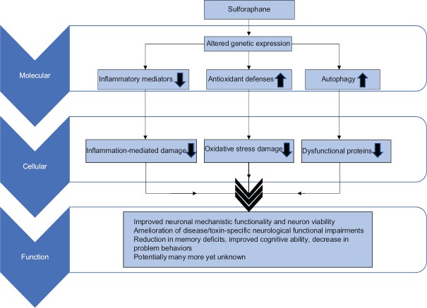
Further investigating A claim of improved cognitive function, Part 3 of Rejuvenation therapy and sulforaphane offered:
“Improving brain function does not depend on neurogenesis as much as it does on synapse formation and factors such as NMDA receptors which decline in density with age.”
A PubMed “sulforaphane NMDA receptors” search turned up a 2019 cell study The glutathione cycle shapes synaptic glutamate activity:
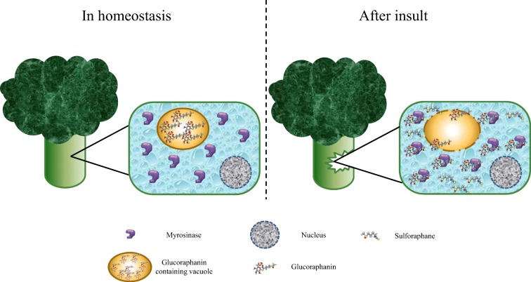
“Sulforaphane is a potent inducer of the Nrf2 transcription factor, has blood–brain barrier penetration, and might expand the size of the glutathione reservoir by our observation that it increases expression of GCL [glutamate cysteine ligase], the rate-limiting step in glutathione biogenesis. Our recent study in human subjects revealed that sulforaphane elevates peripheral glutathione levels and those of other brain metabolites.”
The referenced study was a 2017 Sulforaphane Augments Glutathione and Influences Brain Metabolites in Human Subjects: A Clinical Pilot Study:
“We found that the naturally occurring isothiocyanate sulforaphane increased blood GSH levels in healthy human subjects following 7 days of daily oral administration. In parallel, we explored the potential influence of sulforaphane on brain GSH levels in the anterior cingulate cortex, hippocampus, and thalamus via 7-T magnetic resonance spectroscopy.
A significant positive correlation between blood and thalamic GSH post- and pre-sulforaphane treatment ratios was observed, in addition to a consistent increase in brain GSH levels in response to treatment. The sulforaphane response in brain GSH levels is not influenced by age, sex, or race.
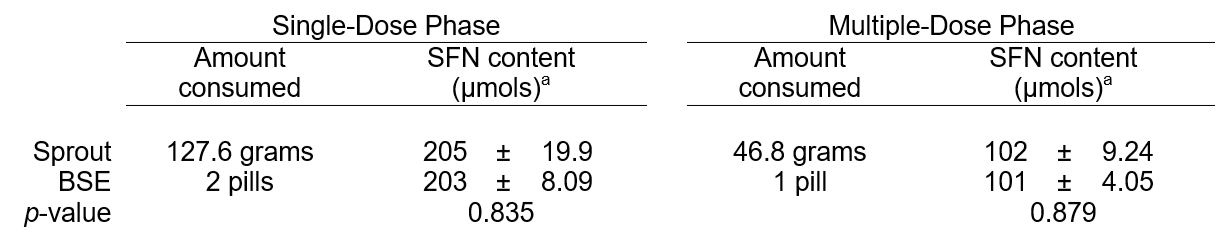
The participants were given 100 µmol sulforaphane as standardized broccoli sprout extract in the form of 2 gel capsules, and instructed to ingest the extract each morning for 1 week.
Following sulforaphane administration, the increase in blood GSH was positively correlated with GABA, Gln [glutamine], Glu [glutamate], and GSH in the THAL [thalamus]. Although these correlations were not significant following multiple comparison, they remain suggestive. Power analysis calculations suggest that a sample size of n = 50 would yield a significant result, and this will be the focus of a future study.
As has been reported for cardiovascular and cerebrovascular diseases, longer treatment duration and/or higher dosages may be warranted. In a submitted study, we will report that peripheral GSH levels may be correlated with cognitive functions.”

One week of consuming sulforaphane wasn’t long enough to achieve much. Not enough subjects and “higher dosages may be warranted” were also thrown in to explain the lack of significant results.
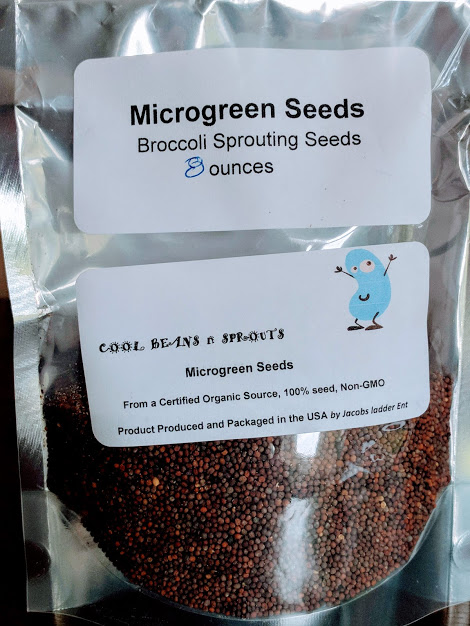
Sulforaphane: Its “Coming of Age” as a Clinically Relevant Nutraceutical in the Prevention and Treatment of Chronic Disease estimated the “100 µmol sulforaphane” dosage to be 17.3 mg. Worst-case estimates made in Estimating daily consumption of broccoli sprout compounds are that since doubling the starting amount of broccoli seeds from one to two tablespoons in Week 6, I’ve consumed 52 mg sulforaphane with microwaving 3-day-old broccoli sprouts every day.
Something happened where the promised “In a submitted study, we will report that peripheral GSH levels may be correlated with cognitive functions” either wasn’t performed or wasn’t published. The follow-on 2019 study became a cell study instead of a 50+ person study.
The study’s thalamus findings provided plausible explanations for why eating a clinically relevant amount of broccoli sprouts every day since at least Week 6, Week 9 was so much different from the others. Sulforaphane changed a blood antioxidant which may have changed four thalamus metabolites.
The thalamus part of our brain is analogous to a switchboard. Signals pass through it to and from other brain areas.
Signals can be routed better when we clean up and upgrade wiring, and lower circuit resistance. Connections within our brains become less inhibited, and external connections concordantly become more apparent.