A case for carnitine supplementation

This 2020 review subject was carnitine, acetyl-L-carnitine, and its other molecular forms:

“Carnitine is necessary to deliver long-chain fatty acids from cytosol into mitochondria. Carnitine homeostasis is maintained by diet and renal absorption, as only a small amount (about 25%) is obtained by endogenous biosynthesis.

Defective fatty acid oxidation occurs with reduced intracellular levels of carnitine, leading to glucose consumption instead of lipid consumption, resulting in hypoglycemia. Non-metabolized lipids accumulate in tissues such as heart, skeletal muscle, and liver, resulting in myopathy and hepatic steatosis.

2000 mg/day is unlikely to provoke unwanted side effects and is safe for humans. In-depth studies are needed to identify a unique method of analysis which can guarantee efficient monitoring of supplement active component amounts.”

https://www.mdpi.com/1420-3049/25/9/2127/htm “The Nutraceutical Value of Carnitine and Its Use in Dietary Supplements”


The review listed animal studies of L-carnitine alone and in combination with:

  • Vitamin D3;
  • Coenzyme Q10;
  • Nicotinamide riboside;
  • Selenium;
  • L-arginine;
  • Anti-histamine drugs cetirizine hydrochloride and chlorpheniramine maleate; and
  • Hypertension drug olmesartan.

Human studies of its effects included:

  • Muscle soreness, damage biomarkers, and cramps;
  • Osteoarthritis knee pain and inflammation markers;
  • Ischemic cerebrovascular injury;
  • Peripheral neuropathy;
  • Nonalcoholic fatty liver disease;
  • Insulin resistance and Type 2 diabetes;
  • Kidney diseases;
  • Inherited diseases phenylketonuria and maple syrup urine;
  • Stress, depression, and anxiety;
  • Male infertility; and
  • Hepatitis C.

Sprouting hulled oats

My Sprouting whole oats trial was a hassle with hulls and a poor germination rate. This week I used hulled oat seeds from a different vendor, and a different study, Degree of oat sprouting, as my model.

  • Oat variety of Avena sativa was a small seed, 7 mm x 2 mm. The model used “huskless oat ‘Gehl'” which is a different species (Avena nuda).
  • 100 seeds weighed 1.5 grams. There were over 1,300 seeds per 20 g batches.
  • Oat sprout batches were processed the same way I do broccoli sprout batches. A new batch started soaking to start germination every 12 hours, then was rinsed three times every 24 hours on a 6 hours – 6 hours – 12 hours cycle. I have an open question to the model’s corresponding coauthor to explain their “4.5‐hr wet steeping, 19‐hr air rest, and 4‐hr steeping, all at 20°C” procedures to start germination, since I didn’t have access to its cited study. The model grew oat sprouts for 1, 2, and 3 days.
  • Temperature in my kitchen was 21°C (70°F) because it’s winter outside. The model grew oat sprouts at 10, 14, 20, 25, and 30°C. Their findings included “Temperatures between 20° and 25°C yielded the most dramatic changes in properties of sprouted oats.”

I evaluated germination results per the model’s Degree of Sprouting finding:

“Length of the coleoptile [shoot] was selected as a criterion of categorization of degree of sprouting. Grains of degree 0 do not show any radicle [root] or coleoptile growth. Degree:

  1. Has visible embryos (small white point), while radicles and coleoptile are not visible;
  2. Shows a developed embryo emerging from the seed coat;
  3. Coleoptile lengths of at least half the oat grain length;
  4. Coleoptile lengths between half and a full grain length; and
  5. Coleoptile longer than a full grain length.”

Most of this trial wasn’t a big deal, adding just a few extra minutes onto what I do three times a day with broccoli sprouts. Here’s what this oat variety’s hulled seeds and 3-day-old sprouts looked like:

The tedious part was evaluating degrees of sprouting. I took as large a bottom-to-top sample as I could tolerate sorting (235 seeds / sprouts, about 17%), with these results:

A 97% germination rate. 🙂 Average weight of three 3-day-old batches was 51.9 grams, for a 260% weight gain. My 5-day-old whole oat sprouts trial had a 22% germination rate and a 221% weight gain.

The model’s Figure 3 Degree of Sprouting finding for 20°C and 25°C at 3 days was hard to read:

Don’t know how > 1% 0 degrees of sprouting at 20°C and 25°C reconciled with their statement “Germinability after 3 days was about 99% at all temperatures.” A numerical table wasn’t provided – yet another question for the corresponding author. Meanwhile, I’ll estimate:

Their hard-to-read Figure 3 also wasn’t completely congruent with their statement:

“Around 20% of grains sprouted at 20° and 25°C had a coleoptile longer than a full grain length (degree of sprouting 5).”


These oat sprouts tasted milder than my previous trial’s. With more than a third at a degree-of-sprouting 5 measurement, they’re sweet, concurrent with the model’s findings that:

“Increased amounts of reducing sugars and ascorbic acid were found particularly in the radicles and coleoptile. Coleoptile and radicle growth (input parameters for the degree of sprouting) and reducing sugars and α‐amylase activity are interdependent.”

Corresponding increased enzyme concentrations produced an aftertaste, though. I ate them along with either food or drink.

Can eating three-day-old oat sprouts of this Montana cultivated variety help with what I’m already doing? Here’s what I expect, given the model was a different oat species, and the Sprouting oats and Oat sprouts analysis studies used different oat cultivars.

1. In order of magnitude: increased antioxidants, GABA, phenolic compounds, protein, amino acids, β-glucan, and polyunsaturated fatty acids. Don’t know about GABA and protein, but the others may help counter inflammation.

2. Increased enzyme intake. The model study used α-amylase as a marker for α-amylase enzymes (catalyze starches), protease enzymes (catalyze proteins), and lipase enzymes (catalyze fats).

Oat sprouts analysis characterized increased α-amylase and lipase activities as undesirable in a sprouted oat flour context. More on enzymes in Part 2 of Sprouting hulled oats.

Week 37 of Changing to a youthful phenotype with broccoli sprouts

1. Been wrong about a few things this past week:

A. I thought in Week 28 that extrapolating A rejuvenation therapy and sulforaphane results to humans would produce personal results by this week. An 8-day rat treatment period ≈ 258 human days, and 258 / 7 ≈ 37 weeks.

There are just too many unknowns to say why that didn’t happen. So I’ll patiently continue eating a clinically relevant 65.5 gram dose of microwaved broccoli sprouts twice every day.

PXL_20201015_105645362

The study’s lead researcher answered:

“Depends, it might take 37 weeks or more for some aspects of ‘youthening’ to become obvious. It might even take years for others.

Who really cares if you are growing younger every day?

For change at the epigenomic/cellular level to travel up the biological hierarchy from cells to organ systems seems to take time. But the process can be repeated indefinitely (so far as we know) so by the second rejuvenation you’re already starting at ‘young’. (That would be every eight to ten years I believe.)”

His framework is in An environmental signaling paradigm of aging.

B. I thought that adding 2% mustard seed powder to microwaved broccoli sprouts per Does sulforaphane reach the colon? would work. Maybe it would, maybe it wouldn’t, but my stomach and gut said that wasn’t for me.

C. I thought I could easily add Sprouting whole oats to my routine. I ran another trial Sprouting hulled oats using oat seeds from a different company and Degree of oat sprouting as a model.

2. Oat sprouts analysis paired studies were very informative, don’t you think? One study produced evidence over 18 germination-parameter combinations (hulled / dehulled seeds of two varieties, for 1-to-9 days, at 12-to-20°C).

Those researchers evaluated what mix of germination parameters would simultaneously maximize four parameters (β-glucan, free phenolic compounds, protease activity, and antioxidant capacity) while minimizing two (enzymes α-amylase and lipase). Then they followed with a study that characterized oat seeds sprouted under these optimal conditions.

I doubted PubMed’s “oat sprout” 20 search results for research 1977 to the present. Don’t know why they didn’t pick up both of these 2020 studies, but I’m sure that .gov obvious hindrances to obtaining relevant information like this won’t be fixed. What other search terms won’t return adequate PubMed results?

3. The blog post readers viewed this week that I made even better was Do delusions have therapeutic value? from May 2019. Sometimes I’ve done good posts describing why papers are poorly researched.

4. I’ve often changed my Week 4 recipe for an AGE-less Chicken Vegetable Soup dinner (half) then the next day for lunch. The biggest change brought about by 33 weeks of behavioral contagion is that I now care more about whether vegetables are available than whether or not they’re organic. Coincidentally, I’ve developed a Costco addiction that may require intervention.

  • 1/2 lemon
  • 4 Roma tomatoes
  • 4 large carrots
  • 6 stalks organic celery
  • 6 mushrooms
  • 6 cloves garlic
  • 6 oz. organic chicken breast fillet
  • 1 yellow squash, alternated with 1 zucchini
  • 1 cup sauvignon blanc
  • 32 oz. “unsalted” chicken broth, which still contains 24% of the sodium RDA

Pour wine into a 6-quart Instant Pot; cut and strain squeezed lemon; cut chicken into 1/4″ cubes and add; start mixture on Sauté. Wash and cut celery and stir in. Wash and cut carrots and stir in.

When pot boils around 8 minutes, add chicken broth and stir. Wash mushrooms, slicing into spoon sizes.

Wash and slice yellow squash / zucchini. Crush and peel garlic, tear but don’t slice. Turn off pot when it boils again around 15 minutes.

Wait 2-3 minutes for boiling to subside, then add yellow squash / zucchini, mushrooms, garlic, whole tomatoes. Let set for 20 minutes; stir bottom-to-top 5 and 15 minutes after turning off, and again before serving.

AGE-less Chicken Vegetable Soup is tasty enough to not need seasoning.

Oat sprouts analysis

A research group published two 2020 studies on sprouting oat seeds. Their first study produced evidence over a range of germination parameters (hulled / dehulled seeds of two varieties, for 1-to-9 days, at 12-to-20°C):

“The aim was to investigate the influence of germination period and temperature on protein profile, bioactive potential (β-glucan and phenolic contents), antioxidant capacity, and on activity of enzymes (α-amylase, protease and lipase) from hulled and dehulled oat varieties. Multi-response optimization was used to identify optimal germination conditions that maximize sprouted oat flour quality.

  • Hulled (variety Barra) and dehulled (variety Meeri) germination was performed in dark at different temperatures (12, 14, 16, 18, and 20 ◦C) and duration (24, 60, 96, 156, and 216 h).
  • Germination at 16 ◦C for 216 h and 20 ◦C for 96 h produced the highest protein accumulation in varieties Barra and Meeri, respectively.
  • Germination for short periods (24–96 h) combined with medium temperatures (12–16 ◦C) retained β-glucan levels, but longer germination times (156–216 h) caused reductions of 47–64%. Endogenous β-glucanases increase activity during germination, causing hydrolysis of β-glucan.
  • Free phenolic compound content was between 1.6-fold and 2.8-fold higher when germination took place at high temperatures (16–18 ◦C) for longer times.
  • Antioxidant capacity was between 1.4 and 4.5-fold higher. High temperatures (16–18 ◦C) and longer germination times (156–216 h) positively influenced antioxidant capacity.

The effect of germination conditions strongly depended on genetic diversity and presence/absence of hull.

Optimal germination conditions maximize contents of β-glucan, free phenolic compounds, protease activity, and antioxidant capacity, and minimize activity of undesirable enzymes α-amylase and lipase. For variety Meeri, that corresponded to 18 ◦C and time 120 h.”

https://www.sciencedirect.com/science/article/abs/pii/S0023643820309440 “Changes in protein profile, bioactive potential and enzymatic activities of gluten-free flours obtained from hulled and dehulled oat varieties as affected by germination conditions” (not freely available)


Their second 2020 study analyzed properties of 4-day-old oat sprouts. Dehulled oat seeds (variety Meeri) were soaked at room temperature for 4 hours, then germinated in darkness at 18°C with humidity ≥ 90%.

“Sprouted oat powder was an excellent source of protein (10.7%), β-glucan (2.1%), thiamine, riboflavin, and minerals (P, K, Mg and Ca). It presented better amino acid and fatty acid compositions, and levels of γ-aminobutyric acid [GABA], free phenolics, and antioxidant capacity than control.

Protein content (g/100 g) and amino acid profile (g/100 g protein). Different letters within a row indicate p ≤ 0.05 statistical differences.

During germination, proteins are partially hydrolyzed increasing availability of free amino acids. Activity of glutamate decarboxylase enzyme is enhanced.

However, no significant reduction of glutamate content was observed. Glutamate is used for GABA and protein synthesis, but it is also produced by protein hydrolysis, glutamine synthetase-glutamate synthase cycle, and GABA transaminase reactions.

Sprouted oat powder exhibited 2.5-fold higher SPC [soluble (free) phenolic compounds] levels. De novo synthesis of phenolic compounds or liberation of phenolic compounds that are linked to macromolecules due to cell wall dismantling during germination could explain enhancement of SPC.

Sprouted oat powder displayed a 3-fold higher antioxidant capacity. Release of bound phenolic compounds and de novo synthesis of avenanthramides might be responsible.

Hydrolysis of β-glucan might also cause an increase in oxygen radical absorbance capacity. β-glucan oligosaccharides exhibit high radical scavenging activity and reducing power, and that could be related with exposure of their active hydroxyl groups and decrease of intermolecular hydrogen bonding during germination.”

https://www.sciencedirect.com/science/article/abs/pii/S0308814620318343 “Sprouted oat as a potential gluten-free ingredient with enhanced nutritional and bioactive properties” (not freely available)


Both studies started germination by:

“Twenty grams of oat seeds were used for germination. Soaking (1:6 ratio, w/v) was performed at room temperature (20 ◦C ±2 ◦C) for 4 h.”

Neither study included estimates of germination rates. I contacted the corresponding coauthor for that information, and they replied:

“The germination rate in hulled oat varieties was around 95% and in
dehulled one around 55-70% depending on the germination conditions.”


Degree of oat sprouting

This 2019 study investigated oat sprout parameters:

Huskless oat ‘Gehl’ cultivated in 2016 in Canada, was used throughout the study. Grains (500 g) were sprouted at different temperatures (10, 14, 20, 25, and 30°C) and for different times (1, 2, and 3 days). Changes in vitamin C, β‐glucan, and reducing sugar were monitored, and α‐amylase activity was studied as a marker for total enzyme activity.

Mass fraction of radicle [root] and coleoptile [shoot] in grain correlated very well with β‐glucan level. A similarly good correlation was found for the much easier applicable degree of sprouting, visual assessment of coleoptile length set into relation to grain size.

Germinability after 3 days was about 99% at all temperatures. Temperatures between 20° and 25°C yielded the most dramatic changes in properties of sprouted oats.

  • At 3 days, α‐amylase activities at 20° and 25°C increased significantly to values one order of magnitude larger than those for other temperatures.
  • β‐glucan content was decreased after 3 days at all temperatures. Degradation was most pronounced at 20°C, almost halving initial β‐glucan content to 3.9%.
  • No ascorbic acid was present in native grain. Upon sprouting, a significant increase in ascorbic acid content was found – except at 30°C – with highest levels at 20°C.

Ascorbic acid content in radicles and coleoptile was four times higher than that in grain without radicles and coleoptile. Oat grains sprouted for 3 days at 20°C had an average degree of sprouting of 3; hence, radicles and coleoptile contributed about 8% of mass. These findings indicate that a fast visual determination of degree of sprouting allows to estimate, for example, ascorbic acid content without doing expensive experiments.

Around 20% of grains sprouted at 20° and 25°C had a coleoptile longer than a full grain length (degree of sprouting 5). Less long coleoptiles developed at other temperatures.

  • For the 3‐day sprouting period, the longest coleoptile was observed for sprouting at 25°C.
  • At 30°C average degree of sprouting was 1.4, and grains showed no practical radicle growth.

Coleoptile and radicle growth (input parameters for the degree of sprouting) and reducing sugars and α‐amylase activity are interdependent. Degree of sprouting could develop into a reliable characterization method for sprouted grains, usable for predicting compositional and nutritional changes of oats during sprouting.”

https://onlinelibrary.wiley.com/doi/full/10.1002/cche.10203 “Sprouting of oats: A new approach to quantify compositional changes”


Relative humidity wasn’t mentioned in this study. I asked the corresponding coauthor about it, since two Sprouting oats studies stated relative humidity as a factor for sprouting oats.

I also asked them to explain their “4.5‐hr wet steeping, 19‐hr air rest, and 4‐hr steeping, all at 20°C” procedures to start germination, since I didn’t have access to the cited study. No reply yet.

This was my model study for Sprouting hulled oats.

Ducks in a row

An oats β-glucan clinical trial

This 2020 human study investigated effects of processing β-glucan:

“Nutritional advantages of oats compared to many other grains include gluten-free nature, high content of polyunsaturated fatty acids, protein composition which complements that of pulses, and substantiated health effects of fibers, specifically oat β-glucan. Novel oat products, which are often semi-solid or liquid, generally need alterations of the physicochemical properties of oats.

The hypothesis in this study [Clinical trial NCT02764931] was that bioprocessing of oat bran with enzyme treatment, causing depolymerization of β-glucan, affects nutritional properties of bran and functional properties of β-glucan in human gastrointestinal tract.

The study meal consisted of oat bran concentrate treated with a commercial food-grade cell wall degrading enzyme preparation at 1 or 50 nkat β-glucanase g-1 dm-1. A control sample was prepared in the same way without added enzymes. Average MW [molecular weight] of β-glucan in:

  • Control oat bran concentrate was >1000 kDa [weight in kilodaltons] (High MW);
  • 1 nkat g-1 dm-1-treated 524 kDa (Medium MW); and
  • 50 nkat g-1 dm-1-treated 82 kDa (Low MW).

Results of this study supported the hypothesis that alteration of oat β-glucan MW with enzymatic treatment affects nutritional properties of oat bran and functional properties of β-glucan in the human gastrointestinal tract:

  • A High MW β-glucan meal resulted in the highest excretion of fecal bile acids, and the lowest excretion of phenolic compounds in urine.
  • A Low MW β-glucan meal resulted in the lowest excretion of fecal bile acids, but the highest excretion of phenolic compounds, especially ferulic acid, in urine.
  • Medium MW β-glucan was similar to High MW β-glucan in that it resulted in high excretion of fecal bile acids and low excretion of phenolic compounds to urine, but mean pressure in the duodenum was closer to Low MW than to High MW meal.

Perceived gut well-being after consumption of each meal did not differ between meals, but varied between genders, which should be further investigated.”

https://www.sciencedirect.com/science/article/abs/pii/S0308814620320811 “Effect of oat β-glucan of different molecular weights on fecal bile acids, urine metabolites and pressure in the digestive tract – A human cross over trial” (not freely available)


I eat 81 grams of steel-cut oats every morning, which is represented by this study’s high-molecular-weight control. Take responsibility for your one precious life.

No β-glucan for dolphins or seagulls

Sprouting oats

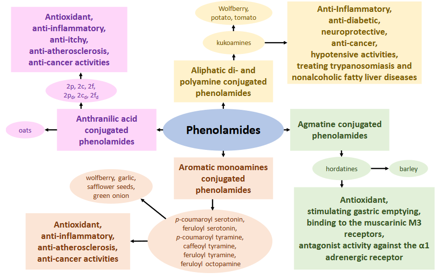
Three 2020 studies investigated properties of sprouted oats. This first study compared one oat cultivar’s seed and sprout contents for phenolic compounds, and evaluated oat sprouts’ protection against developing colon cancer:

“The purpose of this investigation was to evaluate whether sprouted oats (SO) of the Turquesa variety still possessed effective physiologically bioactive compounds, i.e., phenolics, flavonoids, AVAs [avenanthramides], and phytosterols, and whether it exerted antioxidant and anti-inflammatory effects, as well as the capacity to improve relevant intestinal parameters, in an AOM [azoxymethane] / DSS [dextran sulfate sodium]-induced CRC [colorectal cancer] mouse model.

Suboptimal intake of whole grains (38 g/d) was associated with CRC burden across 16 European countries. An optimal intake of 50–100 g/d was considered in our study to establish the dose administered in the AOM/DSS-induced CRC mouse model (75 g/d).

Seeds (100 g) were soaked in distilled water for 12 h then watered daily. Temperature and relative humidity were set at 25 °C and 60%. Germination was performed in darkness for five days. Germination percentage was determined based on total number of fully emerged seedlings.

We reached 100% of germination and a radicle length of 6.47 ± 0.22 cm. Sprouts were dried at 50 °C for 12 h, milled to a particle size of 0.5 mm, and stored at 4 °C until analyses.

Protein and lipid contents were higher in SO, whereas carbohydrate and ash contents were lower. A more than four-fold increase [0.64 mg/g to 2.79 mg/g] in TPC [total phenolic compounds] was obtained after five days.

We identified AVA-D as the most abundant AVA, followed by AVA-L, which had not been reported as one of the three most abundant AVAs in other oat varieties. Of the three most abundant AVAs previously reported, only AVA-B had a higher abundance in germination.

Phytic acid, an antinutritional compound present in oats, was 10 times lower in oat sprouts. Phytic acid has its content decreased by 15%–35% during even a short three-day germination due to activation of phytase activity. Although high doses of phytic acid inhibit absorption of metals and minerals in humans, it has been observed that, in small doses, it can function as a protective factor in several chronic degenerative diseases.

Mice in groups 3 and 4 were gavaged every morning with phenolic-AVA extract (0.084 mg GAE) and 30 mg of SO, respectively. We observed a mild anti-inflammatory effect of SO and AVA treatments, and a reduced adenocarcinoma incidence of 52.5% and 21.3%, respectively.

SO was more efficient in activating the Keap1-Nrf2 signaling pathway compared to treatment with AVA. Oat phenolic compounds together with β-glucans may be acting synergistically, thus offering greater protection for cancer prevention and treatment.”

https://www.mdpi.com/2304-8158/9/2/169/htm “Chemopreventive Effect of the Germinated Oat and Its Phenolic-AVA Extract in Azoxymethane/Dextran Sulfate Sodium (AOM/DSS) Model of Colon Carcinogenesis in Mice”

The supplementary material developed this oat cultivar’s seed and sprout profiles for 138 phenolic compounds. It measured C-type AVAs, but not A-type AVAs.

This was my model study for Sprouting whole oats.


A second study was reviewed in Eat oats today! and repeated here:

“The first evaluation of anti-inflammation effects of A-type AVAs was published from our own group. Fifteen A-type AVAs from commercial sprouted oat products interacted with lipopolysaccharide-induced nitric oxide production and iNOS expression.”

https://pubs.acs.org/doi/full/10.1021/acs.jafc.9b06812 “Quantitative Analysis and Anti-inflammatory Activity Evaluation of the A-Type Avenanthramides in Commercial Sprouted Oat Products” (not freely available)

Oat variety and sprout age weren’t available for the six sprouted oat products tested, so oat seed-to-sprout comparisons weren’t possible. A-type AVA comparisons among products were performed, but weren’t meaningful due to unknown varieties, ages, product processing, and storage.


A third study compared four grains’ sprouted and unsprouted contents:

“Seeds were soaked at 25°C in 1 L of distilled water for 20 (brown rice), 12 (sorghum and millet) and 8 h (oat), respectively. Hydrated grains were allowed to germinate with layering over wet cellulose pads in a humid chamber for 60 h at 25°C (oat seeds) or 30°C (brown rice, sorghum, and millet seeds) with 95% relative humidity.

All seeds derived from brown rice and oat were germinated after 48 h in the humid chamber. Germinated grains were dried at 50°C until reaching a moisture content of 10%. Sample seeds were milled to fine flour, screened through a 100-mesh sieve and stored at 4°C for further analysis.

After 60 h of germination, sprout length in sorghum and millet ranged from 8 to 24 mm, while sprouts obtained from brown rice and oat ranged from 3 to 6 mm.

Compared to raw flours, germinated flours derived from brown rice, sorghum, and millet had lower gelatinization enthalpy, whereas germinated oat flour showed higher gelatinization enthalpy.

During germination, enzymes are activated, catalyzing starch degradation, which may disrupt the double helical structure of starch. Consequently, less energy is required to unravel and melt double helices of starch in germinated flours. The increase in gelatinization enthalpy of germinated oat flour may be due to dissolution of hydrolyzed starch granules during germination.”

https://link.springer.com/article/10.1007%2Fs10068-020-00770-2 “Influence of germination on physicochemical properties of flours from brown rice, oat, sorghum, and millet” (not freely available)


The first study sprouted oats for five days to full germination and a minimum radicle length of 6.25 cm. The third study sprouted oats to full germination in 60 hours and a 3 mm minimum total length.

At the same 25°C, with 60% relative humidity and daily watering, it took 120 hours to achieve full germination. With 95% relative humidity, it took half that time.

Was humidity a relevant difference in oat sprout growth? Would Choyang variety oat sprouts increase their minimum 3 mm total length more than 20 times between Hours 60 and 120 to match the minimum Turquesa radicle length?

This is a count of PubMed “oat sprout” search results, 20 results total:

A “broccoli sprout” search returned 648 results. Is oat sprout research just getting started?

Part 2 of The transgenerational impact of Roundup exposure

This 2020 study followed up The transgenerational impact of Roundup exposure using the Washington State Unversity research group’s most recent methodology in DEET and permethrin cause transgenerational diseases:

“The herbicide glyphosate has been shown to promote epigenetic transgenerational inheritance of pathology and disease in subsequent great-grand offspring (F3 generation). The current study was designed to identify epigenetic biomarkers for glyphosate-induced transgenerational diseases using an epigenome-wide association study.

Pathologies investigated included prostate disease [13 of 44 subjects], kidney disease [11 of 44], obesity [19 of 45], and presence of multiple disease [10 of 45]. Sperm were collected from F3 glyphosate lineage males and used to identify specific differential DNA methylation regions (DMRs) and differential histone retention sites (DHRs).

The number of DHRs were less than the number of DMRs, and DHRs were found to have disease specificity. The combination of DMRs and DHRs is anticipated to facilitate pathology diagnosis.

Low sample number is a limitation in the current analysis. Potential higher variability in data needs to be considered.

This is one of the first observations of DHRs as potential biomarkers for disease. The current study used glyphosate induction of transgenerational disease as a proof of concept such environmental biomarkers can be identified and potentially used as diagnostics for disease susceptibility in the future.”

https://www.tandfonline.com/doi/full/10.1080/15592294.2020.1853319 “Epigenome-wide association study for glyphosate induced transgenerational sperm DNA methylation and histone retention epigenetic biomarkers for disease”


Eat oats today!

This 2020 food chemistry review provided phenolic-compound reasons to eat oats:

“Phenolamides result from the conjugation of hydroxycinnamic acids with amines. These products contain a variety of metabolic, chemical, and functional capabilities due to the large number of possible combinations among the parent compounds.

Of the currently known phenolamides, the most common are avenanthramides (AVAs), which are unique in oats. AVAs possess anti-inflammatory, anti-itch, anti-atherosclerosis, antioxidant, anti-cancer, anti-obesity, anti-fungal, anti-microbial, and neuroprotective properties.

Twenty-nine C-type AVAs have been identified in oats, and twenty-six A-type AVAs.

  • C-type AVAs in commercially available oat products range from 36.49-61.77 mg/kg (fresh weight).
  • A-type AVAs represent approximately 22.5% of total AVA levels in regular oats and 24.7-33.0% in commercial sprouted oats.

Steeping raw groats increased AVA concentrations.”

These reviews were referenced:

“Since publication of these two reviews, a few new studies reported AVAs’ beneficial health effects, mainly related to their anti-inflammatory and anti-cancer activities. AVAs can:

  • Significantly decrease IL-6, IL-8, and MCP-1 in endothelial cells;
  • Inhibit IL-1β- and TNF-α-induced NF-κB activation; as well as
  • Expression of adhesion molecules; and
  • Adhesion of monocytes to endothelial cell monolayer.

In 2020, the first evaluation of anti-inflammation effects of A-type AVAs was published from our own group. Fifteen A-type AVAs from commercial sprouted oat products interacted with lipopolysaccharide-induced nitric oxide production and iNOS expression.

Colloidal oatmeal’s natural components, AVAs, help to restore and maintain skin barrier function. AVAs are safe, well tolerated, and can be effective as adjuvant treatment in atopic dermatitis.

In one mouse model, a C-type AVA was able to mitigate many adverse effects of Alzheimer’s Disease. It restored hippocampal long-term potentiation and synaptic function, enhanced memory function, suppressed pro-inflammatory cytokines TNF-α and IL-6 levels, reduced caspase-3 levels, and increased pS9GSK-3β and IL-10 levels.

AVAs downregulated expression of hTERT and MDR1, pro-survival genes for cancer cells, and COX-2 mRNA and PGE2 levels, known pro-inflammatory markers. AVAs induced apoptosis by activating caspases 8, 3, and 2.”

https://pubs.acs.org/doi/10.1021/acs.jafc.0c02605 “The Chemistry and Health Benefits of Dietary Phenolamides” (not freely available)


Hadn’t thought about sprouting oats before this paper.

Clearing out the 2020 queue of interesting papers

I’ve partially read these 39 studies and reviews, but haven’t taken time to curate them.

Early Life

  1. Intergenerational Transmission of Cortical Sulcal Patterns from Mothers to their Children (not freely available)
  2. Differences in DNA Methylation Reprogramming Underlie the Sexual Dimorphism of Behavioral Disorder Caused by Prenatal Stress in Rats
  3. Maternal Diabetes Induces Immune Dysfunction in Autistic Offspring Through Oxidative Stress in Hematopoietic Stem Cells
  4. Maternal prenatal depression and epigenetic age deceleration: testing potentially confounding effects of prenatal stress and SSRI use
  5. Maternal trauma and fear history predict BDNF methylation and gene expression in newborns
  6. Adverse childhood experiences, posttraumatic stress, and FKBP5 methylation patterns in postpartum women and their newborn infants (not freely available)
  7. Maternal choline supplementation during the third trimester of pregnancy improves infant information processing speed: a randomized, double‐blind, controlled feeding study
  8. Preterm birth is associated with epigenetic programming of transgenerational hypertension in mice
  9. Epigenetic mechanisms activated by childhood adversity (not freely available)

Epigenetic clocks

  1. GrimAge outperforms other epigenetic clocks in the prediction of age-related clinical phenotypes and all-cause mortality (not freely available)
  2. Epigenetic age is a cell‐intrinsic property in transplanted human hematopoietic cells
  3. An epigenetic clock for human skeletal muscle
  4. Immune epigenetic age in pregnancy and 1 year after birth: Associations with weight change (not freely available)
  5. Vasomotor Symptoms and Accelerated Epigenetic Aging in the Women’s Health Initiative (WHI) (not freely available)
  6. Estimating breast tissue-specific DNA methylation age using next-generation sequencing data

Epigenetics

  1. The Intersection of Epigenetics and Metabolism in Trained Immunity (not freely available)
  2. Leptin regulates exon-specific transcription of the Bdnf gene via epigenetic modifications mediated by an AKT/p300 HAT cascade
  3. Transcriptional Regulation of Inflammasomes
  4. Adipose-derived mesenchymal stem cells protect against CMS-induced depression-like behaviors in mice via regulating the Nrf2/HO-1 and TLR4/NF-κB signaling pathways
  5. Serotonin Modulates AhR Activation by Interfering with CYP1A1-Mediated Clearance of AhR Ligands
  6. Repeated stress exposure in mid-adolescence attenuates behavioral, noradrenergic, and epigenetic effects of trauma-like stress in early adult male rats
  7. Double-edged sword: The evolutionary consequences of the epigenetic silencing of transposable elements
  8. Blueprint of human thymopoiesis reveals molecular mechanisms of stage-specific TCR enhancer activation
  9. Statin Treatment-Induced Development of Type 2 Diabetes: From Clinical Evidence to Mechanistic Insights
  10. Rewiring of glucose metabolism defines trained immunity induced by oxidized low-density lipoprotein
  11. Chronic Mild Stress Modified Epigenetic Mechanisms Leading to Accelerated Senescence and Impaired Cognitive Performance in Mice
  12. FKBP5-associated miRNA signature as a putative biomarker for PTSD in recently traumatized individuals
  13. Metabolic and epigenetic regulation of T-cell exhaustion (not freely available)

Aging

  1. Molecular and cellular mechanisms of aging in hematopoietic stem cells and their niches
  2. Epigenetic regulation of bone remodeling by natural compounds
  3. Microglial Corpse Clearance: Lessons From Macrophages
  4. Plasma proteomic biomarker signature of age predicts health and life span
  5. Ancestral stress programs sex-specific biological aging trajectories and non-communicable disease risk

Broccoli sprouts

  1. Dietary Indole-3-Carbinol Alleviated Spleen Enlargement, Enhanced IgG Response in C3H/HeN Mice Infected with Citrobacter rodentium
  2. Effects of caffeic acid on epigenetics in the brain of rats with chronic unpredictable mild stress
  3. Effects of sulforaphane in the central nervous system
  4. Thiol antioxidant thioredoxin reductase: A prospective biochemical crossroads between anticancer and antiparasitic treatments of the modern era (not freely available)
  5. Quantification of dicarbonyl compounds in commonly consumed foods and drinks; presentation of a food composition database for dicarbonyls (not freely available)
  6. Sulforaphane Reverses the Amyloid-β Oligomers Induced Depressive-Like Behavior (not freely available)

Does reprogramming signaling pathways create memories?

This 2020 study investigated genes and signaling pathways for inflammatory memory:

“Fibroblast-like synoviocytes (FLS) play a critical role in pathogenesis of rheumatoid arthritis (RA). Chronic inflammation induces transcriptomic and epigenetic modifications that imparts a persistent catabolic phenotype to the FLS, despite their dissociation from the inflammatory environment.

Sustained activated genes established pro-inflammatory signaling components known to act at multiple levels of NF-κB, STAT and AP-1 signaling cascades. Sustained repressed genes included critical mediators and targets of the BMP [bone morphogenic protein] signaling pathway.

We identified sustained repression of BMP signaling as a unique constituent of the long-term inflammatory memory induced by chronic inflammation.

FLS are synovial tissue-resident and specialized mesenchymal cells critical for homeostasis. Key features of these cells during homeostasis include the production of extracellular matrix components and providing nutrients to the synovial fluid. Healthy synovium is composed of multiple layers of FLS, which forms the synovial lining and sublining through cell–cell contacts.

Inflammatory and pro-resolving mediators are tightly regulated to maintain normal synovium functioning. However, in inflammatory and autoimmune diseases such as rheumatoid arthritis, an imbalance between these signals causes homeostasis disruption leading to synovial tissue damage, cartilage destruction and bone degeneration.”

https://www.ncbi.nlm.nih.gov/pmc/articles/PMC7679373/ “Chronic exposure to TNF reprograms cell signaling pathways in fibroblast-like synoviocytes by establishing long-term inflammatory memory”


These researchers described a positive feedback loop that kept rheumatoid arthritis in place. No feedback diagram or explanation of what sustained a disease condition other than to say:

“Gene expression changes induced by short-term tumor necrosis factor-alpha (TNF-α) treatment were largely sustained in the FLS exposed to chronic inflammation.”

Okay – then what upstream signals sustained TNF-α? What would it take to interrupt that feedback loop? What initiated it?

Studies usually substantiate effects by also developing evidence for causes of opposite effects and of no effects. This study investigated neither reversibility nor no effect, and instead stated:

“Multiple signaling networks are irreversibly modified due to TNF-α-mediated long-term epigenetic and transcriptomic reprogramming. We speculate that sustained repression of BMP signaling may be critically required to ensure the persistently transformed phenotype of RA FLS.”

No evidence was offered for “irreversibly modified.” Anyway, that didn’t fit with:

“We postulate that simultaneous targeting of these activated and repressed signaling pathways may be necessary to combat RA persistence.”

Enduring epigenetic memories? Or continuous toxic stimulation? provided another perspective: “Enduring epigenetic effects may be symptoms rather than causes when toxic conditions persist.”


Been on a Steely Dan kick lately. Probably due to this year’s Royal Scam:

Zinc and broccoli sprouts – a winning combination

This 2019 study deserved better coverage than a one sentence mention in Reversal of aging and immunosenescent trends with sulforaphane:

“Obstructive sleep apnea syndrome is one of the most common breathing disorders in sleep, with a high prevalence of 3–7% and severe consequences. It is characterized by intermittent hypoxia (IH) due to recurrent episodes of partial or complete collapse of the upper airway during sleep, leading to blood hypoxemia, hypercapnia, sleep fragmentation, augmented respiratory efforts, and increased sympathetic activity.

Our study is the first investigation of the combination of BSE [broccoli sprout extract] and Zn [zinc] – Nrf2 and MT [metallothionein] inducers – to protect against IH-induced cardiomyopathy. By effectively activating Nrf2, its downstream targets, and MT, this combination can ameliorate defects associated with IH-induced cardiomyopathy more effectively than monotherapies.

Mice were administered with BSE (equivalent to SFN [sulforaphane] 2 mg/kg) and/or Zn sulfate heptahydrate (5 mg/kg) by gavage from 8 weeks of age at a frequency of once every other day for 8 weeks. Doses used in this study are safe to convert to human doses. [2 mg x .081 x 70 kg = 11 mg sulforaphane; 5 mg x .081 x 70 kg = 28 mg zinc]

  • Heart mass was significantly lower in the IH-BSE/Zn group than in IH and IH-BSE groups. Heart mass / tibia length ratio was significantly lower in the IH-BSE/Zn group than in IH and monotherapy groups.
  • Treatment with BSE and/or Zn can ameliorate myocardial fibrosis associated with IH, to a certain extent, and combination therapy had the best antifibrotic effect among treatments.
  • BSE or Zn can significantly ameliorate myocardial inflammation induced by IH, but the combination provided a better anti-inflammatory effect.
  • We used 3-NT as an indicator of the severity of oxidative stress. 3-NT protein levels were significantly reduced in IH mice for all treatment groups, and reduction was greater in the combination treatment group.
  • Combination was more effective than monotherapies to activate Nrf2-mediated antioxidant function.

  • In Zn-treated and combination treatment groups, MT protein expression was significantly higher than in the IH group, and there was only a slight increase in the IH-BSE group.”

Combination of Broccoli Sprout Extract and Zinc Provides Better Protection Against Intermittent Hypoxia-Induced Cardiomyopathy Than Monotherapy in Mice


One way to improve broccoli sprout compounds’ effects is to eat them with zinc. One way to improve zinc’s actions is to take it with broccoli sprouts.

Week 34 of Changing to a youthful phenotype with broccoli sprouts

1. Thank you to readers of this blog who find the 650+ curations and other posts worth their time. I reread blog posts after you read them, and sometimes improve them for our mutual benefit.

One such post this week was Broccoli sprout compounds include sinapic acid derivatives. Although it was already fairly detailed, it received a half-dozen improvements.

  • Those researchers measured composition changes of 31 compounds (18 sinapic acid derivatives, 8 glucosinolates, and 5 flavonoids) identified in seed-2-4-6-day germination stages of one cultivar. They provided convertible dry weight and fresh weight measurements in mg / g.
  • It complemented the 3-day-old broccoli sprouts have the optimal yields study comparisons of six cultivated varieties’ seed-3-5-7-day germination stage weights and measurements with their origins using a milligram-per-gram-of-seeds scale:

    “To be comparable, the content of these bioactive compounds from 100 fresh sprouts was divided by the weight (g) of 100 seeds, and then this value was compared with their content from one gram seeds.”

  • The sinapic acid study discussed another study for:

    “In a study, diminishing amounts of total phenolic acids in sprouts of three broccoli cultivars was observed only between 3rd and 7th day of germination under photoperiod conditions and only when expressed on fresh weight basis. After recalculating results to dry weight, amounts were increasing during the whole 14-day observation period.”

All studies were scientifically informative. Still, results depended on researchers’ operative paradigms, and human behavior such as unconscious act-outs of unsatisfied needs to feel important.

2. Speaking of which, I viewed a 1:48 video with broccoli sprout experts who disparaged microwaving around the 1:10 mark. I’m not an expert, but I’ve eaten a clinically-relevant dose of microwaved broccoli sprouts every day for 34 weeks now. Read this post’s comment.

Here are a few studies of microwaving’s effects on phenolic, glucosinolate, and flavonoid broccoli compounds. Just for those who value evidence more than opinion.

  • Microwaving broccoli sprouts may not affect phenolic levels found four of five test cases didn’t significantly diminish total phenolic fresh weight contents of whole broccoli. They blended 100 grams broccoli in 200 ml water, halved the purée, then microwaved half on 700W power for 30 seconds. No disclosure of what temperature was achieved, but it was probably < 60°C (140°F). Microwaving significantly increased the glucosinolate hydrolysis product indole-3-carbinol:

    “I3C in broccoli was increased by 3.1, 9.1 and 1.9 folds respectively using blenders 1, 2 and 5 with microwaving.”

  • Microwave broccoli to increase flavonoid levels study design was “Broccoli florets (150 g) were put in a microwave safe bowl with a 1 tablespoon [15 ml] of water” and a 1200W microwave on full power for one minute. Although this may have produced temperatures > 60°C, flavonoid fresh weight contents increased > 30%:

    Microwaving may increase extractability and/or release from binding to other compounds as a result of matrix softening.

  • Microwave broccoli to increase sulforaphane levels demonstrated significant differences for 475W (LL) and 950W (HL) power settings in glucoraphanin and sulforaphane dry weight amounts when broccoli florets were microwaved to the same temperatures. Compare white bar sulforaphane amounts for LL60 and HL60 (both 60°C), annotated as E and F:

    “Microwave treatment causes a sudden collapse of cell structure due to the increase in osmotic pressure difference over vacuole membrane. Microwave irradiation might help to release more conjugated forms of glucosinolates and then get hydrolyzed by released myrosinase.

  • Enhancing sulforaphane content confirmed the above 60°C finding with broccoli florets:

    “The best treatment temperature for maximizing sulforaphane yield was 60 °C. The slightly higher sulforaphane yield than would be predicted from the level of glucoraphanin in raw broccoli requires further investigation. Sulforaphane yield of broccoli after 5 min thermal treatment at 65 °C was even lower than the value obtained for raw broccoli.

3. I see socialistic animal behavior often during beach walks. If one seagull pecks a food morsel, a half-dozen others immediately position themselves to take it. It’s a race to the bottom of existence.

Too bad we humans don’t learn pertinent lessons from others’ experiences, much less our own. Today’s US Thanksgiving provides one example.

Richard Ebeling presented the factual Thanksgiving story a while back. Have you read about collectivism that arrived with the Mayflower in 1620? Do you think we’ve learned what we needed to learn about communism from four centuries ago through today?

4. Seagulls are also inspirational in their flock behavior of joie de vivre predawn flying.

Part 2 of Eat broccoli sprouts for your eyes

I was a little bothered by an unreferenced statement in Eat broccoli sprouts for your eyes that:

“Once AGEs are formed, most are irreversible.”

I searched curated 2020 studies for “revers” and found that recent blog studies favored reversibility of epigenetic changes 12-to-2. Do they reflect my selection bias, or is there something different about AGEs?

Let’s start with this statement:

“Although AGEs are irreversible adducts and cross-links in our tissues, these can be removed through different proteolytic capacities:

  • The ubiquitin proteasome system (UPS) – Ubiquitin is a protein that when conjugated to a protein substrate can facilitate degradation of that substrate by the proteasome. Obsolete or damaged proteins are tagged with ubiquitin and these ubiquitinated substrates are degraded by the proteasome. Operates mainly on soluble substrates.
  • Autophagy – Can operate on insoluble substrates, including organelles such as mitochondria. Autophagy requires macromolecular assemblies and organelles to identify, sequester, and eventually degrade substrates via the lysosome.

Unfortunately, the function of both proteolytic pathways declines with extensive glycative stress and upon aging in many tissues, resulting in intracellular accumulation of protein aggregates (also glycated conjugates) and dysfunctional organelles. This thwarts strategies to lower AGEs accumulation by boosting proteolytic capacities.”

https://www.mdpi.com/2076-3921/9/11/1062/htm “Glyoxalase System as a Therapeutic Target against Diabetic Retinopathy”


So humans can remove irreversible AGE epigenetic changes as long as the individual isn’t too stressed or old? Studies from 2008 to 2012 were cited for the above statement and graphic.

Citation 211 Sulforaphane delays diabetes-induced retinal photoreceptor cell degeneration (not freely available) 2020 findings were instructive:

“SF [sulforaphane] can delay photoreceptor degeneration in diabetes. The underlying mechanism is related to:

  • Inhibition of ER [endoplasmic reticulum] stress;
  • Inflammation; and
  • Txnip [thioredoxin-interacting protein] expression through activation of the AMPK [adenosine 5′-monophosphate (AMP)-activated protein kinase] pathway.

Function of the retina in diabetic mice [DM] as determined by ERG [electroretinography].”

This chart demonstrated that preventing diabetes’ negative effects on retinal function (i.e. controls) was measurably better than trying to fix subjects’ vision after onset of diabetes. Are future choices of humans who give themselves this non-communicable disease also limited to addressing symptoms?

The AMPK pathway was previously mentioned in:

  1. Reversal of aging and immunosenescent trends with sulforaphane:

    “Dihydroxyvitamin D3 and sulforaphane are compounds that safely induce AMPK activation, and may have wide-ranging implications for both normal and pathological aging.”

  2. Part 2 of Reversal of aging and immunosenescent trends with sulforaphane:

    “NQO1 plays a key role in AMPK-induced cancer cell death through the CD38/cADPR/RyR/Ca2+/CaMKII signaling pathway. Expression of NQO1 is elevated by hypoxia / reoxygenation or inflammatory stresses through nuclear accumulation of the NQO1 transcription factor, Nrf2. Activation of the cytoprotective Nrf2 antioxidant pathway by sulforaphane protects immature neurons and astrocytes from death caused by exposure to combined hypoxia and glucose deprivation.”

This first example was vitamin D3’s separate yet connected signaling pathway that acts both additively and synergistically with broccoli sprout compound effects. The second was signaling pathways becoming cascadingly activated from sulforaphane’s main effect, Nrf2 signaling pathway activation.


Eat broccoli sprouts for your eyes

This 2020 review subject concerned a leading cause of blindness:

“Advanced glycation end products (AGEs) are toxic compounds that have adverse effects on many tissues including the retina and lens. AGEs promote formation of reactive oxygen species (ROS), which, in turn, boost production of AGEs, a vicious cycle.

Diabetic retinopathy (DR) is a devastating microvascular complication of diabetes mellitus and the leading cause of blindness in working-age adults. The onset and development of DR is multifactorial. Lowering AGEs accumulation may represent a potential therapeutic approach.

Once AGEs are formed, most are irreversible. Cataracts are perhaps the earliest pathobiology of AGEs:

Nε-(carboxymethyl)-lysine (CML) [a representative AGE] in lens crystallins from diabetic (■) and non-diabetic (♦) subjects as a function of age.

The glyoxalase system is a protective mechanism that slows down synthesis of AGEs by limiting reactive dicarbonyls formed during sugar metabolism. Glutathione (GSH) in the eye is present at concentrations many times blood levels, and is a critical component of the glyoxalase system.

Proteomic analysis identified GLO1 [glyoxalase 1] as a protein differentially expressed in cells treated with sulforaphane. Sulforaphane inhibited AGEs-derived pericyte damage and delayed diabetes-induced retinal photoreceptor cell degeneration.

No AGE inhibitors have reached clinical use. The glyoxalase system and discovery of compounds that enhance this detoxifying activity represent a therapeutic alternative to fight glycation-derived damage.”

https://www.mdpi.com/2076-3921/9/11/1062/htm “Glyoxalase System as a Therapeutic Target against Diabetic Retinopathy”


The above graph – plotting a cataract AGE level against chronological age – represented life stage progression without effective personal agency, without taking responsibility for your one precious life.

Citation 156 was Activation of Nrf2 attenuates carbonyl stress induced by methylglyoxal in human neuroblastoma cells: Increase in GSH levels is a critical event for the detoxification mechanism (not freely available):

“The present study focused on the methylglyoxal (MG) detoxification mechanism. MG treatment resulted in accumulation of modified proteins bearing the structure of AGEs.

This accumulation was suppressed by activation of the Nrf2 pathway prior to MG exposure via pre-treatment with an Nrf2 activator:

Although pre-treatment with the Nrf2 activator did not affect mRNA levels of GLO1, expressions of GCL and xCT mRNA, involved in GSH synthesis, were induced prior to increase in GSH levels.

These results indicated that increase in GSH levels promoted formation of the GLO1 substrate, thereby accelerating MG metabolism via the glyoxalase system and suppressing its toxicity. Promotion of GSH synthesis via the Nrf2/Keap1 pathway is important in MG detoxification.”

Continued in Part 2.


PXL_20201121_113656177