How to hijack science: Ignore its intent and focus on the 0.0001%

This 2018 Belgian review hijacked science to further an agenda:

“We addressed this issue at the LATSIS Symposium ‘Transgenerational Epigenetic Inheritance: Impact for Biology and Society’, in Zürich, 28–30 August 2017, and here provide important arguments why environmental and lifestyle-related exposures in young men should be studied.”

The reviewer DETRACTED from science in the studied area – transgenerational epigenetic inheritance – by ignoring its intent. As shown by A self-referencing study of transgenerational epigenetic inheritance which I also curated today, the purpose of such animal studies is to find the mechanisms in order to help humans.


Putting that study’s graphic into human terms, F3 male great-grandchildren may be adversely affected by their F0 great-grandmothers being poisoned while pregnant with their F1 grandfathers, who – with their F2 fathers – may have also been adversely affected.


What the reviewer asserted without proof:

“The importance of maternal lifestyle, diet and other environmental exposures before and during gestation period is well recognized.”

is NOT TRUE for the studied area.

The evidence disproving this assertion is that NO HUMAN STUDIES scientifically demonstrating causes for transgenerational epigenetic inheritance effects have been published!

EVER!!

There’s a huge gap between “The importance..is well recognized” of anything regarding transgenerational epigenetic inheritance and ZERO human studies.

Why has no one published scientifically adequate human evidence to demonstrate “Transgenerational Epigenetic Inheritance: Impact for Biology and Society” effects on ALL of the F1, F2, and F3 human generations as consequences “of maternal lifestyle, diet and other environmental exposures before and during gestation period?” What are we waiting for?

The reviewer said “young men should be studied” but said nothing about resolving bottlenecks in funding human research of the studied area. Do researchers even have opportunities to make a NON-AGENDA-DRIVEN difference in this field?

With ZERO published human studies, transgenerational epigenetic inheritance research can’t be recharacterized into a female vs. male agenda. The reviewer’s attempt to do so diminished the importance of research into human critical development periods.

This agenda’s viewpoint ignored human correlates of evidence from animal studies like The lifelong impact of maternal postpartum behavior:

“The defect in maternal care induced by gestational stress programs the development of the offspring.”

Will the reviewer’s suggested interventions – such as changing an adult’s lifestyle a long time after their development was altered – somehow make up for what went wrong early in their life, even before they were born?

With the evidence from animal studies such as:

is there any doubt that similar mechanisms may be involved in humans, and that human phenotypes may likewise be intergenerationally and/or transgenerationally transmitted?

The reviewer asserted:

“Studying humans is challenging, because of ethical reasons”

But do “ethical reasons” prohibit non-instigating human studies of stress, the intergenerationally and transgenerationally transmitted effects of which seem to be ubiquitous among humans?

In The Not-Invented-Here syndrome I pointed out another problem that the reviewer’s agenda is less than helpful in resolving:

“How can animal studies like the current study help humans when their models don’t replicate common human conditions? This failure to use more relevant models has follow-on effects such as human intergenerational and transgenerational epigenetic inheritance being denigrated due to insufficient evidence.”


I’ll repeat What is a father’s role in epigenetic inheritance? in closing:

“The review focused on 0.0001% of the prenatal period for what matters with the human male – who he was at the time of a Saturday night drunken copulation – regarding intergenerational and transgenerational epigenetic inheritance of metabolic diseases.

The human female’s role – who she was at conception AND THEN what she does or doesn’t do during the remaining 99.9999% of the prenatal period to accommodate the fetus and prevent further adverse epigenetic effects from being intergenerationally and transgenerationally transmitted – wasn’t discussed.

Who benefits from this agenda’s narrow focus?”

https://academic.oup.com/eep/article/4/2/dvy007/4987171 “POHaD: why we should study future fathers”

A self-referencing study of transgenerational epigenetic inheritance

This 2018 Washington rodent study subject was transgenerational epigenetic inheritance of disease caused by a fungicide that’s been phased out or banned for over a decade:

“This study was designed to help understand how three different epigenetic processes in sperm are correlated with vinclozolin-induced epigenetic transgenerational inheritance of disease.

  1. Most DMRs [differential DNA-methylated regions] identified in this study are unique between the F1, F2, and F3 generations.
  2. The number of lncRNA was much higher than the number of sncRNA [small noncoding RNA, including microRNA]. The overlap between each generation was very low or nonexistent.
  3. The F1 and the F2 generation control versus vinclozolin lineage sperm had negligible DHRs [differential histone retention sites]. This observation suggests that the direct vinclozolin exposure does not alter histone retention or trigger any changes. However, the F3 generation control versus vinclozolin lineage sperm DHRs increased considerably.

It appears that the phenomenon is more complex than just a direct exposure triggering the formation of epimutations that are then simply maintained in the subsequent generations.”


There’s something odd about a study where a third of the 87 cited references list one of the study’s coauthors, who also coauthored A review of epigenetic transgenerational inheritance of reproductive disease. I couldn’t find a satisfactory explanation for the study’s over-the-top self-referencing.

What do you think?

I asked the coauthors why a third of the cited references were self-referencing. The lead author replied:

“The field in epigenetic transgenerational inheritance is expanding, however it is still hard for us to find relevant studies in rodents or human that we can cite. Most of the time DNA methylation, ncRNA and histone modifications are investigated from a direct exposure and/or from a purely mechanistic angle (e.g. DNA methylation of specific genes).

In contrast, transgenerational phenotypes and toxicology by definition excludes direct exposure and must be transmitted through multiple generations (the F3 generation is the first transgenerational one). We are not looking at specific genes but using whole genome sequencing technologies which is a broader approach.

Besides, if you do a pubmed search with the keywords “epigenetics” and “transgenerational”, you will probably find that more than 50% of the studies have been done by Dr Michael K. Skinner. He is also one of the first researcher who started to work on the epigenetic transgenerational inheritance phenomenon 15 years ago. Not citing his previous work is challenging.

We hope to see other labs contributing to this particular field and we will be delighted to cite them. In the meantime, our only option is to reference our previous work.”

I replied:

“Thank you for your reply! It must be exasperating to see other researchers stop their studies short of the F3 generation for no apparent or disclosed reason.

Have you seen even one scientifically adequate human study of transgenerational epigenetic inheritance?”

https://academic.oup.com/eep/article/4/2/dvy010/4987173 “Alterations in sperm DNA methylation, non-coding RNA expression, and histone retention mediate vinclozolin-induced epigenetic transgenerational inheritance of disease”

Immune memory in the brain

This 2018 German rodent study was a proof-of-principle for immune epigenetic memory in the brain:

“Innate immune memory is a vital mechanism of myeloid [bone marrow] cell plasticity that occurs in response to environmental stimuli and alters subsequent immune responses.

Two types of immunological imprinting can be distinguished – training and tolerance. These are epigenetically mediated and enhance or suppress subsequent inflammation, respectively.

Certain immune stimuli train blood monocytes to generate enhanced immune responses to subsequent immune insults. By contrast, other stimuli induce immune tolerance – suppression of inflammatory responses to subsequent stimuli.

Microglia (brain-resident macrophages) are very long-lived cells. This makes them particularly interesting for studying immune memory, as virtually permanent modification of their molecular profile appears possible. Immune memory in the brain is predominantly mediated by microglia.

In a mouse model of Alzheimer’s pathology, immune training exacerbates cerebral β-amyloidosis and immune tolerance alleviates it; similarly, peripheral immune stimulation modifies pathological features after stroke. Our results identify immune memory in the brain as an important modifier of neuropathology.

Immune memory in the brain could conceivably affect the severity of any neurological disease that presents with an inflammatory component, but this will need to be studied for each individual condition.”


The researchers performed multiple experiments to test different hypotheses about how immune-response experiences are remembered. Modifications to histone methylation and acetylation were targeted.

The stimulus dosage needed to produce immune tolerance – “suppression of inflammatory responses to subsequent stimuli” – was usually four times the dosage used for immune training – “enhanced immune responses to subsequent immune insults.”

https://www.nature.com/articles/s41586-018-0023-4 “Innate immune memory in the brain shapes neurological disease hallmarks” (not freely available)

Resiliency in stress responses

This 2018 US Veterans Administration review subject was resiliency and stress responses:

“Neurobiological and behavioral responses to stress are highly variable. Exposure to a similar stressor can lead to heterogeneous outcomes — manifesting psychopathology in one individual, but having minimal effect, or even enhancing resilience, in another.

We highlight aspects of stress response modulation related to early life development and epigenetics, selected neurobiological and neurochemical systems, and a number of emotional, cognitive, psychosocial, and behavioral factors important in resilience.”

The review cited studies I’ve previously curated:


There were two things I didn’t understand about this review. The first was why the paper isn’t freely available. It’s completely paid for by the US taxpayer, and no copyright is claimed. I recommend contacting the authors for a copy.

The second was why the VA hasn’t participated in either animal or human follow-on studies to the 2015 Northwestern University GABAergic mechanisms regulated by miR-33 encode state-dependent fear. That study’s relevance to PTSD, this review’s subject, and the VA’s mission is too important to ignore. For example:

“Fear-inducing memories can be state dependent, meaning that they can best be retrieved if the brain states at encoding and retrieval are similar.

“It’s difficult for therapists to help these patients,” Radulovic said, “because the patients themselves can’t remember their traumatic experiences that are the root cause of their symptoms.”

The findings imply that in response to traumatic stress, some individuals, instead of activating the glutamate system to store memories, activate the extra-synaptic GABA system and form inaccessible traumatic memories.”

I curated the research in A study that provided evidence for basic principles of Primal Therapy. These researchers have published several papers since then. Here are the abstracts from three of them:

Experimental Methods for Functional Studies of microRNAs in Animal Models of Psychiatric Disorders

“Pharmacological treatments for psychiatric illnesses are often unsuccessful. This is largely due to the poor understanding of the molecular mechanisms underlying these disorders. We are particularly interested in elucidating the mechanism of affective disorders rooted in traumatic experiences.

To date, the research of mental disorders in general has focused on the causal role of individual genes and proteins, an approach that is inconsistent with the proposed polygenetic nature of these disorders. We recently took an alternative direction, by establishing the role of miRNAs in the coding of stress-related, fear-provoking memories.

Here we describe in detail our work on the role of miR-33 in state-dependent learning, a process implicated in dissociative amnesia, wherein memories formed in a certain brain state can best be retrieved if the brain is in the same state. We present the specific experimental approaches we apply to study the role of miRNAs in this model and demonstrate that miR-33 regulates the susceptibility to state-dependent learning induced by inhibitory neurotransmission.”

Neurobiological mechanisms of state-dependent learning

“State-dependent learning (SDL) is a phenomenon relating to information storage and retrieval restricted to discrete states. While extensively studied using psychopharmacological approaches, SDL has not been subjected to rigorous neuroscientific study.

Here we present an overview of approaches historically used to induce SDL, and highlight some of the known neurobiological mechanisms, in particular those related to inhibitory neurotransmission and its regulation by microRNAs (miR).

We also propose novel cellular and circuit mechanisms as contributing factors. Lastly, we discuss the implications of advancing our knowledge on SDL, both for most fundamental processes of learning and memory as well as for development and maintenance of psychopathology.”

Neurobiological correlates of state-dependent context fear

“Retrieval of fear memories can be state-dependent, meaning that they are best retrieved if the brain states at encoding and retrieval are similar. Such states can be induced by activating extrasynaptic γ-aminobutyric acid type A receptors (GABAAR) with the broad α-subunit activator gaboxadol. However, the circuit mechanisms and specific subunits underlying gaboxadol’s effects are not well understood.

Here we show that gaboxadol induces profound changes of local and network oscillatory activity, indicative of discoordinated hippocampal-cortical activity, that were accompanied by robust and long-lasting state-dependent conditioned fear. Episodic memories typically are hippocampus-dependent for a limited period after learning, but become cortex-dependent with the passage of time.

In contrast, state-dependent memories continued to rely on hippocampal GABAergic mechanisms for memory retrieval. Pharmacological approaches with α- subunit-specific agonists targeting the hippocampus implicated the prototypic extrasynaptic subunits (α4) as the mediator of state-dependent conditioned fear.

Together, our findings suggest that continued dependence on hippocampal rather than cortical mechanisms could be an important feature of state-dependent memories that contributes to their conditional retrieval.”


Here’s an independent 2017 Netherlands/UC San Diego review that should bring these researchers’ efforts to the VA’s attention:

MicroRNAs in Post-traumatic Stress Disorder

“Post-traumatic stress disorder (PTSD) is a psychiatric disorder that can develop following exposure to or witnessing of a (potentially) threatening event. A critical issue is to pinpoint the (neuro)biological mechanisms underlying the susceptibility to stress-related disorder such as PTSD, which develops in the minority of ~15% of individuals exposed to trauma.

Over the last few years, a first wave of epigenetic studies has been performed in an attempt to identify the molecular underpinnings of the long-lasting behavioral and mental effects of trauma exposure. The potential roles of non-coding RNAs (ncRNAs) such as microRNAs (miRNAs) in moderating or mediating the impact of severe stress and trauma are increasingly gaining attention. To date, most studies focusing on the roles of miRNAs in PTSD have, however, been completed in animals, using cross-sectional study designs and focusing almost exclusively on subjects with susceptible phenotypes.

Therefore, there is a strong need for new research comprising translational and cross-species approaches that use longitudinal designs for studying trajectories of change contrasting susceptible and resilient subjects. The present review offers a comprehensive overview of available studies of miRNAs in PTSD and discusses the current challenges, pitfalls, and future perspectives of this field.”

Here’s a 2017 Netherlands human study that similarly merits the US Veterans Administration’s attention:

Circulating miRNA associated with posttraumatic stress disorder in a cohort of military combat veterans

“Posttraumatic stress disorder (PTSD) affects many returning combat veterans, but underlying biological mechanisms remain unclear. In order to compare circulating micro RNA (miRNA) of combat veterans with and without PTSD, peripheral blood from 24 subjects was collected following deployment, and isolated miRNA was sequenced.

PTSD was associated with 8 differentially expressed miRNA. Pathway analysis shows that PTSD is related to the axon guidance and Wnt signaling pathways, which work together to support neuronal development through regulation of growth cones. PTSD is associated with miRNAs that regulate biological functions including neuronal activities, suggesting that they play a role in PTSD symptomatology.”


See the below comments for reasons why I downgraded this review’s rating.

https://link.springer.com/article/10.1007/s11920-018-0887-x “Stress Response Modulation Underlying the Psychobiology of Resilience” (not freely available)

Are there epigenetic causes for sexual orientation and gender identity?

This US 2018 review lead author was a gynecologic oncologist in private practice:

“Sexual orientation is biologically conferred in the first trimester of pregnancy. Gender identity is biologically conferred during the middle trimester of pregnancy.

Since the genitals differentiate in the first trimester, and the brain becomes imprinted in the latter half of gestation, it is possible for the fetal brain to be imprinted differently than the genitals. As children mature, this innate imprinting expresses as genital anatomy, gender identity, sexual orientation and other physiologic capabilities and natural preferences along a continuum, between masculine and feminine.

The evidence shows that both orientation and identity are biologic features that co-vary with a very large number of other biologic sexually dimorphic traits.”


1. A fetus’ development is influenced by survival reactions to their environment. Although fetal and placental responses to environmental stressors are relevant to sexual orientation and gender identity, the reviewers didn’t explore the subject.

2. Epigenetic adaptations to the prenatal environment involving microRNA were mentioned in a small subsection. But the reviewers didn’t cite relevant studies involving DNA methylation, chromatin and histone modifications for epigenetic causes of and effects on sexual orientation and gender identity.

3. The reviewers included a half-dozen anecdotal quotations from personal correspondence that promoted their narrative. These impressed as appeals to authority rather than evidence for scientific understanding of the subject.

It was insufficient for the review to note “a continuum between masculine and feminine” without also exploring evidence for an individual’s placement on the continuum. The question of possible epigenetic causes for sexual orientation and gender identity remains.

https://www.sciencedirect.com/science/article/pii/S009082581731510X “Biological origins of sexual orientation and gender identity: Impact on health” (not freely available)

Faith-tainted epigenetics

This 2018 Loma Linda review subject was epigenetic interventions for aging:

“Epigenomic markers of aging, global DNA hypomethylation and promoter-specific hypermethylation may be engendered by iron and HCys [homocysteine] retention.

MiR-29/p53 axis may reverse age-related methylomic shifts, stabilizing both the genome and the epigenome, therefore removing a major risk factor of neurodegeneration. Lowering iron and HCys overload can be accomplished via chelation, blood donation and maintaining an adequate omega-6/omega-3 ratio.”


Sometimes it’s difficult to detect researchers’ biases. If a reader didn’t know about the funding sponsor’s mission:

“Each day we seek to extend the teaching and healing ministry of Jesus Christ”

they may view this paper as unbiased rather than as a directed narrative.

Consider the sponsor’s influence from the perspective of someone seeking treatment for Alzheimer’s disease. If a doctor in this review sponsor’s hospital system recommended chelation treatment, hope would be generated for the patient. Adopting the doctor’s belief about the treatment, though, would be contrary to other evidence per this review:

“In 2008, the NIH chelation trial stopped enrolling patients, approximately two years early.

There is no indication for exposing patients with dementia to the risks of chelation therapy because current chelators cannot help them.”

After reading another review that had this sponsor – The lack of oxygen’s epigenetic effects on a fetus – which also reflected the influence of the sponsor’s biases, and had a directed narrative that ignored evidence contradicting the narrative, and involved storytelling, I’m done curating any paper sponsored by this institution.

http://www.nrronline.org/downloadpdf.asp?issn=1673-5374;year=2018;volume=13;issue=4;spage=635;epage=636;aulast=Sfera;type=2 “Epigenetic interventions for brain rejuvenation: anchoring age-related transposons” (click the pdf button)

Methods of detecting additional epigenetic modifications

This 2018 German review subject was detecting DNA modifications that are derivatives of the much-studied 5-methlycytosine:

“The discovery of modified nucleobases arising from 5-methylcytosine (5mC) through consecutive oxidation to give 5-hydroxymethylcytosine (5hmC), 5-formylcytosine (5fC), and 5-carboxylcytosine (5caC) has stimulated intense research efforts regarding the biological functions of these epigenetic marks.

Recent findings revealed that 5hmC and 5fC are stable DNA modifications in the genome, thus suggesting that oxidized 5mC derivatives may function as epigenetic marks in their own right, exhibiting regulatory purposes and participating in DNA replication, transcription, repair, and recombination.

The bisulfite-sequencing method (BS-Seq) has widely been used as the gold standard in determining the methylation status with single-base resolution in genomic DNA. The BS-Seq method, however, has some severe drawbacks, such as:

  • Harsh reaction conditions which might cause undesired DNA damage,
  • Requirements for relatively large amounts of input DNA,
  • Dependence on PCR, and resulting short sequence reads, as well as
  • Reduced sequence complexity due to deamination of all nonmethylated cytosines and
  • Accompanied challenges for primer hybridization.

Most importantly, however, with BS-Seq it is not possible to discriminate between 5mC and 5hmC..Furthermore, since 5fC and 5aC undergo deamination similar to unmodified cytosine, they are indistinguishable from C under bisulfite conditions.”


https://febs.onlinelibrary.wiley.com/doi/abs/10.1002/1873-3468.13058 “Chemoselective labeling and site-specific mapping of 5-formylcytosine as a cellular nucleic acid modification” (click the PDF link)

A flying human tethered to a monkey

Ponder this drone photo of “a flying human tethered to a monkey” ground drawing made over 1,000 years ago as reported by National Geographic and excerpted by the Daily Star:
Flying human tethered to a monkey


1. Aren’t the geoglyph and its description pretty good expressions of our evolved condition? Especially since it’s the interpretation of people who lived more a millennium ago?

With so many information sources freely available now, though, one couldn’t successfully argue that the ancients understood the world better than we do. Our understanding comes from our “flying human” time and efforts, without which we’re as ignorant as our “monkey.”

2. A few aspects of the current comprehension of the differences between our two pictured primates are in Genetic imprinting, sleep, and parent-offspring conflict:

“I remain skeptical of a tendency to ascribe most modern woes to incongruence between our evolved nature and western cultural practices. We did not evolve to be happy or healthy but to leave genetic descendants, and an undue emphasis on mismatch risks conflating health and fitness [genetic rather than physical fitness].”

Our “flying human” can make happiness and health choices that our “monkey” can’t:

“Our genetic adaptations often try to fool us into doing things that enhance fitness at costs to our happiness.

Our genes do not care about us and we should have no compunction about fooling them to deliver benefits without serving their ends.

Contraception, to take one obvious example, allows those who choose childlessness to enjoy the pleasures of sexual activity without the fitness-enhancing risk of conception.”

3. Another aspect of our two primates’ differences is illuminated in a reference to A study of DNA methylation and age:

“Aging is not and cannot be programmed. Instead, aging is a continuation of developmental growth, driven by genetic pathways.

Genetic programs determine developmental growth and the onset of reproduction. When these programs are completed, they are not switched off.

Aging has no purpose (neither for individuals nor for group), no intention. Nature does not select for quasi-programs. It selects for robust developmental growth.”

The epigenetic clock theory of aging cited the same author, and modified his point to say:

“The proposed epigenetic clock theory of ageing views biological ageing as an unintended consequence of both developmental programmes and maintenance programmes.”

Aging decisions are examples of our “flying human” making choices that aren’t available to our “monkey” concerning the structure, direction, and duration of our one precious life.

“What are you doing to reverse epigenetic processes and realize what you want? Do you have ideas and/or behaviors that interfere with taking constructive actions to change your phenotype?”

The Not-Invented-Here syndrome

I have high expectations of natural science researchers. I assume that their studies will improve over time, and develop methods and experiments that produce reliable evidence to inform us of human conditions.

My confidence is often unrealistic. Scientists are people, after all, and have the same foibles as the rest of us.

I anticipate that researchers will keep abreast of others’ work around the world. If other groups in their research areas are developing better methods and exploring hypotheses that discover better applications for humans, why not adopt them in the interest of advancing science?

That’s not what happened with this 2018 UK rodent study. The rat model some of the coauthors have built their reputations on depends on disturbing rat pregnancies by administering glucocorticoids. But both the rat model and a guinea pig model in Do you have your family’s detailed medical histories? demonstrated that physicians who disturb their pregnant human patients in this way may be acting irresponsibly toward their patients’ fetuses and their future generations.

This study didn’t find mechanisms that explained transgenerational epigenetic birth weight effects through the F2 grandchild generation:

“Although the phenotype is transmitted to a second generation, we are unable to detect specific changes in DNA methylation, common histone modifications or small RNA [including microRNA] profiles in sperm.

The inheritance mechanism for the paternally derived glucocorticoid-reprogrammed phenotype may not be linked with the specific germline DNA, sRNA and chromatin modifications that we have profiled here.”


The linked guinea pig model was developed specifically to inform physicians of the consequences through the F3 great-grandchild generation of disturbing human pregnancies with glucocorticoids:

“Antenatal exposure to multiple courses of sGC [synthetic glucocorticoid] has been associated with hyperactivity, impaired attention, and neurodevelopmental impairment in young children and animals. It is imperative that the long-term effects of antenatal exposure to multiple courses of sGC continue to be investigated since the use of a ‘rescue’ (i.e. a second) course of sGC has recently re-introduced the practice of multiple course administration.”


If a study’s purpose is to investigate potential mechanisms of epigenetic inheritance, why not adopt a model that better characterizes common human conditions, regardless of which research group initially developed it?

The prenatal stress model used in The lifelong impact of maternal postpartum behavior is one model that’s more representative of human experiences. Those researchers pointed out in Prenatal stress produces offspring who as adults have cognitive, emotional, and memory deficiencies:

“Corticosterone-treated mice and rats exposed to chronic stress are models that do not recapitulate the early programming of stress-related disorders, which likely originates in the perinatal period.”

Animal models that chemically redirect fetal development also “do not recapitulate the early programming of stress-related disorders.”

Other than research that’s done to warn against disrupted development, how can animal studies like the current study help humans when their models don’t replicate common human conditions? This failure to use more relevant models has follow-on effects such as human intergenerational and transgenerational epigenetic inheritance being denigrated due to insufficient evidence.

Of course there’s insufficient human evidence! Researchers developed and sponsors funded animal study designs that ensured there wouldn’t be wide applicability to humans!! Few derivative human studies have been developed and funded as a result.

https://genomebiology.biomedcentral.com/articles/10.1186/s13059-018-1422-4 “Investigation into the role of the germline epigenome in the transmission of glucocorticoid-programmed effects across generations”

Little evidence for mitochondrial DNA methylation

This 2018 Japanese rodent study used three different techniques to detect mitochondrial DNA methylation:

“Whilst 5-methylcytosine (5mC) is a major epigenetic mark in the nuclear DNA in mammals, whether or not mitochondrial DNA (mtDNA) receives 5mC modification remains controversial.

We used bisulfite sequencing, McrBC digestion analyses and liquid chromatography mass spectrometry, which are distinctly differing methods for detecting 5mC. We analysed mtDNAs from mouse ESCs [embryonic stem cells] and from mouse liver and brain tissues.

Taken together, we propose that 5mC is not present at any specific region(s) of mtDNA and that levels of the methylated cytosine are fairly low, provided the modification occurs. It is thus unlikely that 5mC plays a universal role in mtDNA gene expression or mitochondrial metabolism.”


Bisulfite sequencing infers the presence of CpG (CG above) and non-CpG (CH above) methylation through unconverted residues:

“Synthetic and native mtDNA gave similar patterns, suggesting that the resistance of cytosines to bisulfite conversion is not due to methylation.”


It seems that epigenetic changes to mitochondrial DNA occur primarily through histone modifications. Lysine acetylation is gnarly and dynamic is one paper that detailed aspects of this functionality in mitochondria.

https://www.nature.com/articles/s41598-018-24251-z “Accurate estimation of 5-methylcytosine in mammalian mitochondrial DNA”

The epigenetic clock theory of aging

My 400th curation is a 2018 US/UK paper by coauthors of Using an epigenetic clock to distinguish cellular aging from senescence. They reviewed the current state of epigenetic clock research, and proposed a new theory of aging:

“The proposed epigenetic clock theory of ageing views biological ageing as an unintended consequence of both developmental programmes and maintenance programmes, the molecular footprints of which give rise to DNAm [DNA methylation] age estimators.

It is best to interpret epigenetic age estimates as a higher-order property of a large number of CpGs much in the same way that the temperature of a gas is a higher-order property that reflects the average kinetic energy of the underlying molecules. This interpretation does not imply that DNAm age simply measures entropy across the entire genome.

To date, the most effective in vitro intervention against epigenetic ageing is achieved through expression of Yamanaka factors, which convert somatic cells into pluripotent stem cells, thereby completely resetting the epigenetic clock. In vivo, haematopoietic stem cell therapy resets the epigenetic age of blood of the recipient to that of the donor.

Future epidemiological studies should consider other sources of DNA (for example, buccal cells), because more powerful estimates of organismal age can be obtained by evaluating multiple tissues. Other types of epigenetic modifications such as adenine methylation or histone modifications may lend themselves for developing epigenetic age estimators.”


https://www.nature.com/articles/s41576-018-0004-3 “DNA methylation-based biomarkers and the epigenetic clock theory of ageing” (not freely available)


I curated four other papers cited in this review:

Do you want your quality of life to be under or over this curve?

What are you doing to reverse epigenetic processes and realize what you want?

  • Do you have ideas and/or behaviors that interfere with taking constructive actions to change your phenotype?
  • If you aren’t doing anything, are you honest with yourself about feelings of helplessness?
  • Do your beliefs in fate, or in technology, or in divine interventions justify inactions?

The lifelong impact of maternal postpartum behavior

This 2018 French/Italian/Swiss rodent study was an extension of the work done by the group of researchers who performed Prenatal stress produces offspring who as adults have cognitive, emotional, and memory deficiencies and Treating prenatal stress-related disorders with an oxytocin receptor agonist:

“Reduction of maternal behavior [nursing behavior, grooming, licking, carrying pups] was predictive of behavioral disturbances in PRS [prenatally restraint stressed] rats as well as of the impairment of the oxytocin and its receptor gene expression.

Postpartum carbetocin [an oxytocin receptor agonist unavailable in the US] corrected the reduction of maternal behavior induced by gestational stress as well as the impaired oxytocinergic system in the PRS progeny, which was associated with reduced risk-taking behavior.

Moreover, postpartum carbetocin had an anti-stress effect on HPA [hypothalamic-pituitary-adrenal] axis activity in the adult PRS progeny and increased hippocampal mGlu5 [type 5 metabotropic glutamate] receptor expression in aging.

Early postpartum carbetocin administration to the dam enhances maternal behavior and prevents all the pathological outcomes of PRS throughout the entire lifespan of the progeny..proves that the defect in maternal care induced by gestational stress programs the development of the offspring.“


This chart from Figure 4 summarized the behavioral performance of aged adult male progeny in relation to the experimental variables of:

  1. Stress administered to the mothers three times daily every day during the second half of pregnancy up until delivery; and
  2. The effects on the mothers’ behavior of daily carbetocin administration during postpartum days 1 through 7.

The symbols denote which of these relationships had statistically significant effects:

  • “* p [Pearson’s correlation coefficient] < 0.05 PRS-Saline vs. CONT-Saline;
  • # p < 0.05 PRS-Carbetocin vs. the PRS-Saline group.”

There are many interesting aspects to this study. Ask the corresponding coauthor Dr. Sara Morley-Fletcher at sara.morley-fletcher@univ-lille1.fr for a copy.

One place the paper referenced the researchers’ previous studies was in this context:

“Postpartum carbetocin administration reversed the same molecular and behavioral parameters in the hippocampus, as does adult chronic carbetocin treatment, i.e. it led to a correction of the HPA axis negative feedback mechanisms, stress and anti-stress gene expression, and synaptic glutamate release. The fact that postpartum carbetocin administration [to the stressed mothers in this study] had the same effect [on the PRS infants in this study] as adult carbetocin treatment [to the PRS offspring in the previous study] indicates a short-term effect of carbetocin when administered in adulthood and a reprogramming (long-term) effect lasting until an advanced age when administered in early development.”

This group’s research seems to be constrained to treatments of F0 and F1 generations. What intergenerational and transgenerational effects would they possibly find by extending research efforts to F2 and F3 generations?


As the study may apply to humans:

The study demonstrated that stresses during the second half of pregnancy had lifelong impacts on both the mothers’ and offsprings’ biology and behavior. Studies and reviews that attribute similar human biological and behavioral conditions to unknown causes, or shuffle them into the black box of individual differences, should be recognized as either disingenuous or insufficient etiological investigations.

The study showed that prevention of gestational stress was a viable strategy. The control group progeny’s biology and behavior wasn’t affected by carbetocin administration to their mothers because neither they nor their mothers had experience-dependent epigenetic deficiencies.

The study demonstrated a biological and behavioral cure for the PRS offspring by changing their stressed mothers’ behaviors during a critical period of their development. The above excerpt characterized improving the mothers’ behaviors as a long-term cure for the PRS descendants, as opposed to the short-term cure of administering carbetocin to the PRS children when they were adults.

What long-term therapies may be effective for humans who had their developmental trajectories altered by their mothers’ stresses during their gestation, or who didn’t get the parental care they needed when they needed it?

https://www.sciencedirect.com/science/article/pii/S0161813X18301062 “Reduced maternal behavior caused by gestational stress is predictive of life span changes in risk-taking behavior and gene expression due to altering of the stress/anti-stress balance” (not freely available)

Prenatal stress produces offspring who as adults have cognitive, emotional, and memory deficiencies

This 2018 French/Italian/Swiss rodent study used a prenatally restraint stressed (PRS) model to create problems that could be resolved by various chemicals:

“S 47445 is a positive modulator of glutamate AMPA-type receptors, possessing neurotrophic and enhancing synaptic plasticity effects as well as pro-cognitive and anti-stress properties.

Most of studies examining the antidepressant effects of new molecules are carried out using behavioral tests performed in unstressed animals.

Corticosterone-treated mice and rats exposed to chronic stress are models that do not recapitulate the early programming of stress-related disorders, which likely originates in the perinatal period. The PRS rat model is characterized by a prolonged corticosterone response to stress and by abnormal behavior.

All the behavioral alterations induced by PRS were corrected by chronic S 47445 administration at both doses.”


The paper included a section comparing S 47445 to ketamine:

“Ketamine, however, causes severe cognitive impairment and psychotomimetic [mimics the symptoms of psychosis, reference not freely available] effects that are direct consequences of the prolonged inhibition of NMDA receptors in cortical and hippocampal interneurons, and seriously limit the chronic administration of the drug in the clinical setting. [reference not freely available]

S 47445 by inducing a direct activation of AMPARs displayed an antidepressant activity without the adverse effect of ketamine. Indeed, contrary to ketamine, S 47445 presented no psychotomimetic effects and induced no occurrence of spontaneous epileptic seizures. [reference freely available] Moreover, S 47445 also presented pro-cognitive properties.”

Compare the above with this April 2018 Chicago Tribune story that had opinions with no linked references:

“ketamine, an anesthetic used to sedate both people and animals before surgery. It’s also a notorious street drug, abused by clubgoers seeking a trancelike, hallucinatory high. But in recent years, numerous studies have found that ketamine can be an effective and speedy treatment for people with depression.”

Which coverage better informed us?


Treating prenatal stress-related disorders with an oxytocin receptor agonist was performed by several of this paper’s coauthors. One references to it was:

“We have already reported that depolarization-evoked glutamate release in the ventral hippocampus is negatively correlated with risk-taking behavior of PRS rats, and that such correlation can be corrected by chronic treatment with monoaminergic/ melatoninergic antidepressants or oxytocin receptor agonist. Thus, an impairment of glutamatergic transmission in the ventral hippocampus lies at the core of the pathological phenotype of PRS rats.”

Looking at the above graphic of the experimental design, I’m not sure why the term perinatal (occurring during or pertaining to the phase surrounding the time of birth) was used in the paper’s title and content to describe the stress period. The pregnant females were stressed three times every day during the second half of pregnancy up until delivery, so the prenatal (previous to birth) term was more applicable.


So, how does this study help humans?

One takeaway is to avoid stressing pregnant mothers-to-be if her children will be expected to become adults without cognitive, emotional, and behavioral problems.

The study demonstrated one way prenatal events cause lifelong effects. The PRS model provides another example of why it’s useless to ask adult humans to self-report causes of epigenetic problems in their lives when these originated before birth, during infancy, or in early childhood, well before humans develop sufficient cognitive capability to recognize such situations. It’s incomprehensible that this unreliable paradigm is still given significant weight in stress studies, especially when experimental designs:

“Do not recapitulate the early programming of stress-related disorders, which likely originates in the perinatal period.”

Also, a relevant difference between humans and PRS rats is that we can ourselves individually change our responses to experiential causes of ongoing adverse effects. Standard methodologies can only apply external treatments such as those mentioned above.

https://www.sciencedirect.com/science/article/pii/S0028390818301291 “The reduction in glutamate release is predictive of cognitive and emotional alterations that are corrected by the positive modulator of AMPA receptors S 47445 in perinatal stressed rats” (not freely available) Thanks to coauthors Stefania Maccari and Dr. Jerome Mairesse for providing a copy.

Manufacturing PTSD evidence with machine learning

What would you do if you were a scientist who had strong beliefs that weren’t borne out by experimental evidence?

Would you be honest with yourself about the roots of the beliefs? Would you attempt to discover why the beliefs were necessary for you, and what feelings were associated with the beliefs?

Instead of the above, the researchers of this 2017 New York human study reworked negative findings of two of the coauthors’ 2008 study until it fit their beliefs:

“The neuroendocrine response contributes to an accurate predictive signal of PTSD trajectory of response to trauma. Further, cortisol provides a stable predictive signal when measured in conjunction with other related neuroendocrine and clinical sources of information.

Further, this work provides a methodology that is relevant across psychiatry and other behavioral sciences that transcend the limitations of commonly utilized data analytic tools to match the complexity of the current state of theory in these fields.”


1. The limitations section included:

“It is important to note that ML [machine learning]-based network models are an inherently exploratory data analytic method, and as such might be seen as ‘hypotheses generating’. While such an approach is informative in situations where complex relationships cannot be proposed and tested a priori, such an approach also presents with inherent limitations as a high number of relationships are estimated simultaneously introducing a non-trivial probability of false discovery.”

2. Sex-specific impacts of childhood trauma summarized why cortisol isn’t a reliable biological measurement:

“Findings are dependent upon variance in extenuating factors, including but not limited to, different measurements of:

  • early adversity,
  • age of onset,
  • basal cortisol levels, as well as
  • trauma forms and subtypes, and
  • presence and severity of psychopathology symptomology.”

Although this study’s authors knew or should have known that review’s information, cortisol was the study’s foundation, and beliefs in its use as a biomarker were defended.

3. What will it take for childhood trauma research to change paradigms? described why self-reports of childhood trauma can NEVER provide direct evidence for trauma during the top three periods when humans are most sensitive to and affected by trauma:

“The basic problem prohibiting the CTQ (Childhood Trauma Questionnaire) from discovering likely most of the subjects’ historical traumatic experiences that caused epigenetic changes is that these experiences predated the CTQ’s developmental starting point.

Self-reports were – at best – evidence of experiences after age three, distinct from the experience-dependent epigenetic changes since conception.”

Yet the researchers’ beliefs in the Trauma History Questionnaire’s capability to provide evidence for early childhood traumatic experiences allowed them to make such self-reports an important part of this study’s findings, for example:

“The reduced cortisol response in the ER [emergency room] was dependent on report of early childhood trauma exposure.”

https://www.nature.com/articles/tp201738 “Utilization of machine learning for prediction of post-traumatic stress: a re-examination of cortisol in the prediction and pathways to non-remitting PTSD”

The role of DNMT3a in fear memories

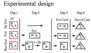
This 2018 Chinese rodent study found:

“Elevated Dnmt3a [a DNA methyltransferase] level in the dorsal dentate gyrus (dDG) of hippocampus was associated with the absence of fear renewal in an altered context after extinction training. Overexpression and knockdown of Dnmt3a in the dDG regulated the occurrence of fear renewal in a bi-directional manner.

We found that renewal of remote fear memory can be prevented, and the absence of renewal was concurrent with an elevated Dnmt3a level.

Our results indicate that Dnmt3a in the dDG is a key regulator of fear renewal after extinction, and Dnmt3a may play a critical role in controlling fear memory return and thus has therapeutic values.”


The study was a collection of five experiments investigating causes and effects of biology and behavior. The researchers used different techniques to achieve their goals. I’ve quoted extensively below to show some background and results.

“Alterations in histone acetylation and DNA methylation are involved in the formation and extinction of long-term memory. DNMTs catalyze the cytosine methylation and are required to establish and maintain genomic methylation.

Dnmt3a and Dnmt3b are de novo DNA methyltransferases. Dnmt1 is the maintenance DNA methyltransferase.

  1. Dnmt3a expression was elevated in the dDG after extinction training followed by a brief memory retrieval (Rec+Ext), which was associated with the absence of fear renewal when tested in an altered context.
  2. Increasing Dnmt3a expression in the dDG using AAV [recombinant adeno-associated virus] expression led to the prevention of fear renewal following a standard extinction training protocol. 
  3. Knockdown of Dnmt3a in the dDG using CRISPR/Cas9 resulted in fear renewal following Rec+Ext protocol.
  4. Renewal of remote fear memory can be prevented using the Rec+Ext protocol.
  5. The absence of renewal was concurrent with an elevated Dnmt3a level.

Current exposure therapy, although effective in many patients, suffers from the inability to generalize its efficacy over time, or is limited by the potential return of adverse memory in the new/novel contexts. These limitations are caused by the context-dependent nature of extinction which is widely viewed as the biological basis of exposure therapy.

Achieving a context-independent extinction may significantly reduce fear renewal to improve the efficacy of exposure therapy. Our current study suggests that the effectiveness of these approaches, and ultimately the occurrence of fear renewal, is determined by the level of Dnmt3a after extinction training, especially in the dDG.

There are two potential mechanisms underlying extinction, one is erasure or updating of the formed memory, and the other is the formation of a new extinction memory which suppresses or competes with the existing memory in a context-dependent manner. While most studies favor the suppression mechanism in the adult, limited studies do suggest that erasure occurs in the immature animals.

We propose that if Dnmt3a level is elevated with extinction training (such as with Rec+Ext protocol), modification to the existing memory occurs and as a consequence extinction does not act as a separate mechanism or form a new memory; but if Dnmt3a level is unaltered with extinction training, a separate extinction memory is formed which acts to suppress or compete with the existing memory.”


The relevant difference between humans and lab rats is that we can ourselves individually change our responses to experiential causes of ongoing adverse effects. Standard methodologies can only apply external treatments such as exposure therapy and manipulating Dnmt3a levels.

https://www.nature.com/articles/s41598-018-23533-w “Dnmt3a in the dorsal dentate gyrus is a key regulator of fear renewal”