Dr. Harold Katcher increased interviews to coincide with release of his book this month. Here’s one in four parts that provides highlights of his rejuvenation research progress:
Previously curated papers of his work include:
Dr. Harold Katcher increased interviews to coincide with release of his book this month. Here’s one in four parts that provides highlights of his rejuvenation research progress:
Previously curated papers of his work include:
This 2021 rodent study investigated:
“We studied long-term dynamics of gut microbiome and short-chain fatty acids (SCFAs) in isogenic mice with distinct microbiota baselines fed with fermentable fiber inulin compared to non-fermentable fiber cellulose.
- We found that inulin produced generally rapid response followed by gradual stabilization to new equilibria, and those dynamics were baseline-dependent.
- Levels of SCFAs such as propionate were associated with abundance of inulin responders, yet inter-individual variation of gut microbiome impedes prediction of SCFAs by machine learning models.
- Our methods and major findings are generalizable to dietary resistant starch.
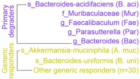
We divided the entire gut microbiota into three eco-groups: 5 primary degraders of inulin; 32 generic responders to inulin intervention; and non-responders. Primary degraders and their competitions are key drivers of baseline-dependent ecological dynamics of microbiota response to dietary fibers.
SCFA concentrations cannot be maintained at its peak, and drop by 35%-40% even under continuous inulin intake until four weeks. 90%-95% SCFAs produced in colonic lumen are absorbed by gut mucosa. The declining phase of SCFAs in our study may be explained by reduced production rate, increased absorption rate, or both.
Our study confirms findings in the literature and advances understanding of effects of dietary fibers on the gut microbiome at the system level:
- The small number of fiber degraders (five for inulin and two for resistant starch) suggested that fiber-induced bacterial shifts are very selective and occur to a restricted number of taxa.
- Absolute abundance of many fiber-degrading bacteria, such as taxa related to genus Bifidobacterium, failed to expand in both fibers. This indicates that fiber-induced bacterial enrichment cannot be simply predicted from in vitro growth, and suggests that dietary response of a gut bacterial taxa depends on the ecological context.
- Personalized fiber-induced response of gut microbiota were largely determined by baseline abundance of fiber degraders and ecological interactions among these degraders.”
https://www.biorxiv.org/content/10.1101/2021.08.20.457175v1.full “Ecological dynamics of the gut microbiome in response to dietary fiber”

2021’s busiest researcher took time out this month to update progress on epigenetic clocks:
Hallmarks of aging aren’t all associated with epigenetic aging.

Interventions that increase cellular lifespan aren’t all associated with epigenetic aging.

Many of his authored or coauthored 2021 papers developed human / mammalian species relative-age epigenetic clocks.

Relative-age epigenetic clocks better predict human results from animal testing.

Previously curated papers that were mentioned or relevant included:
This 2021 rodent study focused on global histone acetylation as a model to understand roles of microbially produced short-chain fatty acids in liver function:
“Despite the utility of germ-free mice in probing complex interactions between gut microbiota and host physiology, germ-free mice are developmentally, physiologically, and metabolically unique when compared with their conventionally housed counterparts. We sought to determine whether antibiotic-mediated microbiota depletion would affect global hepatic histone acetylation states through SCFA-dependent mechanisms, as previously observed in germ-free mice.
The inability of antibiotic-mediated microbiota depletion to recapitulate findings observed in germ-free mice suggests that the transition from a germ-free to a colonized mouse leads to resilient alterations in hepatic histone acetylation states that cannot be altered by further modulating the microbial environment. This finding is distinct from other germ-free phenotypes that are considered to be partially reversible, with clear alterations in their function observed after antibiotic treatment.

Comparing antibiotic-treated and untreated mice that both received CCl4 at 24 and 48 hours after injury, there were almost no histone acetylation differences. This demonstrates that hepatic injury leads to a global shift in histone acetylation that is primarily independent of gut microbiota.
Major chromatin reorganization driven by histone acetylation leads to markers of differentiation, and addition of targeted differentiation signals induces events to stabilize these histone acetylation patterns – a key feature of embryonic development and terminal cellular differentiation. Differences in histone acetylation patterns seen between germ-free and conventionally raised mice may be a developmental-like effect of hepatocytes not yet exposed to microbial by-products.
Results suggest that microbial and dietary modifications to the gut microbiome in conventionally raised mice are not a means to modulate global hepatic histone acetylation. Microbiota-dependent landscaping of the hepatic epigenome appears static in nature, while the hepatic transcriptome is responsive to alterations in the gut microbiota, yet independent of global histone acetylation.
Findings underscore significant differences between these model systems that should be taken into account when considering their relevance to human biology.”
https://aasldpubs.onlinelibrary.wiley.com/doi/10.1002/hep.32043 “Global Microbiota-Dependent Histone Acetylation Patterns Are Irreversible and Independent of Short Chain Fatty Acids” (not freely available) Thanks to Dr. Elliot S. Friedman for providing a copy.
1. By describing “a key feature of embryonic development,” this study provided a gut microbiota-liver analogy of critical periods. If developmental events don’t happen when they are required, it’s probable that their window is missed, and won’t reopen later for a second chance at normalizing.
2. Many studies used a germ-free animal model, such as:
This study provided evidence for a limitation of this model, especially when extrapolating germ-free animal results to humans without similarly testing humans.

This 2021 paper covered a 2016 human clinical trial, and several in vitro and rodent follow-up studies:
“Oat has been widely accepted as a key food for human health. It is becoming increasingly evident that individual differences in metabolism determine how different individuals benefit from diet. Both host genetics and gut microbiota play important roles on metabolism and function of dietary compounds.
Results:
- Avenanthramides (AVAs), the signature bioactive polyphenols of whole-grain (WG) oat, were not metabolized into their dihydro forms, dihydro-AVAs (DH-AVAs), by both human and mouse S9 fractions.
- DH-AVAs were detected in colon and distal regions, but not in proximal and middle regions of the perfused mouse intestine, and were in specific pathogen–free (SPF) mice but not in germ-free (GF) mice.
- A kinetic study of humans fed oat bran showed that DH-AVAs reached their maximal concentrations at much later time points than their corresponding AVAs (10.0–15.0 hours vs. 4.0–4.5 hours, respectively).
- We observed interindividual variations in metabolism of AVAs to DH-AVAs in humans.
- Faecalibacterium prausnitzii was identified as the individual bacterium to metabolize AVAs to DH-AVAs by 16S rRNA sequencing analysis.
- Moreover, as opposed to GF mice, F. prausnitzii–monocolonized mice were able to metabolize AVAs to DH-AVAs.

These findings demonstrate that intestinal F. prausnitzii is indispensable for proper metabolism of AVAs in both humans and mice. We propose that abundance of F. prausnitzii can be used to subcategorize individuals into AVA metabolizers and nonmetabolizers after WG oat intake.
Our findings pave the way to use AVAs and DH-AVAs as exposure biomarkers to reflect WG oat intake, which could more accurately record WG oat intake. Whether production of DH-AVAs is part of the beneficial effect of oats on human health will require further investigation.”
https://academic.oup.com/jn/article/151/6/1426/6165027 “Avenanthramide Metabotype from Whole-Grain Oat Intake is Influenced by Faecalibacterium prausnitzii in Healthy Adults”
Commentary at Faecalibacterium prausnitzii Abundance in Mouse and Human Gut Can Predict Metabolism of Oat Avenanthramides.
This study advanced an understanding of inter-individual variability, rather than usual practices that try to sweep individual differences under a statistical rug. Study designs such as four mentioned in Part 2 of Switch on your Nrf2 signaling pathway could have benefited from a similar approach to their research areas.
Not sure why it took over five years to get this paper published after its clinical trial’s January 21, 2016 completion. Meanwhile, science marched on to study effects of specific F. prausnitzii strains, providing results such as three human studies curated in Gut microbiota strains:
“Only a small number of bacteria with genetic capacity for producing SCFAs were able to take advantage of this new resource and become dominant positive responders. The response, however, was strain specific: only one of the six strains of Faecalibacterium prausnitzii was promoted.”
Resistant starch therapy recommended de-emphasizing relative gut microbiota abundance measurements, because:
“Relative abundances of smaller keystone communities (e.g. primary degraders) may increase, but appear to decrease simply because cross-feeders [like F. prausnitzii] increase in relative abundance to a greater extent. These limitations illustrate the necessity of sufficiently powering resistant starch interventions where microbiome composition is the primary endpoint, collecting critical baseline data and employing appropriate statistical techniques.”
Four humpback whales successively diving for lunch


I was recently asked about taking rapamycin for its effects on mTOR. I replied that diet could do the same thing. Here’s a 2021 review outlining such effects:
“As common, progressive, and chronic causes of disability and death, neurodegenerative diseases (NDDs) significantly threaten human health, while no effective treatment is available. Recent studies have revealed the role of phosphoinositide 3-kinase (PI3K)/Akt (Protein kinase B)/mammalian target of rapamycin (mTOR) in some diseases and natural products with therapeutic potentials.
Growing evidence highlights the dysregulated PI3K/Akt/mTOR pathway and interconnected mediators in pathogenesis of NDDs. Side effects and drug-resistance of conventional neuroprotective agents urge the need for providing alternative therapies.
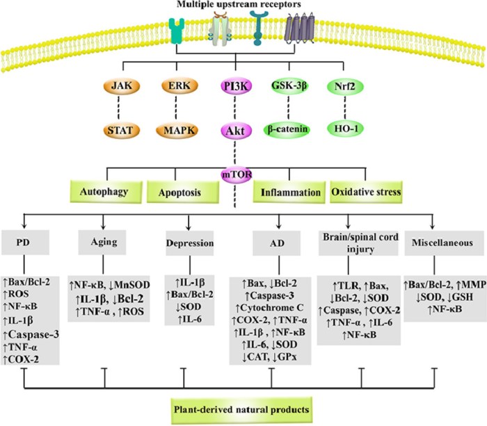
Polyphenols, alkaloids, carotenoids, and terpenoids have shown to be capable of a great modulation of PI3K/Akt/mTOR in NDDs. Natural products potentially target various important oxidative/inflammatory/apoptotic/autophagic molecules/mediators, such as Bax, Bcl-2, p53, caspase-3, caspase-9, NF-κB, TNF-α, GSH, SOD, MAPK, GSK-3β, Nrf2/HO-1, JAK/STAT, CREB/BDNF, ERK1/2, and LC3 towards neuroprotection.
This is the first systematic and comprehensive review with a simultaneous focus on the critical role of PI3K/Akt/mTOR in NDDs and associated targeting by natural products.”
https://www.sciencedirect.com/science/article/abs/pii/S0944711321002075 “Natural products attenuate PI3K/Akt/mTOR signaling pathway: A promising strategy in regulating neurodegeneration” (not freely available) Thanks to Dr. Sajad Fakhri for providing a copy.
Natural products mentioned in this review that I eat in everyday foods are listed below. The most effective ones are broccoli and red cabbage sprouts, and oats and oat sprouts:
Four humpback whales


This 2021 review subject was vasopressin:
“Vasopressin is a ubiquitous molecule playing an important role in a wide range of physiological processes, thereby implicated in pathomechanisms of many disorders. The most striking is its central effect in stress-axis regulation, as well as regulating many aspects of our behavior.
Arginine-vasopressin (AVP) is a nonapeptide that is synthesized mainly in the supraoptic, paraventricular (PVN), and suprachiasmatic nucleus of the hypothalamus. AVP cell groups of hypothalamus and midbrain were found to be glutamatergic, whereas those in regions derived from cerebral nuclei were mainly GABAergic.
In the PVN, AVP can be found together with corticotropin-releasing hormone (CRH), the main hypothalamic regulator of the HPA axis. The AVPergic system participates in regulation of several physiological processes, from stress hormone release through memory formation, thermo- and pain regulation, to social behavior.

AVP determines behavioral responses to environmental stimuli, and participates in development of social interactions, aggression, reproduction, parental behavior, and belonging. Alterations in AVPergic tone may be implicated in pathology of stress-related disorders (anxiety and depression), Alzheimer’s, posttraumatic stress disorder, as well as schizophrenia.
An increasing body of evidence confirms epigenetic contribution to changes in AVP or AVP receptor mRNA level, not only during the early perinatal period, but also in adulthood:
- DNA methylation is more targeted on a single gene; and it is better characterized in relation to AVP;
- Some hint for bidirectional interaction with histone acetylation was also described; and
- miRNAs are implicated in the hormonal, peripheral role of AVP, and less is known about their interaction regarding behavioral alteration.”
https://www.mdpi.com/1422-0067/22/17/9415/htm “Epigenetic Modulation of Vasopressin Expression in Health and Disease”
Find your way, regardless of what the herd does.

This 2021 paper reviewed evidence for immune system effects associated with specific gut areas:
“The intestinal immune system must not only contend with continuous exposure to food, commensal microbiota, and pathogens, but respond appropriately according to intestinal tissue differences. The entire intestine, inclusive of its lymph nodes, is considered a immunosuppressive organ overall compared to most other tissues, indicating that a state of tolerance to food and commensals – yet vigilance toward pathogens – was an evolutionarily stable strategy.
By operating in compartments, the immune system may generate multiple immune outcomes, even with simultaneous opposite goals e.g., tolerance or inflammation. Generation of unique immunologic niches within the intestine is influenced by a combination of tissue intrinsic properties, extrinsic environmental factors, and regionalized immune populations.

Complexity of intrinsic and extrinsic driving forces shaping an intestinal niche makes it very challenging to determine causality in disease development and predicting effective therapeutic approaches. We really only stand at the beginning of understanding this interplay.”
https://www.nature.com/articles/s41385-021-00420-8 “Intestinal immune compartmentalization: implications of tissue specific determinants in health and disease”
I patterned this post after Choosing your future with β-glucan:
“So where do you choose to be? In an 80% survival group who were administered β-glucan before they encountered a serious infection? Or in a < 20% survival group who didn’t take β-glucan?”
and Long-lasting benefits of a common vaccine:
“As inferred by “induction of trained immunity by both Bacillus Calmette-Guerin tuberculosis vaccine and β-glucan” many of these findings also apply to yeast cell wall β-glucan treatments.”
This paper’s food allergy references were interesting. It’s an area that personally requires further work, although avoidance has historically been effective.
This paper briefly mentioned broccoli’s effects in the proximal small intestine. It wasn’t informative per gut compartment with this year’s focus on making my gut microbiota happy, such as what our colonic microbiota can do to reciprocate their host giving them what they want.
This review’s human studies referenced what could be done post-disease like surgery etc. in different gut compartments. Very little concerned an individual taking responsibility for their own one precious life to prevent such diseases in the first place. Its Conclusions section claim was a fallacy:
“..very challenging to determine causality in disease development and predicting effective therapeutic approaches.”

This 2021 review summarized taurine’s beneficial effects on mitochondrial function:
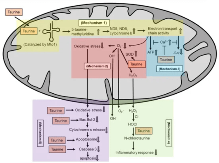
“Taurine supplementation protects against pathologies associated with mitochondrial defects, such as aging, mitochondrial diseases, metabolic syndrome, cancer, cardiovascular diseases and neurological disorders. Potential mechanisms by which taurine exerts its antioxidant activity in maintaining mitochondria health include:
- Conjugates with uridine on mitochondrial tRNA to form a 5-taurinomethyluridine for proper synthesis of mitochondrial proteins (mechanism 1), which regulates the stability and functionality of respiratory chain complexes;
- Reduces superoxide generation by enhancing the activity of intracellular antioxidants (mechanism 2);
- Prevents calcium overload and prevents reduction in energy production and collapse of mitochondrial membrane potential (mechanism 3);
- Directly scavenges HOCl to form N-chlorotaurine in inhibiting a pro-inflammatory response (mechanism 4); and
- Inhibits mitochondria-mediated apoptosis by preventing caspase activation or by restoring the Bax/Bcl-2 ratio and preventing Bax translocation to the mitochondria to promote apoptosis.
An analysis on pharmacokinetics of oral supplementation (4 g) in 8 healthy adults showed a baseline taurine content in a range of 30 μmol to 60 μmol. Plasma content increased to approximately 500 μmol 1.5 h after taurine intake. Plasma content subsequently decreased to baseline level 6.5 h after intake.
We discuss antioxidant action of taurine, particularly in relation to maintenance of mitochondria function. We describe human studies on taurine supplementation in several mitochondria-associated pathologies.”
https://www.mdpi.com/1420-3049/26/16/4913/html “The Role of Taurine in Mitochondria Health: More Than Just an Antioxidant”
I take a gram of taurine at breakfast and at dinner along with other supplements and 3-day-old Avena sativa oat sprouts. Don’t think my other foods’ combined taurine contents are more than one gram, because none are found in various top ten taurine-containing food lists.
As a reminder, your mitochondria came from your mother, except in rare cases.
Another excellent blog post by Josh Mitteldorf, A New Approach to Methylation Clocks, that curated the same study:
“The Levine/Horvath PhenoAge epigenetic clock was calibrated using a combination of metabolic factors that correlate with health, including inflammation, DNA transcription, DNA repair, and mitochondrial activity.
Evolution is not an engineer. Living things are not constructed out of parts that are separately optimized for exactly one function.
Every molecule has multiple functions. Every function is regulated by multiple pathways.
For clock technology, using individual CpGs for a starting point may not be optimal. We suspect that CpGs, like other biological entities, work together closely in teams.
CpGs on a team might vary slightly from one individual to the next. But the team has a function and an identity and a signature that is robust. We expect the team to function more consistently than any of its individual members.
The peer-reviewed version of her paper will be published shortly. Full details of algorithms will be available on GitHub, and script in the R programming language will be released for use of other researchers. If principal component analysis clocks correlate well with previously validated clocks but offer tighter uncertainties, we’ll know we’re on the right track.”
Best wishes for Josh to recover from a bike accident.

This 2021 cat study developed human-comparable epigenetic clocks:
“We aimed to develop and evaluate epigenetic clocks for cats, as such biomarkers are necessary for translating promising anti-aging interventions from humans to cats and vice versa. We also provided the possibility of using epigenetic aging rate of cats to inform on feline health, for which a quantitative measure is presently unavailable. Specifically, we present here DNA methylation-based biomarkers (epigenetic clocks) of age for blood from cats.
Maximum lifespan of cats is 30 years according to the animal age data base (anAge), but most cats succumb to diseases before they are 20 years old. Age is the biggest risk factor for a vast majority of diseases in animals, and cats are no exception.
Interventions to slow aging are being sought. Ideally, testing should occur in species that are evolutionarily close to humans, similar in size, have high genetic diversity, and share the same environment as humans. It has been recognized that domestic dogs fulfill these criteria.
Investigations have yet to be extended to cats although they share similar environments and living conditions with their human owners. Identification of environmental factors and living conditions that affect aging, as well as potential mitigation measures, can be achieved by proxy with cats.
The human-cat clock for relative age exhibited high correlation regardless of whether analysis was applied to samples from both species or only to cat samples. This demonstrated that relative age circumvented skewing that is inherent when chronological age of species with very different lifespans is measured using a single formula.
Evidence is compelling that epigenetic age is an indicator of biological age. These results are consistent with the fact that epigenetic clocks developed for one mammalian species can be employed – to a limited extent – to other species, and reveal association of DNA methylation changes with age.
Human epigenetic age acceleration is associated with a wide array of primary traits, health states, and pathologies. While it is still unclear why age acceleration is connected to these characteristics, it does nevertheless suggest that extension of similar studies to cats may allow for development of epigenetic age acceleration as a surrogate or indicator of feline biological fitness.”
https://link.springer.com/article/10.1007%2Fs11357-021-00445-8 “Epigenetic clock and methylation studies in cats”
As noted earlier this summer in Smoke and die early, while your twin lives on, Dr. Steve Horvath is on a torrid publishing streak this year. He’s made it questionable for study designs based on published science to omit epigenetic clocks.
I titled this post Your pets because I’m too allergic to have cats, dogs, etc. live with me. Maybe this year’s focus on making my gut microbiota happy will change that?
My pets live free:


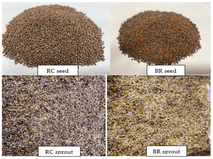
This 2021 study compared properties of red cabbage and broccoli seeds and sprouts:
“Antioxidant and antidiabetic properties and metabolite profiling of ethanol extracts of red cabbage (RC) and broccoli (BR) seeds and sprouts were investigated:
- BR seeds had the highest total phenolic and flavonoid contents;
- BR sprouts had the highest saponin content;
- RC sprouts demonstrated the highest antioxidant capacity;
- BR and RC sprouts showed the most potent inhibition against α-glucosidase and pancreatic lipase; and
- BR seeds demonstrated the lowest AGE inhibition.
In vitro assessment of antidiabetic potential of extracts revealed that sprouts demonstrated better potential as antioxidant, α-glucosidase, and pancreatic lipase inhibitors compared to raw seeds. Amino acids and phenolic compounds were the most improved metabolites in the germination process.
Germination not only enhanced levels of metabolites, but also synthesized new compounds in seeds. Germination effectively enhanced functional properties and metabolite profiles of broccoli and red cabbage seeds, making their sprouts more applicable as functional ingredients.”
https://www.mdpi.com/2076-3921/10/6/852/htm “UHPLC-ESI-QTOF-MS/MS Metabolite Profiling of the Antioxidant and Antidiabetic Activities of Red Cabbage and Broccoli Seeds and Sprouts”
I asked coauthors for sprout ages and pertinent growing conditions for the above-pictured sprouts. I’ll guess > 3-days-old, temperature 25° C, and relative humidity 90%. What would you guess?
Update: Two coauthors replied:
“Red Cabbage and Broccoli were germinated for 6 and 7 days respectively. Temperature ranged between 20-23 °C in the dark.”

“In this episode we speak with Neal Peterson, who has devoted over 40 years to breeding pawpaws (Asimina triloba). We talk about how he first fell in love with this delicious fruit, how he tracked down remnants of the early 20th century pawpaw collections just in time, and selected 7 superior pawpaw cultivars out of 1,500 seedlings.”
Go to https://anchor.fm/plantcunning/ and episode 46.
I first ate a pawpaw while on a Capital Area Hiking Club September 2015 hike on the Susquehanna River. Our hike leader loaded up his backpack and was willing to share when we finished. Didn’t take a photo, but here’s what I picked a year later from a farm:
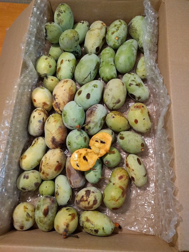
Ugly looking, but beautiful inside.
In subsequent seasons, I found pawpaws in national and state parks. Can’t find pawpaw fruit sales online that I previously bought from, so may revisit these parks next month.
My son and I engaged in Guerrilla Planting. Maybe it’s time to check on those trees’ progress, too?
This 2021 study investigated Vitamins K1 and K2 associations with hospitalization for atherosclerotic cardiovascular disease (ASCVD):
“In this prospective cohort study, both dietary vitamin K1 intake and vitamin K2 intake were inversely related to ASCVD hospitalization risk, and very low vitamin K1 was associated with a higher risk of ASCVD hospitalizations. Given very different food sources, these data support an independent protective effect for both subtypes of vitamin K.
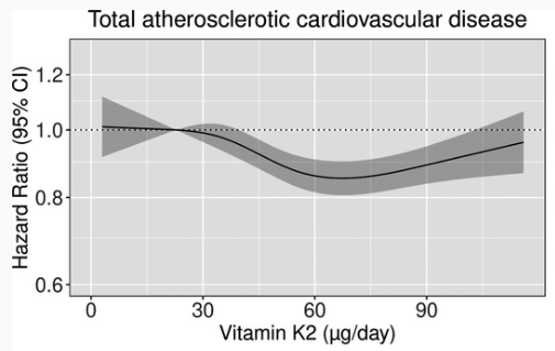
Relatively higher vitamin K2 intake in our cohort permitted discovery of a nonlinear, more U‐shaped association between vitamin K2 intake and ASCVD risk, which, to the best of our knowledge, has not previously been described. This may reflect a competing increase in ASCVD risk associated with overconsumption of vitamin K2‐rich foods (ie, cheese, eggs, butter).
Our study comes with some limitations common to nutritional epidemiology, and has significant strengths:
- A large sample size with up to 23 years of follow‐up, allowing for accumulation of a high number of events;
- Availability of important participant characteristics, enabling appropriate methods to be employed to reduce residual confounding; and
- Minimal loss to follow‐up (<0.3%).”
https://www.ahajournals.org/doi/10.1161/JAHA.120.020551 “Vitamin K Intake and Atherosclerotic Cardiovascular Disease in the Danish Diet Cancer and Health Study”
Daily broccoli / red cabbage / mustard sprouts for Vitamin K1, and a supplement for Vitamin K2 is what I do. Expect more than staying out of hospitals, but don’t know whether previous damage can be repaired.
Looking forward

This 2020 review covered interactions of gut microbiota, intestinal mucus, and dietary fibers. I’ve outlined its headings and subheadings, and ended with its overview:
“I. Dietary fibers and human mucus-associated polysaccharides: can we make an analogy?
I.1 Brief overview of dietary fibers and mucus polysaccharides structures and properties
I.I.1 Dietary fibers
- Dietary fiber intake and health effects
I.I.2 Intestinal mucus polysaccharides
- Structure
- Main functions
I.2 Similarities and differences between dietary fibers and mucus carbohydrates
- Origin and metabolism
- Structure
II. Interactions of dietary fibers and mucus-associated polysaccharides with human gut microbiota
II-1 Substrate accessibility and microbial niches
- Dietary fibers
- Mucus polysaccharides
II-2 Recognition and binding strategies
- Dietary fibers
- Mucus polysaccharides
II-3 Carbohydrate metabolism by human gut microbiota
II-3.1 Specialized carbohydrate-active enzymes
II-3.2 Vertical ecological relationships in carbohydrate degradation
- Dietary fibers
- Mucus polysaccharides
II-3.3 Horizontal ecological relationships in carbohydrate degradation
II.4 Effect of carbohydrates on gut microbiota composition and sources of variability
II.4.1 Well-known effect of dietary fibers on the gut microbiota
II.4.2 First evidences of a link between mucus polysaccharides and gut microbiota composition
III. Gut microbiota, dietary fibers and intestinal mucus: from health to diseases?
[no III.1]
III.2 Current evidences for the relationship between dietary fibers, mucus and intestinal-inflammatory related disorder
III.2.1 Obesity and metabolic-related disorders
- Dietary fibers
- Mucus polysaccharides
III.2.2 Inflammatory bowel diseases
- Dietary fibers
- Mucus polysaccharides
III.2.3 Colorectal cancer
- Dietary fibers
- Mucus polysaccharides
IV. How enteric pathogens can interact with mucus and dietary fibers in a complex microbial background?
IV.1 Mucus-associated polysaccharides: from interactions with enteric pathogens to a cue for their virulence?
IV.1.1 Pathogens binding to mucus
- Binding structures
- Sources of variations
IV.1.2 Mucus degradation by pathogens
- Bacterial mucinases
- Glycosyl hydrolases
IV.1.3 Mucus-based feeding of pathogens
- Primary degraders or cross-feeding strategies
- Importance of microbial background
IV.1.4 Pathogens and inflammation in a mucus-altered context
IV.1.5 Modulation of virulence genes by mucus degradation products
IV.2 How can dietary fiber modulate enteric pathogen virulence?
IV.2.1 Direct antagonistic effect of dietary fibers on pathogens
- Bacteriostatic effect
- Inhibition of cell adhesion
- Inhibition of toxin binding and activity
IV.2.2 Indirect effect of dietary fibers through gut microbiota modulation
- Modulation of microbiota composition
- Modulation of gut microbiota activity
IV.2.3 Inhibition of pathogen interactions with mucus: a new mode of dietary fibers action?
- Binding to mucus: dietary fibers acting as a decoy
- Inhibition of mucus degradation by dietary fibers
V. Human in vitro gut models to decipher the role of dietary fibers and mucus in enteric infections: interest and limitations?
V.1 Main scientific challenges to be addressed
V.2 In vitro human gut models as a relevant alternative to in vivo studies
V.3 In vitro gut models to decipher key roles of digestive secretions, mucus and gut microbiota
V.4 Toward an integration of host responses
V.5 From health to disease conditions

Overview of the potential role of dietary fibers in preventing enteric infections. Reliable and converging data from scientific literature are represented with numbers in circles, while data more hypothetical needing further investigations are represented with numbers in squares.
- Some dietary fibers exhibit direct bacteriostatic effects against pathogens.
- Dietary fiber degradation leads to short-chain fatty acids (SCFAs) production that can modulate pathogens’ virulence.
- By presenting structure similarities with receptors, some dietary fibers can prevent pathogen adhesin binding to their receptors.
- By the same competition mechanism, dietary fibers can also prevent toxins binding to their receptors.
- Dietary fibers are able to promote gut microbiota diversity.
- Dietary fibers may promote growth of specific strains with probiotic properties and therefore exhibit anti-infectious properties.
- Suitable dietary fiber intake prevents microbiota’s switch to mucus consumption, limiting subsequent commensal microbiota encroachment and associated intestinal inflammation.
- Dietary fibers may prevent pathogen cross-feeding on mucus by limiting mucus degradation and/or by preserving diversity of competing bacterial species.
- By preventing mucus over-degradation by switcher microbes, dietary fibers can hamper pathogen progression close to the epithelial brush border, and further restrict subsequent inflammation.”
https://doi.org/10.1093/femsre/fuaa052 “Tripartite relationship between gut microbiota, intestinal mucus and dietary fibers: towards preventive strategies against enteric infections” (not freely available)
There were many links among gut microbiota studies previously curated. For example, Go with the Alzheimer’s Disease evidence found:
“Akkermansia cannot always be considered a potentially beneficial bacterium. It might be harmful for the gut–brain axis in the context of AD development in the elderly.”
The current review provided possible explanations:
“Akkermansia muciniphila could be considered as a species that fulfills a keystone function in mucin degradation. It is a good example of a mucus specialist.”
Points #7-9 of the above overview inferred that insufficient dietary fiber may disproportionately increase abundance of this species. But Gut microbiota strains also found that effects may be found only below species at species’ strain levels.
These reviewers provided copies in places other than what’s linked above. Feel free to contact them for a copy.
Moon bandit

