Train your immune system every day!

This 2019 US review subject was β-glucan:

“β-1,3-Glucans (hereafter referred to as glucan) are natural molecules able to significantly improve our health. In human studies, the tested (and suggested) daily dose remains in the range of 100–500 mg for stimulation of the immune system, whereas for a decrease in cholesterol levels a daily dose of 3 g is recommended.

Glucan does not represent essential nutrients, but it might be successfully used not only for:

  • Improvement of immune functions, but also to improve the general quality of life via
  • Improvements of immune status,
  • Lowering cholesterol,
  • Improving blood glucose levels and
  • Reduction of stress.

ClinicalTrials.gov summarizes 177 [now 207 with 110 completed] β-glucan clinical trials, mostly in cancer, gastrointestinal tract therapy, lowering cholesterol and improvements of immune reactions.

The question is not if glucans will move from food supplement to widely accepted drug, but how soon.

Reactions known to be influenced by glucan are represented in white, reactions where glucan has no confirmed effects are shown in black. The first defensive body response to infection results from formation of the anorexia cytokines (IL-1, IL-6, IL-8, and TNF-α).”

https://www.mdpi.com/1420-3049/24/7/1251/htm “Beta Glucan: Supplement or Drug? From Laboratory to Clinical Trials”

The review is also indexed at https://www.betaglucan.org/i-p/ under “Immunomodulator”


I’m curating this review on Day 12 of a self-quarantine after coming back from Milano, Italy, Monday, February 24, 2020. The previous Friday into Saturday I flew to Milano sitting with a group of elderly Italians who were returning from vacation.

On Saturday my traveling companion and I used the Milano rail and crowded subway system to go downtown. On Sunday we used their crowded rail, crowded bus, and crowded ferry systems to visit Como, Bellagio, and Menaggio.

I don’t think we could have mixed in more with people during transits, touristing, and Carnevale celebrations.

IMG_9539

After returning to our hotel Sunday evening, we heard about the coronavirus outbreak south of Milano and the closing of ten towns. We changed flights and departed for the US early Monday morning.

Neither of us have had any signature symptoms of COVID-19 (fever, shortness of breath, dry cough). Our daily diet the past few years included β-glucan from steel-cut oats (~3 g) and from a 1/3, 1/6 yeast cell wall supplement.

Coincidence?

Each of our futures will depend on what we do Now to prepare.

Take responsibility for your one precious life.

Moonrise at sunrise with Venus

The epigenetics of perinatal stress

This 2019 McGill review discussed long-lasting effects of perinatal stress:

“Epigenetic processes are involved in embedding the impact of early-life experience in the genome and mediating between social environments and later behavioral phenotypes. Since these phenotypes are apparent a long time after early experience, changes in gene expression programming must be stable.

Although loss of methylation in a promoter is necessary for expression, it is not sufficient. Demethylation removes a barrier for expression, but expression might be realized at the right time or context when needed factors or signals are present.

DNA methylation anticipates future transcriptional response to triggers. Comparing steady-state expression with DNA methylation does not capture the full meaning and scope of regulatory roles of differential methylation.

A model for epigenetic programming by early life stress:

  1. Perinatal stress perceived by the brain triggers release of glucocorticoids (GC) from the adrenal in the mother prenatally or the newborn postnatally.
  2. GC activate nuclear glucocorticoid receptors across the body, which epigenetically program (demethylate) genes that are targets of GR in brain and white blood cells (WBC).
  3. Demethylation events are insufficient for activation of these genes. A brain specific factor (TF) is required for expression and will activate low expression of the gene in the brain but not in blood.
  4. During adulthood a stressful event transiently triggers a very high level of expression of the GR regulated gene specifically in the brain.

Horizontal arrow, transcription; circles, CpG sites; CH3 in circles, methylated sites; empty circles, unmethylated CpG sites; horizon[t]al curved lines, mRNA.”

Review points discussed:

  • “Epigenetic marks are laid down and maintained by enzymes that either add or remove epigenetic modifications and are therefore potentially reversible in contrast to genetic changes.
  • Response to early life stress and maternal behavior is also not limited to the brain and involves at least the immune system as well.
  • The placenta is also impacted by maternal social experience and early life stress.
  • Most studies are limited to peripheral tissues such as saliva and white blood cells, and relevance to brain physiology and pathology is uncertain.
  • Low absolute differences in methylation seen in most human behavioral EWAS raise questions about their biological significance.

  • Although post-mortem studies examine epigenetic programming in physiologically relevant tissues, they represent only a final and single stage that does not capture dynamic evolution of environments and epigenetic programming in living humans.”

https://www.ncbi.nlm.nih.gov/pmc/articles/PMC6952743/ “The epigenetics of perinatal stress”


Other reviewers try to ignore times when we were all fetuses and newborns. For example, in the same journal issue was a Boston review of PTSD that didn’t mention anything about earliest times of human lives! Those reviewers speculated around this obvious gap on their way to being paid by NIH.

Why would researchers ignore perinatal stress events that prime humans for later-life PTSD? Stress generally has a greater impact on fetuses and newborns than on infants, and a greater impact on infants than on adults.

Masters of manipulating their host

This 2020 French review subject was parasitical influences on host epigenetic processes:

“Parasites have become masters of manipulating their host cells, exploiting signaling, and metabolic pathways to hijack host gene expression to their own advantage. These intracellular parasites have developed a wide range of strategies that affect transcriptional machineries and epigenetic events in the host cell nucleus.

Parasite effectors regulate host transcription. Secretion of numerous parasite effector proteins are key processes during parasite infection. Parasite effectors deregulate host expression profile which lead to host cell transformation, or escape from the host immune system to allow parasite persistence and survival.”

parasites
The first two of the six strategies discussed are shown above:

  1. “Induction of a host epigenetic enzyme. Parasite infection leads to upregulation of SMYD3, a methyltransferase that activates genes involved in host transformation, through H3K4 trimethylation.
  2. Secreting effector proteins that drive epigenetic repression of host genes. TEEGR activates a host chromatin modifier able to repress transcription of immune system genes through H3K27 trimethylation.”

https://link.springer.com/article/10.1007/s00281-020-00779-z “The clever strategies used by intracellular parasites to hijack host gene expression” (not freely available)


I used a “parasites” paradigm while living in the Washington DC area for three decades to help understand what goes on there. Moved away several years ago, but haven’t changed my thinking that all six of this paper’s parasite strategies had analogous human actions.

Other curated papers that explored the review’s topic include:

Trained immunity responses to bacterial infections

This 2019 Swiss rodent study investigated immune responses to five types of bacterial infections:

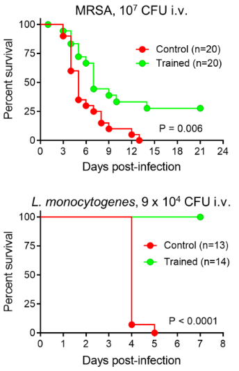
“The innate immune system recalls a challenge to adapt to a secondary challenge, a phenomenon called trained immunity. Trained immunity protected mice from a large panel of clinically relevant bacterial pathogens inoculated systematically and locally to induce peritonitis, enteritis and pneumonia.

Induction of trained immunity remodeled bone marrow and blood cellular compartments, providing efficient barriers against bacterial infections. Protection was remarkably broad when considering the pathogens and sites of infection tested.

We are running experiments to delineate the length of protection conferred by trained immunity. Trained immunity is most typically induced with β-glucan.

Mice were injected with methicillin-resistant Staphylococcus aureus (MRSA). Trained mice survived better than control mice (31% vs. 0% survival) and had 10-fold less bacteria in blood 2 days post-infection.

Mice were challenged with a lethal dose of Listeria monocytogenes. Most strikingly, all trained mice survived infection while all control mice died within 5 days. Bacteria were not detected in blood collected from trained mice 2 and 3 days post-infection.”

https://academic.oup.com/jid/advance-article/doi/10.1093/infdis/jiz692/5691195 “Trained immunity confers broad-spectrum protection against bacterial infections”


One of the coauthors also published:

Clearing out the 2019 queue of interesting papers

I’m clearing out the below queue of 27 studies and reviews I’ve partially read this year but haven’t taken the time to curate. I have a pesky full-time job that demands my presence elsewhere during the day. :-\

Should I add any of these back in? Let’s be ready for the next decade!


Early life

https://link.springer.com/article/10.1007/s12035-018-1328-x “Early Behavioral Alterations and Increased Expression of Endogenous Retroviruses Are Inherited Across Generations in Mice Prenatally Exposed to Valproic Acid” (not freely available)

https://www.sciencedirect.com/science/article/pii/S0166432818309392 “Consolidation of an aversive taste memory requires two rounds of transcriptional and epigenetic regulation in the insular cortex” (not freely available)

https://www.nature.com/articles/s41380-018-0265-4 “Intergenerational transmission of depression: clinical observations and molecular mechanisms” (not freely available)

mother

https://www.ncbi.nlm.nih.gov/pmc/articles/PMC6454089/ “Epigenomics and Transcriptomics in the Prediction and Diagnosis of Childhood Asthma: Are We There Yet?”

https://www.ncbi.nlm.nih.gov/pmc/articles/PMC6628997/Placental epigenetic clocks: estimating gestational age using placental DNA methylation levels”

https://www.ncbi.nlm.nih.gov/pmc/articles/PMC6770436/ “Mismatched Prenatal and Postnatal Maternal Depressive Symptoms and Child Behaviours: A Sex-Dependent Role for NR3C1 DNA Methylation in the Wirral Child Health and Development Study”

https://www.sciencedirect.com/science/article/pii/S0889159119306440 “Environmental influences on placental programming and offspring outcomes following maternal immune activation”

https://academic.oup.com/mutage/article-abstract/34/4/315/5581970 “5-Hydroxymethylcytosine in cord blood and associations of DNA methylation with sex in newborns” (not freely available)

https://physoc.onlinelibrary.wiley.com/doi/full/10.1113/JP278270 “Paternal diet impairs F1 and F2 offspring vascular function through sperm and seminal plasma specific mechanisms in mice”

https://onlinelibrary.wiley.com/doi/full/10.1111/nmo.13751 “Sex differences in the epigenetic regulation of chronic visceral pain following unpredictable early life stress” (not freely available)

https://www.ncbi.nlm.nih.gov/pmc/articles/PMC6811979/ “Genome-wide DNA methylation data from adult brain following prenatal immune activation and dietary intervention”

https://link.springer.com/article/10.1007/s00702-019-02048-2miRNAs in depression vulnerability and resilience: novel targets for preventive strategies”


Later life

https://www.ncbi.nlm.nih.gov/pmc/articles/PMC6543991/ “Effect of Flywheel Resistance Training on Balance Performance in Older Adults. A Randomized Controlled Trial”

https://www.mdpi.com/2411-5142/4/3/61/htm “Eccentric Overload Flywheel Training in Older Adults”

https://www.nature.com/articles/s41577-019-0151-6 “Epigenetic regulation of the innate immune response to infection” (not freely available)

https://link.springer.com/chapter/10.1007/978-981-13-6123-4_1 “Hair Cell Regeneration” (not freely available)

https://www.ncbi.nlm.nih.gov/pmc/articles/PMC6422915/Histone Modifications as an Intersection Between Diet and Longevity”

https://www.sciencedirect.com/science/article/abs/pii/S0306453019300733 “Serotonin transporter gene methylation predicts long-term cortisol concentrations in hair” (not freely available)

https://www.sciencedirect.com/science/article/abs/pii/S0047637419300338 “Frailty biomarkers in humans and rodents: Current approaches and future advances” (not freely available)

https://onlinelibrary.wiley.com/doi/full/10.1111/pcn.12901 “Neural mechanisms underlying adaptive and maladaptive consequences of stress: Roles of dopaminergic and inflammatory responses

https://www.ncbi.nlm.nih.gov/pmc/articles/PMC6627480/ “In Search of Panacea—Review of Recent Studies Concerning Nature-Derived Anticancer Agents”

https://www.sciencedirect.com/science/article/abs/pii/S0028390819303363 “Reversal of oxycodone conditioned place preference by oxytocin: Promoting global DNA methylation in the hippocampus” (not freely available)

https://www.futuremedicine.com/doi/10.2217/epi-2019-0102 “Different epigenetic clocks reflect distinct pathophysiological features of multiple sclerosis”

https://www.ncbi.nlm.nih.gov/pmc/articles/PMC6834159/ “The Beige Adipocyte as a Therapy for Metabolic Diseases”

https://www.sciencedirect.com/science/article/abs/pii/S8756328219304077 “Bone adaptation: safety factors and load predictability in shaping skeletal form” (not freely available)

https://www.nature.com/articles/s41380-019-0549-3 “Successful treatment of post-traumatic stress disorder reverses DNA methylation marks” (not freely available)

https://www.sciencedirect.com/science/article/abs/pii/S0166223619301821 “Editing the Epigenome to Tackle Brain Disorders” (not freely available)

An epigenetic clock review by committee

This 2019 worldwide review of epigenetic clocks was a semi-anonymous mishmash of opinions, facts, hypotheses, unwarranted extrapolations, and beliefs. Diversity of viewpoints among the 21 coauthors wasn’t evident.

1. Citations of coauthors’ works seemed excessive, and they apologized for omissions. However:

  • Challenge 5 was titled “Single-cell analysis of aging changes and disease” and
  • Table 1 “Major biological and analytic issues with epigenetic DNA methylation clocks” had single-cell analysis as the Proposed solution to five Significant issues.

Yet studies such as High-Resolution Single-Cell DNA Methylation Measurements Reveal Epigenetically Distinct Hematopoietic Stem Cell Subpopulations were unmentioned.

2. Some coauthors semi-anonymously expressed faith that using current flawed methodologies in the future – only more thoroughly, with newer equipment, etc. – would yield better results. If all 21 coauthors were asked their viewpoints of Proposed solutions to the top three Significant issues of epigenetic clocks, what would they emphasize when quoted?

3. Techniques were praised:

“Given the precision with which DNA methylation clock age can be estimated and evolving measures of biological, phenotype-, and disease-related age (e.g., PhenoAge, GrimAge)..”

Exactly why these techniques have at times produced inexplicable results wasn’t examined, though. Two examples:

  • In Reversal of aging and immunosenescent trends, Levine PhenoAge methodology estimated that the 51-65 year old subjects’ biological ages at the beginning of the study averaged 17.5 years less than their chronological age. Comparing that to Horvath average biological age of 3.95 years less raised the question: exactly why did PhenoAge show such a large difference?
  • The paper mentioned GrimAge methodology findings about “smoking-related changes.” But it didn’t explain why GrimAge methylation findings most closely associated with smoking history also accurately predicted future disease risk with non-smokers.

Eluding explanations for these types of findings didn’t help build confidence in methodologies.

4. A more readable approach to review by committee could have coauthors – in at least one section – answer discussion questions, as Reversing epigenetic T cell exhaustion did with 18 experts.

https://genomebiology.biomedcentral.com/articles/10.1186/s13059-019-1824-y “DNA methylation aging clocks: challenges and recommendations”

A review of fetal adverse events

This 2019 Australian review subject was fetal adversities:

“Adversity during the perinatal period is a significant risk factor for the development of neurodevelopmental disorders long after the causative event. Despite stemming from a variety of causes, perinatal compromise appears to have similar effects on the developing brain, thereby resulting in behavioural disorders of a similar nature.

These behavioural disorders occur in a sex‐dependent manner, with males affected more by externalizing behaviours such as attention deficit hyperactivity disorder (ADHD) and females by internalizing behaviours such as anxiety. The term ‘perinatal compromise’ serves as an umbrella term for intrauterine growth restriction, maternal immune activation, prenatal stress, early life stress, premature birth, placental dysfunction, and perinatal hypoxia.

The above conditions are associated with imbalanced excitatory-inhibitory pathways resulting from reduced GABAergic signalling. Methylation of the GAD1/GAD67 gene, which encodes the key glutamate‐to‐GABA synthesizing enzyme Glutamate Decarboxylase 1, resulting in increased levels of glutamate is one epigenetic mechanism that may account for a tendency towards excitation in disorders such as ADHD.

The posterior cerebellum’s role in higher executive functioning is becoming well established due to its connections with the prefrontal cortex, association cortices, and limbic system. It is now suggested that disruptions to cerebellar development, which can occur due to late gestation compromises such as preterm birth, can have a major impact on the region of the brain to which it projects.

Activation of the maternal hypothalamic-pituitary adrenal (HPA) axis and placental protection. Psychological stress is perceived by the maternal HPA axis, which stimulates cortisol release from the maternal adrenal gland.

High levels of maternal cortisol are normally prevented from reaching the fetus by the 11β-hydroxysteroid dehydrogenase 2 (HSD11B2) enzyme, which converts cortisol to the much less active cortisone. Under conditions of high maternal stress, this protective mechanism can be overwhelmed, with the gene encoding the enzyme becoming methylated, which reduces its expression allowing cortisol to cross the placenta and reach the fetus.”


The reviewers extrapolated many animal study findings to humans, although most of their own work was with guinea pigs. The “suggest” and “may” qualifiers were used often – 22 and 37 times, respectively. More frequent use of the “appears,” “hypothesize,” “propose,” and “possible” terms was justified.

As a result, many reviewed items such as the above graphic and caption should be viewed as hypothetical for humans rather than reflecting solid evidence from quality human studies.

The reviewers focused on the prenatal (before birth) period more than the perinatal (last trimester of pregnancy to one month after birth) period. There were fewer mentions of birth and early infancy adversities.

https://onlinelibrary.wiley.com/doi/abs/10.1111/jne.12814 “Perinatal compromise contributes to programming of GABAergic and Glutamatergic systems leading to long-term effects on offspring behaviour” (not freely available)

A transgenerational view of the rise in obesity

This 2019 Washington State University rodent study found epigenetically inherited transgenerational effects in great-grand offspring due to their great-grandmothers’ toxicant exposures during pregnancy:

“Previous studies found an increased susceptibility to obesity in F3 generation rats ancestrally exposed to the pesticide DDT, and an increase in a lean phenotype in the F3 generation rats ancestrally exposed to the herbicide atrazine. The present study investigated whether there were common DMR [differential DNA methylated region] and associated genes between the control, DDT, and atrazine lineage male and female adipocytes in order to identify potential novel gene pathways modulated by DNA methylation.

Comparison of epigenetic alterations indicated that there were substantial overlaps between the different treatment lineage groups for both the lean and obese phenotypes. Novel correlated genes and gene pathways associated with DNA methylation were identified, and may aid in the discovery of potential therapeutic targets for metabolic diseases such as obesity.

Given that the first widespread [DDT] exposures to gestating human females started in the 1950s, the majority of the subsequent F3 generation are adults today. Ancestral exposures to environmental toxicants like DDT may have had a role in the dramatic rise in obesity rates worldwide.”


This same research group noted in Transgenerational diseases caused by great-grandmother DDT exposure:

“DDT was banned in the USA in 1973, but it is still recommended by the World Health Organization for indoor residual spray. India is by far the largest consumer of DDT worldwide.

India has experienced a 5-fold increase of type II diabetes over the last three decades with a predisposition to obesity already present at birth in much of the population. Although a large number of factors may contribute to this increased incidence of obesity, the potential contribution of ancestral toxicant exposures in the induction of obesity susceptibility requires further investigation.”

https://www.tandfonline.com/doi/full/10.1080/21623945.2019.1693747 “Adipocyte epigenetic alterations and potential therapeutic targets in transgenerationally inherited lean and obese phenotypes following ancestral exposures”

A GWAS meta-analysis of two epigenetic clocks

This 2019 UK human study conducted a meta-analysis of genome-wide association studies of two epigenetic clocks using 13,493 European-ancestry individuals aged between ten and 98 years:

“Horvath-EAA, described in previous publications as ‘intrinsic’ epigenetic age acceleration (IEAA), can be interpreted as a measure of cell-intrinsic ageing that exhibits preservation across multiple tissues, appears unrelated to lifestyle factors, and probably indicates a fundamental cell ageing process that is largely conserved across cell types.

In contrast, Hannum-EAA, referred to in previous studies as ‘extrinsic’ epigenetic age acceleration (EEAA), can be considered a biomarker of immune system ageing, explicitly incorporating aspects of immune system decline such as age-related changes in blood cell counts, correlating with lifestyle and health-span related characteristics, and thus yielding a stronger predictor of all-cause mortality.

The meta-analysis of Horvath-EAA identified ten independent associated SNPs [single nucleotide polymorphisms], doubling the number reported to date, and highlighted 21 genes involved in Horvath-based epigenetic ageing. Four of the ten Horvath-EAA-associated SNPs are mQTL [methylation quantitative trait loci] for CpGs used in the Horvath/Hannum epigenetic clocks. A possible interpretation of this is that the functional mechanism by which these SNPs influence the rate of biological ageing is via altering methylation levels.

Father’s age at death, a rough proxy for lifespan, was nominally significantly correlated with both EAA measures, and parents’ age at death was additionally correlated with Hannum-EAA. Aside from these, genetic correlations with age-related traits were surprisingly few: it is possible that this could reflect an overly conservative correction for the multiple tests carried out, or low statistical power, rather than a genuine lack of correlations.

Genetic correlation analysis should be restricted to GWAS with a heritability Z-score of 4 or more, on the grounds of interpretability and power, so the Horvath-based results particularly should be interpreted with caution.”


A non-apologetic way to explain the above graphic is that NONE of these 218 “health and behavioral traits” were any more associated with the studied genetic measurements than would be expected by chance!

Fervent believers in the GWAS methodology’s capability to exactly predict individual phenotypes eventually become victims of the scientific method. These GWAS researchers griped about “overly conservative correction, or low statistical power” and other predictable shortfalls, and ended a long limitations statement with:

“While we have identified a number of SNPs and genes significantly associated with EAA, including genes already known to be related to ageing, the analyses presented here fall short of providing a mechanistic explanation for how these variants and genes act to influence biological age.”

Outside of beliefs, it’s hard to understand why research money keeps pouring into the GWAS dead end. If these researchers and their employing institution and sponsors want to make a difference in human lives, they need to get busy in other areas.

These researchers were employed by the same institution that couldn’t be bothered to scrape together six more weeks of funds to study the transgenerational damaging effects of acetaminophen – an analgesic available to billions of people – in Epigenetics research that was designed to fall one step short of wonderful.

https://journals.plos.org/plosgenetics/article?id=10.1371/journal.pgen.1008104 “A meta-analysis of genome-wide association studies of epigenetic age acceleration”

Organismal aging and cellular senescence

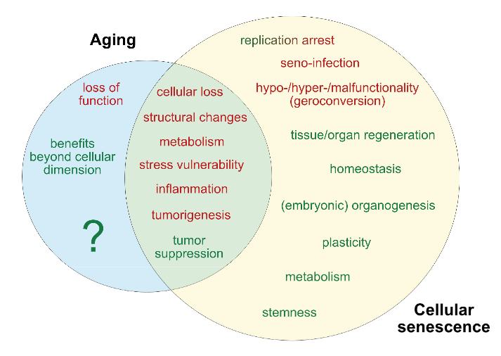
I’ll curate this 2019 German review through its figures:

“With the discovery of beneficial aspects of cellular senescence and evidence of senescence being not limited to replicative cellular states, a redefinition of our comprehension of aging and senescence appears scientifically overdue.

Figure 1. Current determinants and relevant open questions, marking the processes of aging and senescence as discussed in the text. Aspects represented in green are considered as broadly accepted or scientifically consolidated. Novel aspects that are yet unproven, or are under debate, are highlighted in red.

SASP = senescence-associated secretory phenotype. AASP = putative aging-associated secretory phenotype as suggested in the text.

Figure 2. Theories on the causality and purpose of aging. Graphically summarized are four contrasting concepts crystallized from current evidence addressing the inductive driving force of aging. Apart from a stochastic deleteriome, there are arguments for a pseudo-programmed, programmed or at least partially programmed nature of aging.

Figure 3. Comparative representation of the aging and senescence processes highlighting different levels of interaction and putative sites of interventions.

(1) As discussed in the text, causative mechanisms of aging are still not well understood, however, multiple factors including genetic, epigenetic and stress-related effects seem to have an orchestrated role in the progression of aging. Senescence on the other hand, is seen as a programmed response to different kinds of stressors, which proceed in defined stages. Whether, in analogy, aging also follows a defined program or sequential stages is not known.

(2) Senescence involves autocrine and paracrine factors, which are responsible for a ‘seno-infection’ or bystander effect in neighboring cells. There is currently no direct evidence for a similar factor composition propagating the aging process via a kind of ‘gero-infection’.

(3) Accumulation of senescent cells has been described as a hallmark of aging; however, whether they are a causative factor or a consequence of tissue and organismal aging is still unknown. As discussed in the text, it appears possible that aging and senescence mutually influence each other through positive feedback at this level, leading to accelerated tissue damage and aging.

(4,5) Clearance of senescent or aging cells might constitute putative targets for interventional approaches aimed to reduce or reverse the impact of aging and improve cell and tissue homeostasis by inducing a ‘rejuvenation’ process.

Figure 4. Pathological and beneficial functions of aging and senescence, according to current knowledge. In red are represented pathological consequences and in green beneficial functions of aging and senescence.

The impact of aging has mainly been described at the organismal level, since a complete cellular functional profile has not yet been established. Accordingly, whether beneficial consequences of the aging process exist at the cellular level is unclear.”


The assertion of Figure 3 (2) that:

“There is currently no direct evidence for a similar factor composition propagating the aging process via a kind of ‘gero-infection.”

was shown to be false in Reevaluate findings in another paradigm:

“It was demonstrated that increased aging occurred as a result of lack of gonadotropin-releasing hormone and that increased lifespan resulted from its provision during aging.

In this manner:

  1. Aging of hypothalamic microglia leads to
  2. Aging of the hypothalamus, which leads to
  3. Aging elsewhere in the body.

So here we have a multi-level interaction:

  1. Activation of NF-κB leads to
  2. Cellular aging, leading to
  3. A diminished production of GnRH, which then
  4. Acts (through cells with a receptor for it, or indirectly as a result of changes to GnRH-receptor-possessing cells) to decrease lifespan.

So the age state of hypothalamic cells, at least with respect to NF-κB activation, is communicated to other cells via reduced output of GnRH.”


The reviewers’ position on Figure 2 was:

“In our view, recent evidence that

  • Senescence is based on an unterminated developmental growth program and the finding that
  • The concept of post-mitotic senescence requires the activation of expansion, or ‘growth’ factors as a second hit,

favor the assumption that aging underlies a grating of genetic determination similarly to what is summarized above under the pseudo-programmed causative approach.”

Their position on Figure 4’s beneficial effects of aging began with the sentence:

“If we assume that aging already starts before birth, it can be considered simply a developmental stage, required to complete the evolutionary program associated with species-intrinsic biological functions such as reproduction, survival, and selection.”

Cited studies included:

https://www.mdpi.com/2073-4409/8/11/1446 “Dissecting Aging and Senescence-Current Concepts and Open Lessons”

A strawman argument against epigenetic clocks

This 2019 review of epigenetic clocks by Washington cancer researchers ignored the elephant in the room: Their epigenetic drift paradigm is generally inapplicable to humans because the vast majority of our cells don’t divide/proliferate. They repeatedly returned to an argument for randomness as a cause for aging and disease:

“A time-dependent stochastic event process, like epigenetic drift, could lead to cancer formation through the accumulation of random epigenetic alterations that, through chance, eventually alter epigenetic driver gene expression leading to a clone of cells destined to become cancer.

It is plausible that the stochastic process inherent in epigenetic drift can induce aberrant methylation events that accumulate in normal cells and eventually induce cancer formation.

Epigenetic drift relates to a biological process that changes the DNA methylome with age via stochastic gains or losses of DNA methylation. Epigenetic drift can be understood in terms of errors in DNA methylation maintenance during DNA-replication.

The phenomenon of (epi)genetic drift is generally associated with phenotypic neutrality.

For patients who develop cancer around age 80, the most likely initiation time for the founder adenoma cell is predicted to be very early in life, roughly between the ages 15 to 20 years. This unexpected and provocative finding suggests that the optimal age-range for prevention of colorectal cancer may be in adolescence and early adulthood (and ideally through lifelong) dietary and lifestyle interventions.”


The reviewers’ strawman arguments intentionally mischaracterized aspects of the epigenetic clock:

1. The epigenetic clock founder’s actual view on aging was in The epigenetic clock theory of aging:

“The proposed epigenetic clock theory of ageing views biological ageing as an unintended consequence of both developmental programmes and maintenance programmes, the molecular footprints of which give rise to DNAm age estimators.”

The reviewers omitted this intrinsic view of aging, which didn’t fit into the above graphic.

2. Another misrepresentation was:

“In contrast to epigenetic clocks, epigenetic drift refers to a stochastic process that involves both gains and losses of the methylation state of CpG dinucleotides over time.”

A reader of the original 2013 epigenetic clock study would understand that epigenetic clocks measure “both gains and losses of methylation” as in:

“The 193 positively and 160 negatively correlated CpGs get hypermethylated and hypomethylated with age, respectively.”

3. These reviewers omitted recent epigenetic clock significant developments. For example, there was no mention of the GrimAge study, although it was published before this review was submitted.

4. Epigenetic drift as the cause of aging and disease has abundant contrary evidence. These reviewers tossed in a little toward the end of their directed narrative:

“We found only a small number of drift-related CpG island-gene pairs for which drift correlated positively and significantly with gene expression.

The functional consequences of epigenetic drift need to be further elucidated.”

However, they never acknowledged the elephant in the room!

https://cancerres.aacrjournals.org/content/early/2019/11/06/0008-5472.CAN-19-0924 “Epigenetic aging: more than just a clock when it comes to cancer” (not freely available)

Reversing epigenetic T cell exhaustion

This 2019 worldwide discussion among 18 experts concerned T cell exhaustion:

“‘T cell exhaustion’ is a broad term that has been used to describe the response of T cells to chronic antigen stimulation, first in the setting of chronic viral infection but more recently in response to tumours.

Key questions remain about the potential to reverse the epigenetic programme of exhaustion and how this might affect the persistence of T cell populations.”


There were nearly a dozen viewpoints on “What do we mean by T cell exhaustion and/or dysfunction and how would you define this state?” 🙂

Answers to the question “What are the key controversies and outstanding research questions?” included:

  • “What are the cellular signalling and transcriptional pathways that drive the conversion to an exhausted T cell phenotype, and how can the chromatin and transcriptional changes of exhaustion be reversed in individual exhausted cells?
  • Whether and how we can manipulate signalling pathways to both activate and maintain T cell responses remain open questions, as does the question of whether pharmacological manipulations can reverse the epigenetic changes associated with exhaustion versus expand less-exhausted populations.
  • We need to define better the effects of the microenvironment on the induction of T cell exhaustion, the developmental trajectories of exhaustion and the point at which and extent to which exhaustion can be reversed. Understanding the consequences of unleashing T cells from exhaustion will also be crucial to designing the most effective therapeutic interventions.
  • When and how exhausted T cell populations are formed. The original view that they are terminally differentiated descendants of formerly ‘normal’ effector T cells has been challenged.
  • Whether the predysfunctional T cells themselves, or their more differentiated (and phenotypically dysfunctional) progeny, form the ultimate effector pool for control of human tumours.
  • How do the functions and states (subpopulations) of exhausted T cells change over time? Can the epigenetic state of exhaustion be reversed to form true effector or memory T cells, and is this required for improved cancer immunotherapy?
  • There is no definitive marker for exhausted T cells, although TOX may prove to be useful. Transcriptional profiles are informative, but epigenetic changes are more specific and robust. A major clinical question is whether exhausted T cells can be, or indeed need to be, reprogrammed to achieve therapeutic benefit.”

https://www.nature.com/articles/s41577-019-0221-9 “Defining ‘T cell exhaustion'” (not freely available)

Get outside today

This 2019 Finnish review focused on vitamin D’s immune system effects:

“The epigenome of human monocytes is at multiple levels sensitive to vitamin D. These data served as the basis for the chromatin model of vitamin D signaling, which mechanistically explains the activation of a few hundred primary vitamin D target genes.

Vitamin D and its receptor are able to antagonize the pro-inflammatory actions of the transcription factors nuclear factor activated T cells (NF-AT) and nuclear factor κ-light-chain-enhancer of activated B cells (NF-κB) in T cells. In this way, vitamin D reduces autoimmunity, such as the onset and progression of multiple sclerosis, as well as chronic inflammation.

Population-wide recommendations do not take inter-individual variations into account, such as a different molecular response to vitamin D, which are expressed by the vitamin D response index. Instead of population-based recommendations for vitamin D3 supplementation there should be personalized recommendations in order to reach a vitamin D status that is optimized for an individual’s health protection.

Trained immunity implies that immune cells memorize challenges, to which they are exposed in their rather short lifespan, in form of changes of their epigenome leading to subtype specification. The stabilization of the epigenomes of the subtypes of monocytes, macrophages and dendritic cells by vitamin D can prevent or delay the onset of common age-related diseases.”

https://www.ncbi.nlm.nih.gov/pmc/articles/PMC6753645/ “Vitamin D Signaling in the Context of Innate Immunity: Focus on Human Monocytes”


One of the five elements of the clinical trial Reversal of aging and immunosenescent trends was daily 3,000 IU vitamin D3 supplementation for nine months. That study’s monocyte findings included:

“Analysis of CyTOF‐defined immune cell populations revealed the most robust changes to be decreases in total and CD38‐positive monocytes and resulting increases in the lymphocyte‐to‐monocyte ratio (LMR). The changes in mean monocyte populations persisted 6 months after discontinuation of treatment, and the increase in LMR remained highly significant at 18 months as well.”

Reversal of aging and immunosenescent trends

The title of this post is essentially the same as the 2019 human clinical trial:

“Epigenetic aging can be reversed in humans. Using a protocol intended to regenerate the thymus, we observed protective immunological changes, improved risk indices for many age‐related diseases, and a mean epigenetic age approximately 1.5 years less than baseline after 1 year of treatment.

This is to our knowledge the first report of an increase, based on an epigenetic age estimator, in predicted human lifespan by means of a currently accessible aging intervention.

Analysis of CyTOF‐defined immune cell populations revealed the most robust changes to be decreases in total and CD38‐positive monocytes and resulting increases in the lymphocyte‐to‐monocyte ratio (LMR). The changes in mean monocyte populations persisted 6 months after discontinuation of treatment, and the increase in LMR remained highly significant at 18 months as well.

Example of treatment‐induced change in thymic MRI appearance. Darkening corresponds to replacement of fat with nonadipose tissue. White lines denote the thymic boundary. Volunteer 2 at 0 (a) and 9 (b) months”

https://onlinelibrary.wiley.com/doi/full/10.1111/acel.13028 “Reversal of epigenetic aging and immunosenescent trends in humans”


Here’s a 2017 interview with the clinical trial lead author:

“You might also say that what also happened was to just postpone death from infectious diseases to after 60-65 years of age, which means that the same basic problem still remains.”


The popular press botched the facts as they usually do. I won’t link the UK Independent article because they couldn’t be bothered to even define epigenetic clock correctly.

A science journal article did a better job of explaining the study to readers. However, they often used hyperbole instead of trying to promote understanding.

Josh Mitteldorf’s blog post 1st Age Reversal Results—Is it HGH or Something Else? provided the most informative explanations:

“In 2015, Fahy finally had funding and regulatory approval to replicate his one-man trial in a still-tiny sample of ten men, aged 51-65. That it took so long is an indictment of everything about the way aging research is funded in this country; and not just aging – all medical research is prioritized according to projected profits rather than projected health benefits.”


Further thoughts in Reversal of aging and immunosenescent trends with sulforaphane and Part 2 of Reversal of aging and immunosenescent trends with sulforaphane.

Effects of advanced glycation end products on quality of life and lifespan

This 2018 Chinese review concerned advanced glycation end products (AGE) mobility interventions:

“Only a limited number of studies have focused on measuring the effects of low AGEs levels or AGEs inhibitors on mobility, although many observational human studies and in vitro studies have reported the correlation of AGEs with and the contribution of AGEs to mobility, particular in diseases such as:

  • osteoporosis,
  • cartilage degradation,
  • osteoarthritis and
  • sarcopenia.

There is insufficient information from previous animal and human studies for use as a reference to determine the intervention period. Although serum AGEs levels can be easily affected by a lower AGEs diet or AGEs inhibitors, it may take longer to see the changes in certain organs or tissues, as a result of a reduction in AGEs accumulation.”

“Effect of AGEs on apoptosis signalling. AP-1, activator protein 1; ERK, extracellular signal-regulated protein kinases; IGF-I, insulin-like growth factor I; IL-6, interleukin-6; JAK, Janus kinase; JNK, c-Jun N-terminal kinases; MEK, mitogen-activated protein kinase; NF-κB, nuclear factor kappa B; p38 MAPK, p38 mitogen-activated protein kinase; RAGE, receptor for AGEs; STAT3, signal transducers and activators of transcription 3; TGF-β, transforming growth factor-β”

https://www.ncbi.nlm.nih.gov/pmc/articles/PMC6180645/ “Role of advanced glycation end products in mobility and considerations in possible dietary and nutritional intervention strategies”


Citations aren’t validations of the reference’s quality and strength of evidence. This review would have benefited from not citing reviews that contained misrepresentations, such as one mentioned in Wikipedia is a poor source of information on advanced glycation end products (AGEs).

I came across this review as a result of it citing the excellent 2008 rodent study Oral Glycotoxins Determine the Effects of Calorie Restriction on Oxidant Stress, Age-Related Diseases, and Lifespan which found:

“Higher levels of oxidant AGEs in offspring of Reg-F0 dams may be attributable to placental transmission from mothers with high AGE levels. These high intrauterine AGE levels may predispose the offspring to the development of chronic inflammation and diseases in adulthood, such as insulin resistance and diabetes.

Increasing the intake of AGEs in the diet erases the benefits of CR [calorie restriction]. OS [oxidant stress] can be reduced, and healthspan increased, in mice fed a diet that is restricted in the content of AGEs.

The beneficial effects of a CR diet may be partly related to reduced oxidant intake rather than decreased energy intake.”