People will forgive you for being wrong, but they will never forgive you for being right – especially if events prove you right while proving them wrong. Thomas Sowell
This 2019 US/UK human cell study by the founder of the epigenetic clock method investigated epigenetic aging:
“It is widely assumed that extension of lifespan is a result of retardation of ageing. While there is no counter-evidence to challenge this highly intuitive association, supporting empirical evidence to confirm it is not easy to acquire.
The scarcity of empirical evidence is due in part to the lack of a good measure of age that is not based on time. In this regard, the relatively recent development of epigenetic clocks is of great interest.
At the cellular level more is known, but from the perspective of what epigenetic ageing is not, rather than what it is. While we still do not know what cellular feature is associated with epigenetic ageing, we can now remove:
which represent cellular features that are all not linked to epigenetic ageing.”
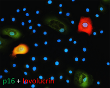
The study used several agents, including rapamycin, to investigate the hypotheses. Rapamycin isn’t a panacea, however:
“The ability of rapamycin to suppress the progression of epigenetic ageing is very encouraging for many reasons not least because it provides a valuable point-of-entry into molecular pathways that are potentially associated with it. Evidently, the target of rapamycin, the mTOR complex is of particular interest.
The convergence of the GWAS observation with the experimental system described here is a testament of the strength of the skin & blood clock in uncovering biological features that are consistent between the human level and cellular level. It lends weight to the emerging view that the mTOR pathway may be the underlying mechanism that supports epigenetic ageing.”
The limitation section ended with:
“It is important to note that it is inadvisable (actively discouraged) to directly extrapolate the studies here, especially in terms of the magnitude of age suppression, to potential effects of rapamycin on humans.”
https://www.aging-us.com/article/101976/text “Rapamycin retards epigenetic ageing of keratinocytes independently of its effects on replicative senescence, proliferation and differentiation”
This 2019 Washington State University rodent study from Dr. Michael Skinner’s lab found:
“A cascade of epigenetic alterations initiated in PGCs [primordial germ cells of F3 males] appears to be required to alter epigenetic programming during spermatogenesis to modify the sperm epigenome involved in transgenerational epigenetic inheritance phenomenon.
Following fertilization there is a DNA methylation erasure to generate stem cells in the early embryo, which then remethylate in a cell type-specific manner. DNA methylation erasure is thought to, in part, reset deleterious epigenetics in the germline. However, imprinted gene DNA methylation sites and induced transgenerational epimutations appear to be protected from this DNA methylation erasure.
A germline with an altered epigenome has the capacity to alter the early embryo’s stem cell’s epigenome and transcriptome that can subsequently impact epigenomes and transcriptomes of all derived somatic cells. Therefore, an altered sperm epigenome has the capacity to transmit phenotypes transgenerationally. Experiments have demonstrated that epigenetic inheritance can also be transmitted through the female germline.
Previously, agricultural fungicide vinclozolin was found to promote transgenerational inheritance of sperm differential DNA methylation regions (DMRs) termed epimutations that help mediate this epigenetic inheritance. The current study was designed to investigate developmental origins of transgenerational DMRs during gametogenesis.
The current study with vinclozolin-induced transgenerational inheritance demonstrates that sperm DMRs also originate during both spermatogenesis and earlier stages of germline development, but at distinct developmental stages. Fetal exposure initiates a developmental cascade (i.e., distinct developmental origins) of aberrant epigenetic programming, and does not simply induce a specific number of DMRs that are maintained throughout development.”
“Following fertilization, the hypothesis is that transgenerational epimutations modify early embryonic transcriptomes and epigenomes to re-establish the cascade for the next generation.
As the individual develops, all somatic cells have altered epigenomes and transcriptomes to promote disease susceptibility later in life.”
Researchers: adopt these hypotheses, and apply them to human studies.
1. Don’t get off track by requiring that the same phenotype must be observed in each generation for there to be transgenerational epigenetic inheritance, because:
“Fetal exposure..does not simply induce a specific number of DMRs that are maintained throughout development.”
Animal transgenerational studies have shown that epigenetic inheritance mechanisms may both express different phenotypes for each generation, and entirely skip a phenotype in one or more generations!
2. Don’t limit your study designs to F1 children as did:
4. Continue studies on to F3 great-grandchildren who had no direct exposure to altering stimulus. Keep in the forefront of your research proposals that there are probably more than 10,000,000 F3 descendants of DES-exposed women just in the US!
This 2019 McGill paper reviewed human and animal studies on brain-shaping influences from the fetal period through childhood:
“In neonates, regions of the methylome that are highly variable across individuals are explained by the genotype alone in 25 percent of cases. The best explanation for 75 percent of variably methylated regions is the interaction of genotype with different in utero environments.
A meta-analysis including 45,821 individuals with attention-deficit/hyperactivity disorder and 9,207,363 controls suggests that conditions such as preeclampsia, Apgar score lower than 7 at 5 minutes, breech/transverse presentations, and prolapsed/nuchal cord – all of which involve some sort of poor oxygenation during delivery – are significantly associated with attention-deficit/hyperactivity disorder. The dopaminergic system seems to be one of the brain systems most affected by perinatal hypoxia-ischemia.
Exposure to childhood trauma activates the stress response systems and dysregulates serotonin transmission that can adverselyimpact brain development. Smaller cerebral, cerebellar, prefrontal cortex, and corpus callosum volumes were reported in maltreated young people as well as reduced hippocampal activity.
Environmental enrichment has a series of beneficial effects associated with neuroplasticity mechanisms, increasing hippocampal volume, and enhancing dorsal dentate gyrus-specific differences in gene expression. Environmental enrichment after prenatal stress decreases depressive-like behaviors and fear, and improves cognitive deficits.”
The reviewers presented strong evidence until the Possible Factors for Reversibility section, which ended with the assertion:
“All these positive environmental experiences mentioned in this section could counterbalance the detrimental effects of early life adversities, making individuals resilient to brain alterations and development of later psychopathology.”
The review’s penultimate sentence recognized that research is seldom done on direct treatments of causes:
“The cross-sectional nature of most epigenetic studies and the tissue specificity of the epigenetic changes are still challenges.”
Cross-sectional studies won’t provide definitive data on cause-and-effect relationships.
The question yet to be examined is: How can humans best address these early-life causes to ameliorate their lifelong effects?
Starting the fifth year of this blog with a 2018 presentation by the founder of the epigenetic clock method describing the state of the art up through July 2018. The webinar was given on the release day of The epigenetic clock now includes skin study.
Segments before the half-hour mark provide an introduction to the method and several details about the concurrently-released study. The Q&A section starts a little before the hour mark.
This 2018 Austrian human study subject was various associations of prenatal testosterone levels to fetal development:
“The available evidence suggests, albeit not conclusively, that prenatal testosterone levels may be one cause for the association of sexual orientation with handedness. Associations among women were consistent with predictions of the Geschwind–Galaburda theory (GGT), whereas those among men were consistent with predictions of the callosal hypothesis. However, research on the associations between sexual orientation and handedness appears to be compromised by various methodological and interpretational problems which need to be overcome to arrive at a clearer picture.
The GGT posits that high prenatal testosterone levels cause a delay in the fetal development of the left cerebral hemisphere which results in a right-hemisphere dominance and hence in a tendency for left-handedness. According to the GGT, high prenatal testosterone levels entail not only a masculinization of the female fetus, but also a feminization of the male fetus (contrary to neurohormonal theory). Overall, the male fetus is subjected to higher levels of intrauterine testosterone than the female fetus. The GGT is thus consistent with the higher prevalence of left-handedness among men than among women.
The callosal hypothesis applies to men only and assumes, in line with neurohormonal theory, that low prenatal testosterone levels are associated with later homosexuality. According to the CH, high prenatal testosterone enhances processes of cerebral lateralization through mechanisms of axonal pruning, thereby resulting in stronger left-hemisphere dominance and a smaller corpus callosum. Consistent with this, women have a larger corpus callosum than men.”
The study’s Limitations section included the following:
“Limitations of the current study pertain to the self-report nature of our data. Behavioral data may provide differing results from those obtained here.
Assessment of sexual orientation relied on a single-item measure. Utilization of rating scales (e.g., the Kinsey Sexual Orientation Scale) or of multi-item scales, and assessing different components of sexual orientation, would have allowed for a more fine-grained analysis and for a cross-validation of sexual orientation ratings with sexual attraction.
Albeit both our samples were large, the proportions of bisexual and homosexual individuals were, expectedly, only small, as were effects of lateral preferences. Thus, in analysis we could not differentiate bisexual from homosexual individuals. Bisexual and homosexual individuals may differ with regard to the distribution of lateral preferences.
Some effect tests in this study have been underpowered. Independent replications with even larger samples are still needed.”
The largest unstated limitation was no fetal measurements. When a fetus’ epigenetic responses and adaptations aren’t considered, not only can the two competing hypotheses not be adequately compared, but causes for the studied phenotypicprogramming and other later-life effects will also be missed.
“A single dimension is able to measure a person’s liability to mental disorder, comorbidity among disorders, persistence of disorders over time, and severity of symptoms.”
The coauthors partially based this on:
“Repeated diagnostic interviews carried out over 25 years, when the research participants were 11, 13, 15, 18, 21, 26, 32, and 38 years old, and include information about seven diagnostic groups: anxiety, depression, attention deficit hyperactivity disorder, conduct disorder, substance dependence, bipolar disorder, and schizophrenia.”
“Dunedin and other studies show that most people have at least one episode of mental illness during their lifetime.”
What compels people to manufacture “universal” truths? Aren’t such beliefs poor substitutes for feeling? For understanding historical, factual, personal truths?
What if the price we pay for avoiding and pressuring down our feelings is: A wasted life?
What if the grand hypothesis worth proving is: For one’s life to have meaning, each individual has to regain their feelings?
This 2018 Chinese animal review subject was prenatal and perinatal anesthesia’s adverse epigenetic effects on a fetus/neonate:
“Accumulating evidence from rodent and primate studies has demonstrated that in utero or neonatal exposure to commonly used inhaled and intravenous general anesthetics is associated with neural degeneration and subsequent neurocognitive impairments, manifested in learning and memory disabilities.
So far, conflicting data exist about the effect of anesthetic agents on neurodevelopment in humans and no definite conclusion has been given yet.”
The inhibitors in the above graphic counter anesthesia’s effects on the fetus/neonate, summarized as:
“Epigenetic targeting of DNA methyltransferases and/or histone deacetylases may have some therapeutic value.”
Do physicians consider possible epigenetic alterations of a newborn’s chromatin structure and gene expression when they administer anesthesia to mothers during childbirth?
This 2017 UC Irvine human review subject provided details of how fetalhypothalamic-pituitary-adrenal components and systems develop, and how they are epigenetically changed by the mother’s environment:
“The developmental origins of disease or fetal programming model predicts that intrauterine exposures have life-long consequences for physical and psychological health. Prenatal programming of the fetal hypothalamic-pituitary-adrenal (HPA) axis is proposed as a primary mechanism by which early experiences are linked to later disease risk.
Development of the fetal HPA axis is determined by an intricately timed cascade of endocrine events during gestation and is regulated by an integrated maternal-placental-fetal steroidogenic unit. Mechanisms by which stress-induced elevations in hormones of maternal, fetal, or placental origin influence the structure and function of the emerging fetal HPA axis are discussed.
Human gestational physiology and fetal HPA axis development differ even from that of closely related nonhuman primates, thereby limiting the generalizability of animal models. This review will focus solely on studies of prenatal stress and fetal HPA axis development in humans.”
1. Every time I read a prenatal study I’m in awe of all that has to go right – and at the appropriate times and sequences – for a fetus to be undamaged. Add in what needs to happen at birth, during infancy, and throughout early childhood, and it seems impossible for any human to escape epigenetic damage.
2. The reviewers referenced animal studies and human research performed with postnatal subjects, despite the disclaimer:
This review will focus solely on studies of prenatal stress and fetal HPA axis development in humans.”
This led to blurring of what had been studied or not with human fetuses regarding the subject.
3. These reviewers uncritically listed many dubious human studies that had both stated and undisclosed severe limitations on their findings. Other reviewers offer informed analysis of cited studies, as Sex-specific impacts of childhood trauma summarized with cortisol:
“Findings are dependent upon variance in extenuating factors, including but not limited to, different measurements of:
presence and severity of psychopathology symptomology.”
4. The paper would have been better had it stayed on topic with its title “Developmental origins of the human hypothalamic-pituitary-adrenal axis.” Let other reviews cover animals, post-natal humans, and questionable evidence.
5. I asked the reviewers to provide a searchable file to facilitate using their work as a reference.
“Genome-wide technology has facilitated epigenome-wide association studies (EWAS), permitting ‘hypothesis-free’ examinations in relation to adversity and/or mental health problems. Results of EWAS are in fact conditional on several a priori hypotheses:
EWAS coverage is sufficient for complex psychiatric problems;
Peripheral tissue is meaningful for mental health problems; and
The assumption that biology can be informative to the phenotype.
1. CpG sites were chosen as potentially biologically informative based on consultation with a consortium of DNA methylation experts. Selection was, in part, based on data from a number of phenotypes (some medical in nature such as cancer), and thus is not specifically targeted to brain-based, stress-related complex mental health phenotypes.
2. The assumption is often that distinct peripheral tissues are interchangeable and equally suited for biomarker detection, when in fact it is highly probable that peripheral tissues themselves correspond differently to environmental adversity and/or disease state.
3. Analyses result in general statements such as ‘neurodevelopment’ or the ‘immune system’ being involved in the aetiology of a given phenotype. Whether these broad categories play indeed a substantial role in the aetiology of the mental health problem is often hard to determine given the post hoc nature of the interpretation.”
The reviewers mentioned in item #2 the statistical flaw of assuming that measured entities are interchangeable with one another. They didn’t mention that the problem also affected item #1 methodologies of averaging CpG methylation measurements in fixed genomic bins or over defined genomic regions, as discussed in:
The reviewers offered suggestions for reducing the impacts of these three hypotheses. But will doing more of the same, only better, advance science?
Was it too much to ask of researchers whose paychecks and reputations depended on a framework’s paradigm – such as the “biomarker” mentioned a dozen and a half times – to admit the uselessness of gathering data when the framework in which the data operated wasn’t viable? They already knew or should have known this.
“When phenotypic variation results from alleles that modify phenotypic variance rather than the mean, this link between genotype and phenotype will not be detected.”
“Blood-based EWAS may yield limited information relating to underlying pathological processes for disorders where brain is the primary tissue of interest.”
The truth about complex traits and GWAS added another example of how this framework and many of its paradigms haven’t produced effective explanations of “the aetiology of the mental health problem”
“The most investigated candidate gene hypotheses of schizophrenia are not well supported by genome-wide association studies, and it is likely that this will be the case for other complex traits as well.”
Researchers need to reevaluate their framework if they want to make a difference in their fields. Recasting GWAS as EWAS won’t make it more effective.
This 2018 German review provided short summaries of 44 studies on the contribution of histone modifications to allergies. An overall summary of their search results was:
“There are at least two levels at which the role of histone modifications is manifested.
One is the regulation of cells that contribute to the allergic inflammation (T cells and macrophages) and those that participate in airway remodeling.
The other is the direct association between histone modifications and allergic phenotypes.
Inhibitors of histone-modifying enzymes may potentially be used as anti-allergic drugs. Furthermore, epigenetic patterns may provide novel tools in the diagnosis of allergic disorders.”
This type of search is what’s expected of researchers who will perform either:
A meta-analysis of studies selected from the search results; or
Their own study.
These reviewers didn’t indicate that they were proceeding along either path.
The review was fine for the purpose of presenting current studies of the subject. But this was just the preparatory stage of research.
This 2018 German review was comprehensive for its subject, epigenetic control of variation and stochasticity in metabolic disease. I’ll focus on one aspect, phenotypic variation:
“Phenotypic [Mendelian] variation can result both from gain- and loss-of-function mutations. Because of the extreme interconnectivity of cell regulatory networks, even at the cellular level, predicting the impact of a sequence variant is difficult as the resultant variation acts:
In the context of all other variants and
Their potential additive, synergistic and antagonistic interactions.
This phenomenon is known as epistasis.
∼98.5% of our genome is non-protein-coding: it is pervasively transcribed, and its transcripts can support regulatory function. Among the best functionally characterized non-coding RNAs (ncRNAs) arising from these sequences are microRNAs (miRNAs).
Environmental [non-Mendelian] variation or ‘stimuli’ occurring during critical windows of susceptibility can elicit lifelong alterations in an individual’s phenotype. Intergenerational metabolic reprogramming [in fruit flies] results from global alterations in chromatin state integrity, particularly from reduced H3K27me3 and H3K9me3 [histone] domains.
The broad variation of fingerprints in humans is thought to depend to a large degree on stochastic variation in mechanical forces. These clear examples of inducible multi-stable or stochastic variation highlight how little we know about the landscape of potential phenotypic variation itself.
Consensus estimates of heritability for obesity and T2D are ∼70% and ∼35% respectively. The remaining, unexplained component is known to involve gene–environment interactions as well as non-Mendelian players.”
Although the above graphic displays transgenerational inheritance for humans, the reviewers didn’t cite any human studies that adequately demonstrated causes for and effects of transgenerational epigenetic inheritance.
I’ve read the cited Swedish and Dutch studies. Their designs, methods, and “correlate with” / “was associated with” results didn’t provide incontrovertible evidence from the F0 great-grandparents, F1 grandparents, F2 parents, and F3 children. It’s necessary to thoroughly study each generation to confirm definitive transgenerational epigenetic inheritance causes and effects.
As noted in How to hijack science: Ignore its intent and focus on the 0.0001%, there aren’t any such published studies to cite. Researchers urgently need to do this human research, and stop using these poor substitutes [1] to pretend there are already adequately evidenced transgenerational epigenetic inheritance human results.
I downgraded the review for treating research of this and other subjects as faits accomplis. It’s opposite ends of the evidential spectrum to state “how little we know about the landscape of potential phenotypic variation,” and in the same review, speciously extrapolate animal experiments into putative human results.
[1] As an example of the poor substitutes for evidence, a researcher referred me to the 2013 “Transgenerational effects of prenatal exposure to the 1944–45 Dutch famine” which is freely available at https://obgyn.onlinelibrary.wiley.com/doi/full/10.1111/1471-0528.12136 as a study finding human transgenerational epigenetic inheritance.
The Methods section showed:
The study’s non-statistical data was almost all unverified self-reports by a self-selected sample of the F2 generation, average age 37.
No detailed physical measurements or samples were taken of them, nor of the F1 generation, nor of the F0 generation, all of which are required as baselines for any transgenerational epigenetic inheritance findings.
No detailed physical measurements or samples were taken of the F3 generation, which is the generation that may provide transgenerational evidence if the previous generations also have detailed physical baselines.
The study’s researchers drew enough participants (360) such that their statistics package allowed them to impute and assume into existence a LOT of data. But the scientific method constrained them to make factual statements of what the evidence actually showed. They admitted:
“In conclusion, we did not find a transgenerational effect of prenatal famine exposure on the health of grandchildren in this study.”
Yet this study is somehow cited for evidence of human transgenerational epigenetically inherited causes and effects!
This 2018 Loma Linda review subject was gestational hypoxia:
“Of all the stresses to which the fetus and newborn infant are subjected, perhaps the most important and clinically relevant is that of hypoxia. This review explores the impact of gestational hypoxia on maternal health and fetal development, and epigenetic mechanisms of developmental plasticity with emphasis on the uteroplacental circulation, heart development, cerebral circulation, pulmonary development, and the hypothalamic-pituitary-adrenal axis and adipose tissue.
An understanding of the specific hypoxia-induced environmental and epigenetic adaptations linked to specific organ systems will enhance the development of target-specific inhibition of DNA methylation, histone modifications, and noncoding RNAs that underlie hypoxia-induced phenotypicprogramming of disease vulnerability later in life.
A potential stumbling block to these efforts, however, relates to timing of the intervention. The greatest potential effect would be accomplished at the critical period in development for which the genomic plasticity is at its peak, thus ameliorating the influence of hypoxia or other stressors.
With future developments, it may even become possible to intervene before conception, before the genetic determinants of the risk of developing programmed disease are established.”
Table 3 “Antenatal hypoxia and developmental plasticity” column titles were Species | Offspring Phenotypes of Disorders and Diseases | Reference Nos.
This review was really an ebook, with 94 pages and 1,172 citations in the pdf file. As I did with Faith-tainted epigenetics, I read it with caution toward recognizing 1) the influence of the sponsor’s biases, 2) any directed narrative that ignored evidence contradicting the narrative, and 3) any storytelling.
One review topic that was misconstrued was transgenerational epigenetic inheritance of hypoxic effects. The “transgenerational” term was used inappropriately by several of the citations, and no cited study provided evidence for gestational hypoxic effects through the F3 great-grandchild generation.
“One substance that fetuses are frequently exposed to is caffeine, which is a non-selective adenosine receptor antagonist. We discovered that in utero alteration in adenosine action leads to adverse effects on embryonic and adult murine hearts. We find that cardiac A1ARs [a type of adenosine receptor] protect the embryo from in utero hypoxic stress, a condition that causes an increase in adenosine levels.
After birth in mice, we observed that in utero caffeine exposure leads to abnormal cardiac function and morphology in adults, including an impaired response to β-adrenergic stimulation. Recently, we observed that in utero caffeine exposure induces transgenerational effects on cardiac morphology, function, and gene expression.”
The timing of in utero caffeine treatment leads to differences in adult cardiac function, gene expression, and phenotype. Exposure to caffeine from E6.5–9.5 leads the F1 generation to develop dilated cardiomyopathy with decrease % FS and increased Myh7 expression. In utero caffeine exposure from E10.5–13.5 leads to a hypertrophic cardiomyopathy in the F2 generation along with increased % FS and decreased Myh7 expression
Why was this review and its studies omitted? It was on target for both gestational hypoxia and transgenerational epigenetic inheritance of hypoxic effects!
It was alright to review smoking, cocaine, methamphetamine, etc., but the most prevalent drug addiction – caffeine – couldn’t be a review topic?
The Loma Linda review covered a lot, but I had a quick trigger due to the sponsor’s bias. I started to lose “faith” in the reviewers after reading the citation for the review’s last sentence that didn’t support the statement.
My “faith” disappeared after not understanding why a few topics were misconstrued and omitted. Why do researchers and sponsors ignore, misrepresent, and not continue experiments through the F3 generation to produce evidence for and against transgenerational epigenetic inheritance? Where was the will to follow evidence trails regardless of socially acceptable beverage norms?
The review acquired the taint of storytelling with the reviewers’ assertion:
“..timing of the intervention. The greatest potential effect would be accomplished at the critical period in development for which the genomic plasticity is at its peak, thus ameliorating the influence of hypoxia or other stressors.”
Contradictory evidence was in the omitted caffeine study’s graphic above which described two gestational critical periods where an “intervention” had opposite effects, all of which were harmful to the current fetus’ development and/or to following generations. Widening the PubMed link’s search parameters to “caffeine hypoxia” and “caffeine pregnancy” returned links to human early life studies that used caffeine in interventions, ignoring possible adverse effects on future generations.
This is my final curation of any paper sponsored by this institution.
This 2018 Alabama rodent study investigated the epigenetic effects on developing breast cancer of timing a sulforaphane-based broccoli sprouts diet. Timing of the diet was as follows:
Conception through weaning (postnatal day 28), named the Prenatal/maternal BSp (broccoli sprouts) treatment (what the mothers ate starting when they were adults at 12 weeks until their pups were weaned; the pups were never on a broccoli sprouts diet);
Postnatal day 28 through the termination of the experiment, named the Postnatal early-life BSp treatment (what the offspring ate starting at 4 weeks; the mothers were never on a broccoli sprouts diet); and
Postnatal day 56 through the termination of the experiment, named the Postnatal adult BSp treatment (what the offspring ate starting when they were adults at 8 weeks; the mothers were never on a broccoli sprouts diet).
“The experiment was terminated when the mean tumor diameter in the control mice exceeded 1.0 cm.
Our study indicates a prenatal/maternal BSp dietary treatment exhibited maximal preventive effects in inhibiting breast cancer development compared to postnatal early-life and adult BSp treatments in two transgenic mouse models that can develop breast cancer.
Postnatal early-life BSp treatment starting prior to puberty onset showed protective effects in prevention of breast cancer but was not as effective as the prenatal/maternal BSp treatment. However, adulthood-administered BSp diet did not reduce mammary tumorigenesis.
The prenatal/maternal BSp diet may:
Primarily influence histone modification processes rather than DNA methylation processes that may contribute to its early breast cancer prevention effects;
Exert its transplacental breast cancer chemoprevention effects through enhanced histone acetylation activator markers due to reduced HDAC1 expression and enzymatic activity.
This may be also due to the importance of a dietary intervention window that occurs during a critical oncogenic transition period, which is in early life for these two tested transgenic mouse models. Determination of a critical oncogenic transition period could be complicated in humans, which may partially explain the controversial findings of the adult BSp treatment on breast cancer development in the tested mouse models as compared the previous studies. Thus long-term consumption of BSp diet is recommended to prevent cancers in humans.”
“The dietary concentration for BSp used in the mouse studies was 26% BSp in formulated diet, which is equivalent to 266 g (~4 cups) BSp/per day for human consumption. Therefore, the concentration of BSp in this diet is physiological available and represents a practical consumption level in the human diet.
Prior to the experiment, we tested the potential influences of this prenatal/maternal BSp regimen on maternal and offspring health as well as mammary gland development in the offspring. Our results showed there was no negative effect of this dietary regimen on the above mentioned factors (data not shown) suggesting this diet is safe to use during pregnancy.”
I downgraded the study’s rating because I didn’t see where the above-labelled “Broccoli Sprout Seeds” content of the diet was defined. It’s one thing to state:
“SFN as the most abundant and bioactive compound in the BSp diet has been identified as a potent HDAC inhibitor that preferably influences histone acetylation processes.”
and describe how sulforaphane may do this and may do that, and include it in the study’s title. It’s another thing to quantify an animal study into findings that can help humans.
The study’s food manufacturer offers dietary products to the public without quantifying all of the contents. Good for them if they can stay in business by serving customers who can’t be bothered with scientific evidence.
What’s the difference between the above-labelled “Broccoli Sprout Seeds” and broccoli seeds? Where was the evidence that “Broccoli Sprout Seeds” and SPROUTED “Broccoli Sprout Seeds” were equivalent to the point of claiming:
“Equivalent to 266 g (~4 cups) BSp/per day for human consumption. Therefore, the concentration of BSp in this diet is physiological available and represents a practical consumption level in the human diet.”
To help humans, this animal study had to have more details than the food manufacturer provided. The researchers should have either tasked the manufacturer to specify the “Broccoli Sprout Seeds” content, or contracted out the analysis if they weren’t going to do it themselves.
Regarding timing of a broccoli sprouts diet for humans, the study didn’t provide evidence for recommending:
“Thus long-term consumption of BSp diet is recommended to prevent cancers in humans.”
We’ll start with a 2018 epigenetic clock human study from Finland:
“We evaluated the association between maternal antenatal depression and a novel biomarker of aging at birth, namely epigenetic gestational age (GA) based on fetal cord blood methylation data. We also examined whether this biomarker prospectively predicts and mediates maternal effects on early childhood psychiatric problems.
Maternal history of depression diagnosed before pregnancy and greater antenatal depressive symptoms were associated with child’s lower epigenetic GA. Child’s lower epigenetic GA, in turn, prospectively predicted total and internalizing problems and partially mediated the effects of maternal antenatal depression on internalizing problems in boys.”
Listening to a podcast by one of the coauthors, although the researchers’ stated intent was to determine the etiology of the findings, I didn’t hear any efforts to study the parents in sufficient detail to be able to detect possible intergenerational and transgenerational epigenetic inheritance causes and effects. There were the usual “associated with” and “it could be this, it could be that” hedges, which were also indicators of the limited methods employed toward the study’s limited design.
Why was an opportunity missed to advance human research in this area? Are researchers satisfied with non-causal individual differences non-explanations instead of making efforts in areas that may produce etiological findings?
The second 2018 epigenetic clock human study was from Alabama:
“We estimated measures of epigenetic age acceleration in 830 Caucasian participants from the Genetics Of Lipid Lowering Drugs and diet Network (GOLDN) considering two epigenetic age calculations.
Both DNA methylation age estimates were highly correlated with chronological age. We found that the Horvath and Hannum measures of epigenetic age acceleration were moderately correlated.
The Horvath age acceleration measure exhibited marginal associations with increased postprandial [after eating a meal] HDL [high-density lipoprotein], increased postprandial total cholesterol, and decreased soluble interleukin 2 receptor subunit alpha (IL2sRα). The Hannum measure of epigenetic age acceleration was inversely associated with fasting HDL and positively associated with postprandial TG [triglyceride], interleukin-6 (IL-6), C-reactive protein (CRP), and tumor necrosis factor alpha (TNFα).
The third 2018 epigenetic clock human study was a meta-analysis of cohorts from the UK, Italy, Sweden, and Scotland:
“The trajectories of Δage showed a declining trend in almost all of the cohorts with adult sample collections. This indicates that epigenetic age increases at a slower rate than chronological age, especially in the oldest population.
Some of the effect is likely driven by survival bias, where healthy individuals are those maintained within a longitudinal study, although other factors like underlying training population for the respective clocks may also have influenced this trend. It may also be possible that there is a ceiling effect for Δage whereby epigenetic clock estimates plateau.”