Taurine’s effects on healthspan and lifespan, Part 2

Four 2023 papers that cited Part 1, starting with a review of hypothetical parameters for taurine clinical trials that aren’t going to happen because:

  • Drug companies can’t make money from a research area that’s cheap, not patentable, and readily accessible.
  • Government sponsors are likewise not incentivized to act in the public’s interest per their recent behavior.

“We propose the rationale that an adequately powered randomized-controlled-trial (RCT) is needed to confirm whether taurine can meaningfully improve metabolic and microbiome health, and biological age.

taurine hypothetical trial

Using long-term survival as a primary outcome is desirable but difficult; any demonstrable difference in this outcome will require a substantial sample size with prolonged follow-up (e.g., 5 years or longer) if the effect size is relatively small (or modest at best). Biological age based on DNA methylation biomarkers according to the Levine PhenoAge or newer biological age models is increasingly being recognized as an important dynamic health parameter, and hence it can also be used as a surrogate outcome in assessing benefits of taurine supplementation.

The recent taurine trial on nonhuman primates used an equivalent dose that was between 3 and 6 g per day for an 80-kg person, and this could represent a reasonable dose range for any human RCTs. We believe that a 6-month or longer interventional period matching what was successfully done on nonhuman primates will be an acceptable time frame in assessing potential efficacy of taurine on human metabolic health in a RCT.”

https://www.sciopen.com/article/10.26599/1671-5411.2023.11.004 “Flattening the biological age curve by improving metabolic health: to taurine or not to taurine, that’s the question”

A six-month duration and a 6 grams per day dose were in the above table’s desirable features column, but epigenetic clock measurements weren’t included as an outcome. I’d guess that its omission reflected disagreements among coauthors, because the desirability of using epigenetic clocks as surrogate measures of human healthspan and lifespan was mentioned several times.


Another review:

“As described in the first half of this review, recent advances in omics analysis technology have led to research to detect the causative gene of dilated cardiomyopathy. It has been found that rare mutations in the taurine transporter gene contribute to the development of dilated cardiomyopathy in humans. It is unlikely that a taurine-deficient diet is a factor in dilated cardiomyopathy, but taurine intake may have positive cardiovascular effects.

The second half summarizes the relationship between taurine and healthspan and lifespan. It is difficult to summarize the effect of age in whole body taurine content, which may vary in species, strain, sex, and age of animal models. Future human studies will clarify the relationship between dietary taurine intake and healthy life expectancy.”

https://www.sciencedirect.com/science/article/pii/S1347861323000749 “Taurine deficiency associated with dilated cardiomyopathy and aging”


A human study investigated brain chemicals that fluctuate with our circadian rhythm:

“We conducted a MRS study at 7 T, where occipital NAD content, lactate, and other metabolites were assessed in two different morning and afternoon diurnal states in healthy participants. Salivary cortisol levels were determined to confirm that the experiment was done in two circadian different physiological conditions.

Although no significant differences in NAD+, NADH, and NAD+/NADH were detected between the morning and afternoon sessions, there was a significant variance difference in NAD+/NADH, with a higher variance of NAD+/NADH redox ratio in the morning.

None of the over 30 measured brain metabolites were significantly affected by the circadian rhythm (CR) except for taurine, which decreased in the afternoon. Further CR studies should consider the prospective measurement of taurine levels in different regions of the human brain, and explore how taurine supplements could impact brain CR metabolism in health and diseases.”

https://www.frontiersin.org/articles/10.3389/fphys.2023.1285776/full “Effect of circadian rhythm on NAD and other metabolites in human brain”

I omitted findings regarding this study’s pathetic Balloon Analogue Risk Task (BART) test. Older studies that drew spurious findings from this video game include:


A rodent study modeled human childhood cataracts:

“Our analysis identified targets that are required for early normal differentiation steps and altered in cataractous lenses, particularly metabolic pathways involving glutathione and amino acids. Glutathione and taurine were spatially altered, and both taurine and the ratio of reduced glutathione to oxidized glutathione, two indicators of redox status, were differentially compromised in lens biology.

1-s2.0-S2213231723002707-ga1_lrg

Dietary amino acid supplementation has been shown to prevent cataract development, and dietary intake of taurine was protective in a glutathione depletion-derived opacity model. This opens up the possibility that dietary supplementation of taurine could be used as a strategy to prevent human congenital cataracts.

Our findings shed light on molecular mechanisms associated with congenital cataracts, and point out that unbalanced redox status due to reduced levels of taurine and glutathione, metabolites already linked to age-related cataracts, could be a major underlying mechanism behind lens opacities that appear early in life.”

https://www.sciencedirect.com/science/article/pii/S2213231723002707 “Unbalanced redox status network as an early pathological event in congenital cataracts”


PXL_20240103_191340418

Take acetyl-L-carnitine if you are healthy

Eight 2023 acetyl-L-carnitine / L-carnitine papers, starting with three healthy human studies:

“Thirty healthy volunteers aged between 19 and 52 years were divided randomly into two equal groups, one of which received 1000 mg of L-carnitine (LC) per day over a 12-week period. Total cholesterol and HDL-C increased significantly after supplementation. LC could be useful in impeding development of heart diseases in subjects with low HDL-C.”

https://journaljammr.com/index.php/JAMMR/article/view/5166 “L-Carnitine Increases High Density Lipoprotein-Cholesterol in Healthy Individuals: A Randomized Trial”

Rationale for dose selection wasn’t provided, and the possibility of limited results due to poor study design wasn’t mentioned.


“This study examined effects of 12 weeks of LC supplementation on bone mineral density (BMD) and selected blood markers involved in bone metabolism of postmenopausal women participating in a resistance training (RT) program. Participants’ diets were supplemented with either 1 g of LC-L-tartrate and 3 g of leucine per day (LC group) or 4 g of leucine per day as a placebo (PLA group), in a double-blind fashion.

Because the study protocol consisted of both exercise and supplementation, some favorable changes in the BMD could be expected. However, it was not possible to detect them in the short study period. No significant modification in BMDs of the spine, hip, and total skeleton and no differences between groups in one-repetition maximum could be due to the relatively short duration of the RT intervention.”

https://nutritionandmetabolism.biomedcentral.com/articles/10.1186/s12986-023-00752-1 “Effect of a 3-month L-carnitine supplementation and resistance training program on circulating markers and bone mineral density in postmenopausal women: a randomized controlled trial”

Same comments as the first study regarding no rationale for dose selection, and no mention that limited results were possibly due to an inadequate dose.


In a letter to the editor, a researcher took issue with a study’s methodology:

“Based on finding that intravenous provision with carnitine alone does not increase muscle carnitine accretion, and on the above-reevaluated data, it appears that the basis for carnitine with caffeine being able to increase muscle carnitine levels, and thereby manipulation of muscle metabolism and exercise performance, is uncertain.

Carnitine bioavailability in any group would have been 9.5%. This assessment would be in line with previously recorded values of 5%–18% carnitine bioavailability. It is firmly believed that low carnitine bioavailability is attributable to the inability of kidneys to reabsorb carnitine when the threshold concentration for tubular reabsorption (about 40–60 μmol/L) has passed this value.

The authors’ proposed long-term use of carnitine supplementation as an aid to improve fat oxidation in type II diabetes also seems to lack provision.”

https://physoc.onlinelibrary.wiley.com/doi/10.14814/phy2.15736 “LTE: Does caffeine truly raise muscle carnitine in humans?”


Two genetic studies:

“Our findings suggest that humans have lost a gene involved in carnitine biosynthesis. Hydroxytrimethyllysine aldolase (the second enzyme of carnitine biosynthesis) activity of serine hydroxymethyl transferase partially compensates for its function.”

https://www.researchsquare.com/article/rs-3295520/v1 “One substrate-many enzymes virtual screening uncovers missing genes of carnitine biosynthesis in human and mouse”


“Reported prevalence of primary carnitine deficiency (PCD) in the Faroe Islands of 1:300 is the highest in the world. The Faroese PCD patient cohort has been closely monitored and we now report results from a 10-year follow-up study of 139 PCD patients.

PCD is an autosomal recessive disorder that affects the function of organic cation transporter 2 (OCTN2) high-affinity carnitine transporters, that localizes to the cell membrane and transport carnitine actively inside the cell. Without proper functioning OCTN2 carnitine transporters, renal reabsorption of carnitine is impaired, and as a consequence, patients suffering from PCD have low plasma levels of carnitine. This can disturb cellular energy production and cause fatigue, but also in extreme cases lead to cellular dysfunction and severe symptoms of coma and sudden death.

PCD patients seem to adhere well to L-carnitine treatment, even though they have to ingest L-carnitine tablets at least three times a day. Overall mean L-carnitine dosage was 66.3 mg/kg/day.”

https://onlinelibrary.wiley.com/doi/10.1002/jmd2.12383 “Patients with primary carnitine deficiency treated with L-carnitine are alive and doing well—A 10-year follow-up in the Faroe Islands”

The average daily dose is (66.3 mg x 70 kg) = 4,641 mg. A third of this dose would be about 1.5 g.

The first study of Acetyl-L-carnitine dosing also suggested dosing L-carnitine three times a day because of 10-20% bioavailability.


A study with unhealthy humans:

“This retrospective study analyzed medical records of adult patients between March 2007 and April 2019, with presenting complaints of fatigue and lethargy. Acetyl-L-carnitine has physiological functions similar to L-carnitine but has higher bioavailability and antioxidant properties. This study confirmed that a triple combination therapy with γ-linolenic acid, V. vinifera extract, and acetyl-L-carnitine can improve arterial stiffness in patients.

Our study had some limitations:

  1. The study population may not be representative of the entire Korean adult population.
  2. The study did not have a medication-free control group. Instead, the comparison group comprised patients with medication compliance <80%.
  3. Drop-out rate of the triple-combination therapy (46.2%, 147/318) was relatively high, indicating the possibility of bias due to loss to follow-up.
  4. The study did not consider lifestyle factors such as smoking, diet, and physical activity level, which may affect arterial stiffness.
  5. The study did not examine interactions among drugs comprising the combination therapy, although all drugs are known to positively impact blood vessels.”

https://onlinelibrary.wiley.com/doi/10.1111/jch.14708 “Efficacy of γ-linolenic acid, Vitis vinifera extract, and acetyl-L-carnitine combination therapy for improving arterial stiffness in Korean adults: Real-world evidence”

This study’s acetyl-L-carnitine dose was 500 mg three times a day.


Wrapping up with two rodent studies:

“Acetyl L-carnitine (ALCAR) has proved useful in treatment of different types of chronic pain with excellent tolerability. The present work aimed at evaluating the anti-hyperalgesic efficacy of ALCAR in a model of persistent visceral pain associated with colitis.

The acetyl group in the ALCAR molecule can enhance cholinergic signalling by promoting synthesis of neurotransmitter acetylcholine, which plays an important role in both the enteric and central nervous systems. Acetylcholine signalling has significant antinociceptive effects in development of visceral pain, so it has been proposed as a therapeutic target.

ijms-24-14841-g001

ALCAR significantly reduced establishment of visceral hyperalgesia in DNBS-treated animals, though the interventive protocol showed a greater efficacy than the preventive one.

  • The interventive protocol partially reduced colon damage in rats, counteracting enteric glia and spinal astrocyte activation resulting from colitis.
  • The preventive protocol effectively protected enteric neurons from inflammatory insult.

These findings suggest the putative usefulness of ALCAR as a food supplement for patients suffering from inflammatory bowel diseases.”

https://www.mdpi.com/1422-0067/24/19/14841 “Anti-Hyperalgesic Efficacy of Acetyl L-Carnitine (ALCAR) Against Visceral Pain Induced by Colitis: Involvement of Glia in the Enteric and Central Nervous System

This study cited multiple animal studies that found acetyl-L-carnitine was effective for different types of pain. I’ve taken it every day for nineteen years, and haven’t noticed that effect.


“Repetitive mild traumatic brain injuries (rmTBI) may contribute to development of neurodegenerative diseases through secondary injury pathways. Acetyl-L-carnitine (ALC) shows neuroprotection through anti-inflammatory effects, and via regulation of neuronal synaptic plasticity by counteracting post-trauma excitotoxicity. This study aimed to investigate mechanisms implicated in etiology of neurodegeneration in rmTBI mice treated with ALC.

ALC is an endogenously produced carnitine metabolite present in tissue and plasma, and readily crosses the blood brain barrier, unlike its unacetylated form. ALC is also a commonly available nutritional supplement, with a known safety profile, and had been well-studied for its role in aiding β-oxidation of long chain fatty acids in the mitochondria.

While some studies have shown promise for improving clinical and psychometric outcomes in individuals with probable Alzheimer’s disease (AD) and mild cognitive impairment, other studies that included participants with moderate AD progression were less conclusive. It may be that this lack of improvement is related to a therapeutic window of opportunity. Once neurodegenerative mechanisms have commenced, a reversal of these processes is not attainable.

There is currently a lack of evidence for safe therapeutics that can be administered long-term to reduce the risk of individuals developing cognitive and neuropsychological deficits after rmTBIs. Prophylactic ALC treatment in a paradigm of neurotrauma may be a way to maximize its therapeutic potential.

While brain structures display differential vulnerability to insult as evidenced by location specific postimpact disruption of key genes, this study shows correlative mRNA neurodegeneration and functional impairment that was ameliorated by ALC treatment in several key genes. ALC may mitigate damage inflicted in various secondary neurodegenerative cascades – confirmed by improvements in behavioral and cognitive function – and contribute to functional protection following rmTBI.”

https://www.frontiersin.org/articles/10.3389/fphar.2023.1254382/full “Repetitive mild traumatic brain injury-induced neurodegeneration and inflammation is attenuated by acetyl-L-carnitine in a preclinical model”

I read many traumatic brain injury papers earlier this year, but only curated two in Brain endothelial cells. I came away thinking that there’s no permanent recovery from TBIs, as just symptoms are effectively treated.

Most TBIs happen to old people who have diminished brain reserves. I didn’t see studies that factored in evidence of what happened earlier in injured people’s lives that created TBI susceptibility but wasn’t remembered.

Unlike other years, I haven’t watched any football this season. It’s unsettling that transient entertainment value continues to take precedence over permanent effects on players’ lives.


PXL_20231223_175628957

36 holes in your roof

An August 2023 interview with Dr. Dale Bredesen, who has reversed Alheizmer’s disease in many people, which will never be acknowledged by the corrupt paradigm:

“How much do you want me to go into things that are relatively controversial and how much do you want me to stick with kind of the more standard line?

For Alzheimer’s we noticed initially there are 36 different potential contributors. You need to patch as many as possible to have an effect.

All of these things, your estradiol level, your progesterone level, pregnenolone, free T3, TSH, Vitamin D, testosterone, these things are all critical. They all feed into the equation.

You have over a hundred trillion contacts in your brain. Will you be able to keep them? Or do you not have what it takes to keep them, and you have to downsize?

The reality is Alzheimer’s disease should be a rare disease. If everybody would get on appropriate prevention or early reversal, we could make it a rare disease.”

https://brokenscience.org/podcasts-ep-5/ “Dale Bredesen – Reversing Alzheimer’s Fate”


See A therapy to reverse cognitive decline for previous curation of Dr. Bredesen’s work.

Transgenerational transmission of stress

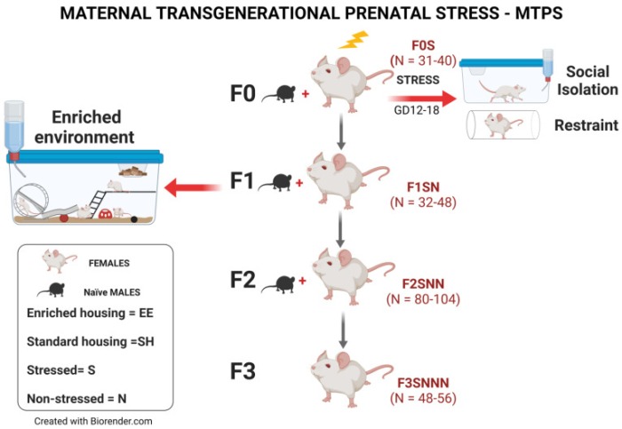
This 2023 rodent study found that effects of stress during mid-late gestation were epigenetically transmitted to the first, second, and third female generations:

“We investigated effects of gestational chronic variable stress (CVS) in rats using restraint and social isolation stress in the parental F0 generation. Only the F0 pregnant dams were subjected to stress.

When a pregnant female experiences adversity, impacts of that stress affect exposed somatic tissues (F0 generation), the fetuses (F1 generation), and the fetuses’ germline (F2 generation). A true transgenerational inheritance arises when germline epimutations are transmitted to unexposed F3 offspring.

A subset of F1 rats was housed in an enriched environment (EE) to mitigate adverse effects of CVS. F2 offspring reared in EE had increased birth weights, but their uterine gene expression patterns remained comparable to those of stressed animals.

ijms-24-03734-g001

We provide evidence that psychological and psychosocial CVS alters inflammatory status and endocrine markers in uteri of adult dams through transgenerational programming of the female germline. EE therapy in prenatally stressed F1 offspring had no beneficial effects on uterine expression of inflammatory and endocrine markers for them or their future offspring.”

https://www.mdpi.com/1422-0067/24/4/3734 “Environmental Enrichment Promotes Transgenerational Programming of Uterine Inflammatory and Stress Markers Comparable to Gestational Chronic Variable Stress”


PXL_20230611_100728709

No exit

This 2023 rodent study investigated aging processes and gut microbiota in crowded conditions:

“Our study provides clear evidence that high-density crowding accelerates the aging process of Brandt’s voles. We also found that ‘high-density microbiota’ promote the aging-related phenotype in voles.

Because we minimized effects of direct fighting on mortality of voles, observed changes in lifespan in this study should mostly represent the natural aging processes of voles.

high-density survival

High density increased the level of stress hormone corticosterone, which disrupted gut microbiota composition by:

  • Decreasing abundance of anti-aging or anti-inflammatory bacterial species; and
  • Increasing the proportion of pathogenic bacteria.

This caused an increase in DNA oxidation and inflammation through upregulation of NF-kB and COX-2 pathways.

Although high-density relief and butyric acid administration interventions could reverse aging-related processes of adult voles, it remains unclear whether they could reverse the aging process in terms of lifespan.

Our results suggest that gut microbiota play a significant role in mediating aging-related processes of voles under high-density conditions, and can be used as a potential therapeutic target for treating stress-related diseases in humans.”

https://onlinelibrary.wiley.com/doi/10.1002/advs.202205346 “Gut Microbiota is Associated with Aging-Related Processes of a Small Mammal Species under High-Density Crowding Stress”


I came across this study by it citing Reversing hair greying for effects of stress interventions.

PXL_20230505_185253518.MP

What do we know about human aging from mouse models?

Here is a 2021 rodent study and relevant parts from 3 of its 26 citing papers:

“A long line of evidence has established the laboratory mouse as the prime model of human aging. However, relatively little is known about detailed behavioral and functional changes that occur across their lifespan, and how this maps onto the phenotype of human aging.

To better understand age-related changes across the lifespan, we characterized functional aging in male C57BL/6J mice of five different ages (3, 6, 12, 18, and 22 months of age) using a multi-domain behavioral test battery. Assessment of functional aging in humans and mice: age-related patterns were determined based on representative data (Table 2), and then superimposed onto survival rate. (A) Body weight, (B) locomotor activity, (C) gait velocity, (D) grip strength, (E) trait anxiety, (F) memory requiring low attention level, and (G) memory requiring high attention level.

fnagi-13-697621-g012

These functional alterations across ages are non-linear, and patterns are unique for each behavioral trait. Physical function progressively declines, starting as early as 6 months of age in mice, while cognitive function begins to decline later, with considerable impairment present at 22 months of age.

Functional aging of male C57BL/6J mice starts at younger relative ages compared to when it starts in humans. Our study suggests that human-equivalent ages of mice might be better determined on the basis of its functional capabilities.”

https://www.frontiersin.org/articles/10.3389/fnagi.2021.697621/full “Functional Aging in Male C57BL/6J Mice Across the Life-Span: A Systematic Behavioral Analysis of Motor, Emotional, and Memory Function to Define an Aging Phenotype”


“Studies in mice show that physical function (i.e., locomotor activity, gait velocity, grip strength) begins to deteriorate around post-natal day (PND) 180, but cognitive functions (i.e., memory) do not exhibit impairment until roughly PND 660. Our results should be considered within the context of behavior changing throughout vole adulthood. Caution should be taken to avoid categorizing the oldest age group in our study as ‘elderly’ or ‘geriatric.'”

https://journals.plos.org/plosone/article?id=10.1371/journal.pone.0276897 “Behavioral trajectories of aging prairie voles (Microtus ochrogaster): Adapting behavior to social context wanes with advanced age”


“We used adult mice ranging in age from 5-6 months, not enough to modify experimental autoimmune encephalomyelitis progression. Mice are considered adult after 8 weeks; however, rapid growth for most biological processes is observed until 3 months of age, while past 6 months, mice might be affected by senescence.”

https://www.frontiersin.org/articles/10.3389/fimmu.2022.1036680/full “Age related immune modulation of experimental autoimmune encephalomyelitis in PINK1 knockout mice”


“Locomotor activity and gait velocity of 12 months old male C57BL/6 correlates with an elderly human being aged 60 or older, supporting that the ~15 months old mice we used in our study were aged mice at the time of tissue collection.”

https://www.mdpi.com/1422-0067/23/20/12461 “Genomic Basis for Individual Differences in Susceptibility to the Neurotoxic Effects of Diesel Exhaust”


PXL_20221122_200643133

If you were given a lens to see clearly, would you accept it?

Two papers, starting with a 2022 rodent study of maternal behaviors’ effects on offspring physiologies:

Early life adversity (ELA) is a major risk factor for development of pathology. Predictability of parental care may be a distinguishing feature of different forms of ELA.

We tested the hypothesis that changes in maternal behavior in mice would be contingent on the type of ELA experienced, directly comparing predictability of care in limited bedding and nesting (LBN) and maternal separation (MS) paradigms. We then tested whether predictability of ELA environment altered expression of corticotropin-releasing hormone (Crh), a sexually-dimorphic neuropeptide that regulates threat-related learning.

MS was associated with increased expression of Crh-related genes in males, but not females. LBN primarily increased expression of these genes in females, but not males.”

https://www.sciencedirect.com/science/article/pii/S2352289522000595 “Resource scarcity but not maternal separation provokes unpredictable maternal care sequences in mice and both upregulate Crh-associated gene expression in the amygdala”


I came across this first study by it citing a republished version of 2005 epigenetic research from McGill University:

“Early experience permanently alters behavior and physiology. A critical question concerns the mechanism of these environmental programming effects.

We propose that epigenomic changes serve as an intermediate process that imprints dynamic environmental experiences on the fixed genome, resulting in stable alterations in phenotype. These findings demonstrate that structural modifications of DNA can be established through environmental programming and that – in spite of inherent stability of this epigenomic marker – it is dynamic and potentially reversible.”

https://www.tandfonline.com/doi/full/10.31887/DCNS.2005.7.2/mmeaney “Environmental programming of stress responses through DNA methylation: life at the interface between a dynamic environment and a fixed genome”


This post commemorates the five-year anniversary of Dr. Arthur Janov’s death. Its title is taken from my reaction to his comment on Beyond Belief: Symptoms of hopelessness. Search his blog for mentions of the second paper’s coauthors, Drs. Meaney and Szyf.


Our lives are substantially a product of our parents’ actualized and unsatisfied needs. Our children and their children are reflections of us with our problems (unfelt needs) or elucidations (felt needs).

What if the price we pay for avoiding and pressuring down our feelings is: A wasted life?

What if the grand hypothesis worth proving is: For one’s life to have meaning, each individual has to regain their feelings?

PXL_20221010_104026908.NIGHT

Non-patentable boron benefits

To follow up Is boron important to health? I’ll highlight a 2022 review of boron intake:

“Boron is essential for activity of several metabolic enzymes, hormones, and micronutrients. It is important for growth and maintenance of bone, reduction in inflammatory biomarkers, and increasing levels of antioxidant enzymes.

The average person’s daily diet contains 1.5 to 3 milligrams of boron. Boron intakes of 1–3 mg/day have been shown to improve bone and brain health in adults when compared to intakes of 0.25–0.50 mg/day.

One week of 10 mg/d boron supplementation resulted in a 20% reduction in inflammatory biomarkers TNF-α, as well as significant reductions (nearly 50%) in plasma concentrations of hs-CRP and IL-6. Calcium fructoborate, a naturally occurring, plant-based boron-carbohydrate complex, had beneficial effects on osteoarthritis (OA) symptoms. A double-blind study in middle-aged patients with primary OA found that all groups except the placebo group saw a reduction in inflammatory biomarkers after 15 days of food supplementation with calcium fructoborate.

Dietary boron intake significantly improves brain function and cognitive functioning in humans. Electroencephalograms showed that boron pharmacological intervention after boron deficiency improved functioning in older men and women, such as less drowsiness and mental alertness, better psychomotor skills (for example, motor speed and dexterity), and better cognitive processing (e.g., attention and short-term memory). Boron compounds can help with both impaired recognition and spatial memory problems.

We discussed the role of boron-based diet in memory, boron and microbiome relation, boron as anti-inflammatory agents, and boron in neurodegenerative diseases. Boron reagents will play a significant role to improve dysbiosis.”

https://www.mdpi.com/1420-3049/27/11/3402/htm “The Role of Microbiome in Brain Development and Neurodegenerative Diseases”


PXL_20220814_101418757

Taurine week #6: Stress

Two 2022 rodent studies of taurine’s associations with long-term stress, starting with a chronic restraint stress model:

“We show that chronic restraint stress can lead to hyperalgesia accompanied by changes in gut microbiota that have significant gender differences. Corresponding changes of bacteria can further induce hyperalgesia and affect different serum metabolism in mice of the corresponding sex.

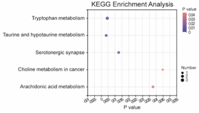
Different serum metabolites between pseudo-germ-free mice receiving fecal microbiota transplantation from the chronic restraint stress group and those from the control group were mainly involved in bile secretion and steroid hormone biosynthesis for male mice, and in taurine and hypotaurine metabolism and tryptophan metabolism for female mice.

Effects of gut microbiota transplantation on serum metabolomics of female host: Taurine and hypotaurine metabolism, tryptophan metabolism, serotonergic synapse, arachidonic acid metabolism, and choline metabolism in cancer were the five identified pathways in which these different metabolites were enriched.

1-s2.0-S1043661822000743-gr11_lrg

Taurine and hypotaurine play essential roles in anti-inflammation, anti-hypertension, anti-hyperglycemia, and analgesia. Taurine can be used as a diagnostic index for fibromyalgia syndrome and neuropathic pain.

These findings improve our understanding of sexual dimorphism in gut microbiota in stress-induced hyperalgesia and the effect of gut microbiota on blood metabolic traits. Follow-up research will investigate causal relationships between them.”

https://www.sciencedirect.com/science/article/pii/S1043661822000743 “Gut microbiota and its role in stress-induced hyperalgesia: Gender-specific responses linked to different changes in serum metabolites”

Human equivalents:

  • A 7-8 month-old mouse would be a 38-42 year-old human.
  • A 14-day stress period is about two years for humans.

A second study used a chronic social defeat stress model:

“The level of taurine in extracellular fluid of the cerebral medial prefrontal cortex (mPFC) was significantly reduced in mice with chronic social defeat stress (CSDS)-induced depression. We found that taurine supplementation effectively rescued immobility time during a tail suspension assay and improved social avoidance behaviors in CSDS mice.

Male C57BL/6 J mice (∼ 23 g) and male CD-1 mice aged 7–8 months (∼ 45 g) were used. CD-1 mice were screened for aggressive behavior during social interactions for three consecutive days before the start of the social defeat sessions. Experimental C57BL/6 J mice were subjected to physical interactions with a novel CD-1 mouse for 10 min once per day over 10 consecutive days.

We found significant reductions in taurine and betaine levels in mPFC interstitial fluid of CSDS mice compared with control mice.

csds taurine betaine

We additionally investigated levels of interstitial taurine in chronic restraint stress (CRS) mice, another depressive animal model. After 14 days of CRS treatment, mice showed typical depression-like behaviors, including decreased sucrose preference and increased immobility time. mPFC levels of interstitial taurine were also significantly decreased in CRS mice.

Taurine treatment protected CSDS mice from impairments in dendritic complexity, spine density, and proportions of different types of spines. Expression of N-methyl D-aspartate receptor subunit 2A, an important synaptic receptor, was largely restored in the mPFC of these mice after taurine supplementation.

These results demonstrated that taurine exerted an antidepressive effect by protecting cortical neurons from dendritic spine loss and synaptic protein deficits.”

https://link.springer.com/article/10.1007/s10571-022-01218-3 “Taurine Alleviates Chronic Social Defeat Stress-Induced Depression by Protecting Cortical Neurons from Dendritic Spine Loss”

Human equivalents:

  • A 7-8 month-old mouse would be a 38-42 year-old human.
  • A 500 mg/kg taurine dose injected intraperitoneally is (.081 x 500 mg) x 70KG = 2.835 g.
  • A 10-day stress period is about a year and a half for humans.

Don’t think aggressive humans would have to be twice as large to stress those around them. There may be choices other than enduring a year and a half of that.

Taurine week #5: Blood

Two 2022 papers investigated taurine’s effects in blood, starting with a review of platelets:

“Taurine is the most abundant free amino acid in the human body, with a six times higher concentration in platelets than any other amino acid. It is highly beneficial for the organism, has many therapeutic actions, and is currently approved for heart failure treatment in Japan. Only the lack of large-scale phase 3 clinical trials restricts taurine use as a therapeutic agent in several other pathologies for treatment of which it has been shown to be effective (hypertension, atherosclerosis, stroke, neurodegenerative diseases, metabolic diseases, e.g., diabetes mellitus, and others).

Because taurine was seen as a non-patentable nutrient, the pharmaceutical industry has not shown much interest in its research. Considering that taurine and its analogues display permissible side effects, along with the need of finding new, alternative antithrombotic drugs with minimal side effects and long-term action, the potential clinical relevance of this fascinating nutrient and its derivatives requires further consideration.”

https://www.mdpi.com/2077-0383/11/3/666/htm “Taurine and Its Derivatives: Analysis of the Inhibitory Effect on Platelet Function and Their Antithrombotic Potential”

Figure 1 provided details of taurine and its derivatives’ effects on various processes involved in platelet activation and aggregation.


A second paper was a rodent study:

“To evaluate chronic effects of taurine on cholesterol levels, we analyzed mice fed a taurine-rich diet for 14–16 weeks. Long-term feeding of taurine lowered plasma cholesterol and bile acids without significantly changing other metabolic parameters, but hardly affected these levels in the liver.

ijms-23-01793-g004

Taurine upregulates transcriptional activity of Cyp7a1 by suppressing FGF21 production in the liver. Bile acids are converted from blood cholesterol by CYP7A1, and more efficiently enter enterohepatic circulation via taurine conjugation.

This study shows that long-term feeding of taurine lowers both plasma cholesterol and bile acids, reinforcing that taurine effectively prevents hypercholesterolemia.”

https://www.mdpi.com/1422-0067/23/3/1793/htm “Long-Term Dietary Taurine Lowers Plasma Levels of Cholesterol and Bile Acids”

A human equivalent of this male C57BL/6J mouse 16-week taurine intervention is roughly 17 years. That strain’s male maximum lifespan is around 800 days, and human maximum lifespan is currently 122.5 years.


PXL_20220520_160922433

The oligosaccharide stachyose

Two 2022 stachyose papers to follow on to Don’t take Beano if you’re stressed, which studied raffinose. Stachyose is in the raffinose oligosaccharide group with similar characteristics, and its content is usually larger in legumes. First is a rodent study:

“Stress can activate the hypothalamic–pituitary–adrenal (HPA) axis and elevate glucocorticoids in the body (cortisol in humans and corticosterone in rodents). Glucocorticoid receptors are abundant in the hippocampus, and play an important role in stress-induced cognition alteration.

Corticosterone is often used to model cognitive impairment induced by stress. Long-term potentiation (LTP) deficit and cognitive impairment always coexist in stress models, and LTP impairment is often considered as one mechanism for stress-induced cognitive deficits.

N-methyl-D-aspartate (NMDA) receptors play critical roles both in normal synaptic functions and excitotoxicity in the central nervous system. D-serine, a coactivator of NMDA receptors, plays an important role in brain function.

In this study, we focused on effects of stachyose, on LTP impairment by corticosterone, gut flora, and the D-serine pathway.

tileshop.fcgi

Data in this study showed that 7-consecutive-day intragastric (i.g.) administration of stachyose had protective effect. There was little effect via intracerebroventricular (i.c.v.) and intraperitoneal (i.p.) administration.

To disturb gut flora, a combination of non-absorbable antibiotics (ATB) were applied. Results showed that ATB canceled the protective effect of stachyose without affecting LTP in control and corticosterone-treated mice, suggesting that stachyose may display its protective effects against LTP impairment by corticosterone via gut flora.

Further study is needed to uncover the relation between gut flora and the D-serine metabolic pathway.”

https://www.frontiersin.org/articles/10.3389/fphar.2022.799244/full “Stachyose Alleviates Corticosterone-Induced Long-Term Potentiation Impairment via the Gut–Brain Axis”

One of this study’s references was Eat oats and regain cognitive normalcy.


A stachyose clinical trial is expected to complete this month:

“In the stachyose intervention group, each person took 5 g of stachyose daily before breakfast. Administration method was 100 ml of drinking water dissolved and taken orally for two months. Each person in the placebo control group took the same amount of maltodextrin daily. Stool samples of the 36 subjects were collected weekly.

Primary outcome measures:

  1. Expression of microRNA; and
  2. Structure of gut microbiota.”

https://clinicaltrials.gov/ct2/show/NCT05392348 “Regulatory Effect of Stachyose on Gut Microbiota and microRNA Expression in Human”


PXL_20220518_093025150

Gut microbiota knowledge through 2021

I’ll curate this 2022 review of what’s known and unknown about our trillions of gut microbiota through its topic headings:

“Most microbial taxa and species of the human microbiome are still unknown. Without revealing the identity of these microbes as a first step, we cannot appreciate their role in human health and diseases.

A. Understanding the Microbiome Composition and Factors That Shape Its Diversity
Effect of Diet Composition on the Microbiome Diversity

  • Macronutrients and Microbiome Diversity
  • Nutrient and Mineral Supplements and Microbiome Diversity

Stress

Drugs

Race and Host Genetics

Aging

Lifestyle

  • Exercise
  • Smoking
  • Urbanization

B. Understanding the Microbiome Function and Its Association With Onset and Progression of Many Diseases

Microbiome Association With Inflammatory and Metabolic Disorders

  • Chronic Inflammation in GIT and Beyond
  • Development of Malignant Tumors
  • Obesity
  • Coronary Artery Disease
  • Respiratory Diseases

Microbiome Role in Psychiatric, Behavioral, and Emotional Disorders

C. Understanding the Microbiome Function as Mediated by Secreted Molecules

D. Conclusion and Future Directions – A pioneering study aimed to computationally predict functions of microbes on earth estimates the presence of 35.5 million functions in bacteria of which only 0.02% are known. Our knowledge of its functions and how they mediate health and diseases is preliminary.”

https://www.frontiersin.org/articles/10.3389/fmicb.2022.825338 “Recent Advances in Understanding the Structure and Function of the Human Microbiome”


I took another test last month at the 14-month point of treating my gut microbiota better. Compared with the 7-month top level measurements, what stood out was an increase in relative abundance from 1% to 7% in the Verrucomicrophia phylum that pretty much exclusively comprises species Akkermansia muciniphilia in humans:

top 5 phylum 2-2022

This review termed Akkermansia muciniphilia relative increases as beneficial. Go with the Alzheimer’s Disease evidence didn’t.

Preventing human infections with dietary fibers inferred that insufficient dietary fiber may disproportionately increase abundance of this species. But I already eat much more fiber than our human ancestors’ estimated 100 grams of fiber every day, so lack of fiber definitely didn’t cause this relative increase.

Resistant starch therapy observed:

“Relative abundances of smaller keystone communities (e.g. primary degraders) may increase, but appear to decrease simply because cross-feeders increase in relative abundance to a greater extent.”

I’ll wait for further evidence while taking responsibility for my own one precious life.

Didn’t agree with this review’s statements regarding microbial associations with fear. These reviewers framed such associations as if gut microbiota in the present had stronger influences on an individual’s fear responses than did any of the individual’s earlier experiences. No way.

I came across this review by it citing The microbiome: An emerging key player in aging and longevity, which was Reference 25 of Dr. Paul Clayton’s blog post What are You Thinking?

Also didn’t agree with some of the doctor’s post:

  • Heterochronic parabiosis of young and old animals is wildly different from fecal transfer. Can’t really compare them to any level of detail.
  • Using a rodent young-to-old fecal microbiota transplant study to imply the same effects would happen in humans? Humans don’t live in controlled environments, so why would a young human individual’s gut microbiota necessarily have healthier effects than an old individual’s?
  • Another example was the penultimate paragraph: “By adding a mix of prebiotic fibers to your diet and maintaining a more youthful and less inflammatory microbiome you will have less inflammation, less endotoxaemia and less inflammageing. You will therefore live healthier and longer.” I’m okay with the first sentence. Equivalating the first sentence to both healthspan and lifespan increases in the second sentence wasn’t supported by any of the 45 cited references.

Signaling pathways and disordered proteins

This 2022 review explored the title subject:

“Cell signaling imposes many demands on proteins that comprise these pathways, including abilities to form active and inactive states, and to engage in multiple protein interactions. Signaling often requires amplifying signals, regulating or tuning responses to signals, combining information sourced from multiple pathways, all while ensuring process fidelity.

Sensitivity, adaptability, and tunability are possible, in part, due to inclusion of intrinsically disordered regions in many proteins involved in cell signaling.  This review highlights the critical role of intrinsically disordered proteins for signaling:

  • In widely diverse organisms (animals, plants, bacteria, fungi);
  • In every category of cell signaling pathway (autocrine, juxtacrine, intracrine, paracrine, and endocrine); and
  • At each stage (ligand, receptor, transducer, effector, terminator) in the cell signaling process.

Function of the glucocorticoid receptor is regulated in part by its intrinsically disordered C-terminal tail. Prior to activation, the glucocorticoid receptor resides in cytosol:

glucocorticoid receptor

Intrinsic disorder in the glucocorticoid receptor not only enables multiple allosteric regulatory interactions to impact function, but also allows deployment of different surfaces of the protein to enable binding to many different sets of macromolecules, and regulation of these interactions via mRNA splicing and phosphorylation.

Combinations of alternative translation initiation and alternative mRNA splicing result in production of multiple glucocorticoid receptor isoforms from one gene. Various isoforms exhibit distinctive tissue distribution patterns and altered transcriptional regulatory profiles.

Greater than 90% of transcription factors either contain intrinsically disordered regions of proteins or are entirely intrinsically disordered. The many advantages conferred by disorder to cell signaling cascades means that:

  1. Understanding signaling required definition of roles disorder plays in each pathway;
  2. Many more examples of disordered proteins in cell signaling pathways are likely to be discovered; and
  3. More mechanisms by which disorder functions remain to be elucidated.”

https://biosignaling.biomedcentral.com/articles/10.1186/s12964-022-00821-7 “Intrinsically disordered proteins play diverse roles in cell signaling”


Cells in vivo seldom act on their own impetus. I would have liked discussion – or at least mention – of bidirectional signals between genes / cells / tissues / organs / organism / environment. This review’s topic of cell signaling pathways excluded “interactions of complex, interconnected systems spanning hierarchical levels” as explored in An environmental signaling paradigm of aging.

Eat broccoli sprouts for depression, Part 2

Here are three papers that cited last year’s Part 1. First is a 2021 rodent study investigating a microRNA’s pro-depressive effects:

“Depressive rat models were established via chronic unpredicted mild stress (CUMS) treatment. Cognitive function of rats was assessed by a series of behavioral tests.

Nrf2 CUMS

Nrf2 was weakly expressed in CUMS-treated rats, whereas Nrf2 upregulation alleviated cognitive dysfunction and brain inflammatory injury.

Nrf2 inhibited miR-17-5p expression via binding to the miR-17-5p promoter. miR-17-5p was also found to limit wolfram syndrome 1 (Wfs1) transcription.

We found that Nrf2 inhibited miR-17-5p expression and promoted Wfs1 transcription, thereby alleviating cognitive dysfunction and inflammatory injury in rats with depression-like behaviors. We didn’t investigate the role of Nrf2 in other depression models (chronic social stress model and chronic restraint stress model) and important brain regions other than hippocampus, such as prefrontal cortex and nucleus accumbens. Accordingly, other depression models and brain regions need to be designed and explored to further validate the role of Nrf2 in depression in future studies.”

https://link.springer.com/article/10.1007/s10753-021-01554-4 “Nrf2 Alleviates Cognitive Dysfunction and Brain Inflammatory Injury via Mediating Wfs1 in Rats with Depression‑Like Behaviors” (not freely available)

This study demonstrated that activating the Nrf2 pathway inhibited brain inflammation, cognitive dysfunction, and depression. Would modulating one microRNA and one gene in vivo without Nrf2 activation achieve similar results?


A 2021 review focused on the immune system’s role in depression:

“Major depressive disorder is one of the most common psychiatric illnesses. The mean age of patients with this disorder is 30.4 years, and the prevalence is twice higher in women than in men.

Activation of inflammatory pathways in the brain is considered to be an important producer of excitotoxicity and oxidative stress inducer that contributes to neuronal damage seen in the disorder. This activation is mainly due to pro-inflammatory cytokines activating the tryptophan-kynurenine (KP) pathway in microglial cells and astrocytes.

Elevated levels of cortisol exert an inhibitory feedback mechanism on its receptors in the hippocampus and hypothalamus, stopping stimulation of these structures to restore balance. When this balance is disrupted, hypercortisolemia directly stimulates extrahepatic enzyme 2,3-indolimine dioxygenase (IDO) located in various tissues (intestine, placenta, liver, and brain) and immune system macrophages and dendritic cells.

Elevation of IDO activities causes metabolism of 99% of available tryptophan in the KP pathway, substantially reducing serotonin synthesis, and producing reactive oxygen species and nitrogen radicals. The excitotoxicity generated produces tissue lesions, and activates the inflammatory response.”

https://academic.oup.com/ijnp/article/25/1/46/6415265 “Inflammatory Process and Immune System in Major Depressive Disorder”

This review highlighted that stress via cortisol and IDO may affect the brain and other parts of the body.


A 2022 review elaborated on Part 1’s findings of MeCP2 as a BDNF inhibitor:

“Methyl-CpG-binding protein 2 (MeCP2) is a transcriptional regulator that is highly abundant in the brain. It binds to methylated genomic DNA to regulate a range of physiological functions implicated in neuronal development and adult synaptic plasticity.

Ability to cope with stressors relies upon activation of the hypothalamic–pituitary–adrenal (HPA) axis. MeCP2 has been shown to contribute to early life stress-dependent epigenetic programming of genes that enhance HPA-axis activity.

We describe known functions of MeCP2 as an epigenetic regulator, and provide evidence for its role in modulating synaptic plasticity via transcriptional regulation of BDNF or other proteins involved in synaptogenesis and synaptic strength like reelin. We conclude that MeCP2 is a promising target for development of novel, more efficacious therapeutics for treatment of stress-related disorders such as depression.”

https://www.mdpi.com/2073-4409/11/4/748/htm “The Role of MeCP2 in Regulating Synaptic Plasticity in the Context of Stress and Depression”


Osprey lunch

PXL_20220221_192924474

Your lungs and Nrf2 activity

Two 2021 papers of Nrf2 activation effects on lung diseases, with the first a McGill University review:

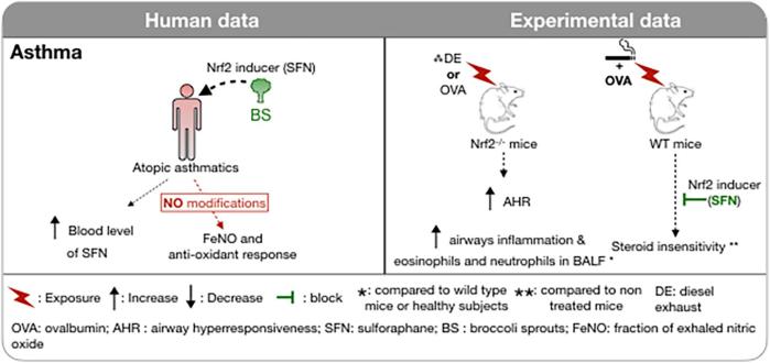
“Oxidative stress and subsequent activation of Nrf2 have been demonstrated in many human respiratory diseases. The purpose of this review is to summarize involvement of Nrf2 and its inducers in acute respiratory distress syndrome, chronic obstructive pulmonary disease (COPD), asthma, and lung fibrosis in both human and experimental models.

fphys-12-727806-g004

These inducers have proven particularly effective at reducing severity of oxidative stress-driven lung injury in various animal models. In humans, these compounds offer promise as potential therapeutic strategies for management of respiratory pathologies associated with oxidative stress, but there is thus far little evidence of efficacy through human trials.

Perhaps, by analogy with biologics, patients with demonstrated deficient antioxidant responses to their disease should be selected for study in future clinical trials.”

https://www.frontiersin.org/articles/10.3389/fphys.2021.727806/full “Role of Nrf2 in Disease: Novel Molecular Mechanisms and Therapeutic Approaches – Pulmonary Disease/Asthma”


A second paper was a human/rodent study of COPD:

“We investigated Nrf2 expression and epigenetic regulation, and mechanisms by which the Nrf2 signaling pathway in ferroptosis is related to COPD. These findings elucidated pathways of ferroptosis in bronchial epithelial cells in COPD, and revealed Nrf2 as a potential target for COPD treatment.

COPD_A_340113_t0001

DNA hypermethylation at specific CpG sites of the Nrf2 promoter in primary epithelial cells and in clinical lung tissues is correlated with decreased Nrf2 expression, which is related to COPD occurrence and development.”

https://www.ncbi.nlm.nih.gov/pmc/articles/PMC8684379/ “Hypermethylation of the Nrf2 Promoter Induces Ferroptosis by Inhibiting the Nrf2-GPX4 Axis in COPD”


Similar to this second paper’s CpG findings, Eat broccoli sprouts for your heart found:

“Sulforaphane (SFN) reduced Ang II‐induced CpG hypermethylation and promoted Ac‐H3 [histone H3 acetylation] accumulation in the Nrf2 promoter region, accompanied by inhibition of global DNMT [DNA methyltransferase] and HDAC [histone deacetylase] activity, and a decreased protein expression of key DNMT and HDAC enzymes. Overall, DNA methylation and histone deacetylation are considered to inhibit gene transcription with a synergistic effect.

Nrf2 can also be regulated independently of Keap1. Evidence indicates that SFN may indirectly activate Nrf2 by affecting activity of several upstream kinases.”

However, this second paper didn’t measure DNMT and HDAC inhibition, although their therapeutic effects in reducing oxidative injury and inflammation may have been present.

PXL_20211225_193439146