Reevaluate findings in another paradigm

It’s challenging for people to change their framework when their paychecks or mental state or reputations depend on it not changing.

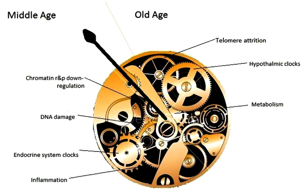
I’ll use The hypothalamus and aging as an example. This review was alright for partial fact-finding up through 2018. Its facts were limited, however, to what fit into the reviewers’ paradigm.

The 2015 An environmental signaling paradigm of aging provided examples of findings that weren’t considered in this 2018 review. It also presented a framework that better incorporated what was known in 2015.


Here’s how they viewed the same 2013 study, Hypothalamic programming of systemic ageing involving IKK-β, NF-κB and GnRH (not freely available).

Paradigm: “The hypothalamus is hypothesized to be a primary regulator of the process of aging of the entire body.”

Study assessment:

“Age-associated inflammation increase is mediated by IκB kinase-β (IKK-β) and nuclear factor κB (NF-κB) in microglia and, subsequently, nearby neurons through microglia–neuron interaction in the mediobasal hypothalamus. Apparently, blocking hypothalamic or brain IKK-β or NF-κB activation causes delayed aging phenotype and improved lifespan.

Aging correlates with a decline in hypothalamic gonadotropin-releasing hormone (GnRH) expression in mice. Mechanistically, activated IKK-β and NF-κB significantly down-regulates GnRH transcription. GnRH therapy through either hypothalamic third ventricularor or subcutaneous injection leads to a significant recovery of neurogenesis in the hypothalamus and hippocampus, and a noticeable improvement of age-related phenotype in skin thickness, bone density, and muscle strength when applied in middle-aged mice.”


Paradigm: Environmental signaling model of aging

Study assessment:

“A link between inflammation and aging is the finding that inflammatory and stress responses activate NF-κB in the hypothalamus and induce a signaling pathway that reduces production of GnRH by neurons. GnRH decline contributes to aging-related changes such as bone fragility, muscle weakness, skin atrophy, and reduced neurogenesis. Consistent with this, GnRH treatment prevents aging-impaired neurogenesis, and decelerates aging in mice.

Zhang et al. report that there is an age-associated activation of NF-κB and IKK-β. Loss of sirtuins may contribute both to inflammation and other aspects of aging. But this explanation, also given by Zhang et al., merely moves the question to why there is a loss of sirtuins.

The case is particularly interesting when we realize that the aging phenotype can only be maintained by continuous activation of NF-κB – a product of which is production of TNF-α.

Reciprocally, when TNF-α is secreted into the inter-cellular milieu, it causes activation of NF-κB. In their study, Zhang et al. noted that activation of NF-κB began in microglia (the immune system component cells found in the brain), which secreted TNF-α, resulting in a positive feedback loop that eventually encompassed the entire central hypothalamus.

The net result of this is a diminution in production of gonadotropin-releasing factor which accounted for a shorter lifespan. Provision of GnRH eliminated that effect, while either preventing NF-κB activation (or that of the IKK-β upstream activator) or by providing gonadotropin-releasing factor directly into the brain, or peripherally, extending lifespan by about 20%.

In spite of the claim of Zhang et al. that the hypothalamus is the regulator of lifespan in mice, their experiments show that only some aspects of lifespan are controlled by the hypothalamus, as preventing NF-κB activation in this organ did not stop aging and death. Similar increased NF-κB activation with age has been seen in other tissues as well, and said to account for dysfunction in aging adrenal glands.

It was demonstrated that increased aging occurred as a result of lack of gonadotropin-releasing hormone, and that increased lifespan resulted from its provision during aging. In this manner:

  1. Aging of hypothalamic microglia leads to
  2. Aging of the hypothalamus, which leads to
  3. Aging elsewhere in the body.

So here we have a multi-level interaction:

  1. Activation of NF-κB leads to
  2. Cellular aging, leading to
  3. Diminished production of GnRH, which then
  4. Acts (through cells with a receptor for it, or indirectly as a result of changes to GnRH-receptor-possessing cells) to decrease lifespan.

So the age state of hypothalamic cells, at least with respect to NF-κB activation, is communicated to other cells via the reduced output of GnRH.”


Not using the same frameworks, are they?

In 2015, this researcher told the world what could be done to dramatically change the entire aging research area. He and other researchers did so recently as curated in Part 3 of Rejuvenation therapy and sulforaphane which addressed hypothalamus rejuvenation.

An environmental signaling paradigm of aging

To follow up A rejuvenation therapy and sulforaphane, the study’s lead laboratory researcher – Dr. Harold Katcher – provided evidence for an environmental signaling paradigm of aging in this 2015 paper:

“The age-phenotype of a cell or organ depends on its environment and not its history.

Organ dysfunction is not the cause of aging, but is the result of its milieu. Therefore, the aged milieu is the cause. Though it has been thought that the aging immune system is the cause of aging, it can seen to be the result of aging.

The systemic milieu of an organism sets the age-phenotype of its cells, tissues and organs. Cells and organs secrete factors into blood, which are determined by the age-phenotype and repair-states of those cells and organs. The presence and concentrations of these blood-borne factors determine the age-phenotype of cells and organs.

Here we must be a bit more speculative. Changes in concentrations of factors present in blood, rather than their presence or absence, determines age-phenotype.

Interactions between disparate levels of the body’s hierarchy establish a consensus age-phenotype for cells and organs, and this largely occurs via the bloodstream. There appear to be positive factors that promote youthful age-phenotypes and negative factors that promote the aged phenotypes.

We readily consider development as a ‘program’, and it seems clear that we must consider post-adult development as ‘programmed’ as well. But if there is a program it is neither in genes nor chromatin, but in interaction of complex, interconnected systems spanning hierarchical levels.

If these aforementioned principles are correct, it should be easy to verify. If so, whole organism rejuvenation might require little more than:

  • Changing concentrations of all age-determining molecules of the bloodstream and various stem cell niche environments to youthful levels;
  • For a time sufficient to cause rejuvenation at the cellular level.

Once cells start secreting factors appropriate to their new, younger age-phenotypes, cognate changes should propagate through hierarchical levels.

The analogy to workings of a mechanical clock is not very exact. ‘Gears’ represent individual aging clocks, both cellular and organic (shown at different levels within the mechanism) which interact, ultimately resulting in organismic age, i.e. ‘body clock’, represented by the ‘hour hand’ (no minute hand is shown).

In mammals, readout of the clock corresponds to age-related composition of blood plasma. In this model, moving the hour hand backwards should result in a turning back of composite clocks as well – a result obtained when induction to pluripotence is used to reset cellular clocks.

Apart from being slowed down or sped up, the body clock can also be reset. Organisms, organs, and their cells can be reset to different age-phenotypes depending on their environment.

We know that old transplanted tissues and organs can regain function and live for the entire life of the younger host at least in rodents. We must suppose that age-phenotype changes must have taken place at the cellular level to allow this.

Rejuvenation cannot be explained on the basis that aging represents accumulation of irreparable cellular damage.

None of these principles are rigorously established as such, but all are supported by experimental evidence.”

http://www.eurekaselect.com/130538/article “Towards an Evidence-based Model of Aging”


Here are some of his responses to comments on the blog post that first curated his current research:

“We’ve (scientists), spent the past 70 years trying to definitively prove the commonsense ‘wear and tear’ theories and have not succeeded. So I tried something different, looking at results of experiments.

This is not based on ‘theory’ (say mitochondrial aging or ‘wear and tear’) but on experimental evidence. Theory comes in explaining our results, not achieving them. There is a theory becoming clear, one very different from the commonsense view of ‘wear and tear’ aging.

We haven’t examined immune response. All that we know for sure is that chronic inflammation of aging stopped. I can definitively say that chronic inflammation due to aging can be reversed with factors present in young blood.

There are amazing things that Big Pharma won’t touch as there’s not enough profit in them (they can’t be patented). So I guess we’re somewhat the same, but we know what to do and have proven it – for us, it’s not money. However, money allows you to do things.

Being 75 myself puts a time-frame around the project. We plan to propose its use for diseases of aging – eventually, everyone will use it. It will end up changing humanity. As people already seem to have too much free time to begin with, what will people do with those extra years they will be given?”


Sections 3 “Aging Manifestations that Have Hitherto Been Proposed as the Causes of Aging are the Consequences of Aging” and 10 “Several Factors ‘Conspire’ to Promote Inflammation in Old Mammalian Bodies, Inflammation Leads to Several Diseases of Aging and Perhaps to Aging Itself” were especially informative.

The former section discussed cells that were capable of making repairs but didn’t make repairs, with aging being the consequence of this behavior. The latter reviewed topics such as senescence, IL-6, NF-κB, and C-reactive protein in terms of feedback loops.

See Reevaluate findings in another paradigm for comparisons of Section 6 with another view of hypothalamic aging.

We believe what we need to believe

While getting ready for bed tonight, I mused about how my younger brother had such an idealized postmortem view of our father. As he expressed six years ago in an obituary for our high school Literature teacher:

“I’ll remember my favorite teacher and how much he’s meant to my life. My father and Martin Obrentz were the two people who made me care about the things that make me the person I am today.”

Believe what you need to believe, David. But like I said five years ago in Reflections on my four-year anniversary of spine surgery:

“I don’t remember that my three siblings ever received a paddling or belting, although they were spanked. Even before he retired, 17 years before he died, the Miami-Dade County public school system stopped him and the rest of their employees from spanking, whipping, beating, and paddling children.”


It’s extremely important for a child to have a witness to their adverse childhood experiences. Otherwise, it’s crazy-making when these experiences aren’t acknowledged as truths by anyone else.

Especially by those who saw but disavow what they saw.

It didn’t really drum into my conscious awareness until tonight that I had such a witness. It wasn’t my mother, of course, since she directed most of my being whipped with a belt, and beaten with a paddle that had holes in it to produce welts. She has denied and deflected my childhood experiences of her ever since then.

It wasn’t my siblings, regrettably for all of us. It wasn’t our Miami neighbors.

When I was twenty, I ran across a guy 300 miles north in Gainesville, Florida, named David Eisenberg, if I remember correctly. A couple of weeks after we met, he asked if my father was Fred Rice, Dean of Boys, West Miami Junior High School. He said he had been beaten by my father several times!

Those weren’t early childhood memories like mine. Those were experiences of a young man during grades 7-9 that he remembered more than a decade later.

I was shocked. It came at a time when I wasn’t ready to face facts about my life, though. I needed fantasies, beliefs to smother what I felt.


I don’t expect that the impacts of my childhood experiences will ever go away. After three years of Primal Therapy that ended a decade ago, at least mine don’t completely control my life anymore.

Dr. Arthur Janov put self-narratives of several patients’ experiences into his May 2016 book Beyond Belief which I partially curated in February 2017. It was partial because I couldn’t read much past Frank’s horrendous story in pages 89 – 105, “The Myth of a Happy Childhood.”

Aging as an unintended consequence

The coauthors of 2018’s The epigenetic clock theory of aging reviewed progress that’s been made todate in understanding epigenetic clock mechanisms.

1. Proven DNA methylation features of epigenetic clocks:

  1. “Methylation of cytosines is undoubtedly a binary event.
  2. The increase in epigenetic age is contributed by changes of methylation profiles in a very small percent of cells in a population.
  3. The clock ticks extremely fast in early post-natal years and much slower after puberty.
  4. Clock CpGs have specific locations in the genome.
  5. It applies to prenatal biological samples and embryonic stem cells.

While consistency with all the five attributes does not guarantee veracity of a model, inconsistency with any one will signal the unlikely validity of a hypothesis.”

2. Regarding what epigenetic clocks don’t measure:

“The effects of

  • Telomere maintenance,
  • Cellular senescence,
  • DNA damage signaling,
  • Terminal differentiation and
  • Cellular proliferation

have all been tested and found to be unrelated to epigenetic ageing.”

3. Regarding cyclical features:

Both the epigenetic and circadian clocks are present in all cells of the body, but their ticking rates are regulated. Both these clocks lose synchronicity when cells are isolated from tissues and grown in vitro.

These similarities compel one to ponder potential links between them.”

This was among the points that Linear thinking about biological age clocks missed.

4. The reviewers discussed 3 of the 5 treatment elements in Reversal of aging and immunosenescent trends:

“It is not known at this stage whether the rejuvenating effect is mediated through the regeneration of the thymus or a direct effect of the treatment modality on the body. Also, it is not known if the effect is mediated by all three compounds or one or two of them.

What we know at this stage does not allow the formation of general principles regarding the impact of hormones on epigenetic age, but their involvement in development and maintenance of the body argue that they do indeed have a very significant impact on the epigenetic clock.”

Not sure why they omitted 3000 IU vitamin D and 50 mg zinc, especially since:

“It is not known if the effect is mediated by all three [five] compounds or one or two of them.”

5. They touched on the specialty of Aging as a disease researchers with:

“Muscle stem cells isolated from mice were epigenetically much younger independently of the ages of the tissue / animal from which they were derived.

The proliferation and differentiation of muscle stem cells cease upon physical maturation. These activities are initiated in adult muscles only in response to injury.

6. The reviewers agreed with those researchers in the Conclusion:

“Epigenetic ageing begins from very early moments after the embryonic stem cell stage and continues uninterrupted through the entire lifespan. The significance of this is profound as the question of why we age has been attributed to many different things, most commonly to ‘wear-and-tear.’

The ticking of the epigenetic clock from the embryonic state challenges this perspective and supports the notion that ageing is an unintended consequence of processes that are necessary for

  • The development of the organism and
  • Tissue homeostasis thereafter.”


https://journals.sagepub.com/doi/10.1177/1535370220918329 “Current perspectives on the cellular and molecular features of epigenetic ageing” (not freely available)

Do early experiences of hunger affect our behavior, thoughts, and feelings today?

Reposted from five years ago.


A 2015 worldwide human study Hunger promotes acquisition of nonfood objects found that people’s current degree of hungriness affected their propensity to acquire nonfood items.

The researchers admitted that they didn’t demonstrate cause and effect with the five experiments they performed, although the findings had merit. News articles poked good-natured fun at the findings with headlines such as “Why Hungry People Want More Binder Clips.”

The research caught my eye with these statements:

“Hunger’s influence extends beyond food consumption to the acquisition of nonfood items that cannot satisfy the underlying need.

We conclude that a basic biologically based motivation can affect substantively unrelated behaviors that cannot satisfy the motivation.


The concept of the quotes relates to a principle of Dr. Arthur Janov’s Primal Therapy – symbolic satisfaction of needs. Two fundamentals of Primal Therapy:

  1. The physiological impacts of our early unmet needs drive our behavior, thoughts, and feelings.
  2. The painful impacts of our unfulfilled needs impel us to be constantly vigilant for some way to fulfill them.

Corollary principles of Primal Therapy:

  • Our present efforts to fulfill our early unmet needs will seldom be satisfying. It’s too late.
  • We acquire substitutes now for what we really needed back then.
  • Acquiring these symbols of our early unmet needs may – at best – temporarily satisfy derivative needs.

But the symbolic satisfaction of derived needs – the symptoms – never resolves the impacts of early unfulfilled needs – the motivating causes:

  • We repeat the acquisition behavior, and get caught in a circle of acting out our feelings and impulses driven by these conditions.
  • The unconscious act-outs become sources of misery both to us and to the people around us.

As this study’s findings showed, there’s every reason for us to want researchers to provide a factual blueprint of causes for our hunger sensation effects, such as “unrelated behaviors that cannot satisfy the motivation.

Hunger research objectives could include answering:

  • What enduring physiological changes occurred as a result of past hunger?
  • How do these changes affect the subjects’ present behaviors, thoughts, and feelings?

Hunger research causal evidence for the effect of why people acquire items that cannot satisfy the underlying needmay include studying where to start the timelines for the impacts of hunger. The impacts potentially go back at least to infancy when we were completely dependent on our caregivers.

Infants can’t get up to go to the refrigerator to satisfy their hunger. All a hungry infant can do is call attention to their need, and feel pain from the deprivation of their need.

Is infancy far back enough, though, to understand the beginnings of potential impacts of hunger?

The epigenetics of perinatal stress

This 2019 McGill review discussed long-lasting effects of perinatal stress:

“Epigenetic processes are involved in embedding the impact of early-life experience in the genome and mediating between social environments and later behavioral phenotypes. Since these phenotypes are apparent a long time after early experience, changes in gene expression programming must be stable.

Although loss of methylation in a promoter is necessary for expression, it is not sufficient. Demethylation removes a barrier for expression, but expression might be realized at the right time or context when needed factors or signals are present.

DNA methylation anticipates future transcriptional response to triggers. Comparing steady-state expression with DNA methylation does not capture the full meaning and scope of regulatory roles of differential methylation.

A model for epigenetic programming by early life stress:

  1. Perinatal stress perceived by the brain triggers release of glucocorticoids (GC) from the adrenal in the mother prenatally or the newborn postnatally.
  2. GC activate nuclear glucocorticoid receptors across the body, which epigenetically program (demethylate) genes that are targets of GR in brain and white blood cells (WBC).
  3. Demethylation events are insufficient for activation of these genes. A brain specific factor (TF) is required for expression and will activate low expression of the gene in the brain but not in blood.
  4. During adulthood a stressful event transiently triggers a very high level of expression of the GR regulated gene specifically in the brain.

Horizontal arrow, transcription; circles, CpG sites; CH3 in circles, methylated sites; empty circles, unmethylated CpG sites; horizon[t]al curved lines, mRNA.”

Review points discussed:

  • “Epigenetic marks are laid down and maintained by enzymes that either add or remove epigenetic modifications and are therefore potentially reversible in contrast to genetic changes.
  • Response to early life stress and maternal behavior is also not limited to the brain and involves at least the immune system as well.
  • The placenta is also impacted by maternal social experience and early life stress.
  • Most studies are limited to peripheral tissues such as saliva and white blood cells, and relevance to brain physiology and pathology is uncertain.
  • Low absolute differences in methylation seen in most human behavioral EWAS raise questions about their biological significance.

  • Although post-mortem studies examine epigenetic programming in physiologically relevant tissues, they represent only a final and single stage that does not capture dynamic evolution of environments and epigenetic programming in living humans.”

https://www.ncbi.nlm.nih.gov/pmc/articles/PMC6952743/ “The epigenetics of perinatal stress”


Other reviewers try to ignore times when we were all fetuses and newborns. For example, in the same journal issue was a Boston review of PTSD that didn’t mention anything about earliest times of human lives! Those reviewers speculated around this obvious gap on their way to being paid by NIH.

Why would researchers ignore perinatal stress events that prime humans for later-life PTSD? Stress generally has a greater impact on fetuses and newborns than on infants, and a greater impact on infants than on adults.

A blood plasma aging clock

This 2019 Stanford human study developed an aging clock using blood plasma proteins:

“We measured 2,925 plasma proteins from 4,331 young adults to nonagenarians [18 – 95] and developed a novel bioinformatics approach which uncovered profound non-linear alterations in the human plasma proteome with age. Waves of changes in the proteome in the fourth, seventh, and eighth decades of life reflected distinct biological pathways, and revealed differential associations with the genome and proteome of age-related diseases and phenotypic traits.

To determine whether the plasma proteome can predict chronological age and serve as a “proteomic clock,” we used 2,858 randomly selected subjects to fine-tune a predictive model that was tested on the remaining 1,473 subjects. We identified a sex-independent plasma proteomic clock consisting of 373 proteins. Subjects that were predicted younger than their chronologic age based on their plasma proteome performed better on cognitive and physical tests.

The 3 age-related crests were comprised of different proteins. Few proteins, such as GDF15, were among the top 10 differentially expressed proteins in each crest, consistent with its strong increase across lifespan. Other proteins, like chordin-like protein 1 (CHRDL1) or pleiotrophin (PTN), were significantly changed only at the last two crests, reflecting their exponential increase with age.

We observed a prominent shift in multiple biological pathways with aging:

  • At young age (34 years), we observed a downregulation of proteins involved in structural pathways such as the extracellular matrix. These changes were reversed in middle and old ages (60 and 78 years, respectively).
  • At age 60, we found a predominant role of hormonal activity, binding functions and blood pathways.
  • At age 78, key processes still included blood pathways but also bone morphogenetic protein signaling, which is involved in numerous cellular functions, including inflammation.

These results suggest that aging is a dynamic, non-linear process characterized by waves of changes in plasma proteins that are reflective of a complex shift in the activity of biological processes.”

https://www.biorxiv.org/content/10.1101/751115v1.full “Undulating changes in human plasma proteome across lifespan are linked to disease”


A non-critical review of the study was published by the Life Extension Advocacy Foundation. Frequent qualifiers like “could,” “may,” and “possible” were consistent with the confirmation biases of their advocacy.

There were several misstatements of what the study did, including the innumerate:

  1. “used around half of the participant data to build a “proteomic clock”
  2. tested it on the other half of the participants
  3. a total of 3000 proteins”

Per the above study quotation, the numbers were actually:

  1. Closer to two thirds (2,858 ÷ 4,331), not “around half”;
  2. The other third (1,473 ÷ 4,331), not “the other half”; and
  3. 2,925 not 3000.

The final paragraph and other parts of the review bordered on woo. Did a review of the findings have to fit LEAF’s perspective?


In contrast, Josh Mitteldorf did his usual excellent job of providing contexts for the study with New Aging Clock based on Proteins in the Blood, emphasizing comparisons with epigenetic clock methodologies:

“For some of the proteins that feature prominently in the clock, we have a good understanding of their metabolic function, and for the most part they vindicate my belief that epigenetic changes are predominantly drivers of senescence rather than protective responses to damage.

Wyss-Coray compared the proteins in the new (human) proteome clock with the proteins that were altered in the (mouse) parabiosis experiments, and found a large overlap [46 proteins change in the same direction and define a conserved aging signature]. This may be the best evidence we have that the proteome changes are predominantly causal factors of senescence.

46 plasma proteins

Almost all the proteins identified as changing rapidly at age 78 are increasing. In contrast, a few of the fastest-changing proteins at age 60 are decreasing (though most are increasing). GDF15 deserves a story of its own.

The implication is that a more accurate clock can be constructed if it incorporates different information at different life stages. None of the Horvath clocks have been derived based on different CpG sites at different ages, and this suggests an opportunity for a potential improvement in accuracy.”

A commentator linked the below study:

https://www.sciencedirect.com/science/article/pii/S0092867419308323 “GDF15 Is an Inflammation-Induced Central Mediator of Tissue Tolerance” (not freely available)

which prompted his response:

“Thanks, Lee! This is just the kind of specific information that I was asking for. It would seem we should construct our clocks without GDF15, which otherwise might loom large.”

Epigenetic transgenerational inheritance extends to the great-great-grand offspring

This 2019 rodent study by the Washington State University labs of Dr. Michael Skinner continued to F4 generation great-great-grand offspring, and demonstrated that epigenetic inheritance mechanisms are similar to imprinted genes:

“Epigenetic transgenerational inheritance potentially impacts disease etiology, phenotypic variation, and evolution. An increasing number of environmental factors from nutrition to toxicants have been shown to promote the epigenetic transgenerational inheritance of disease.

Imprinted genes are a special class of genes since their DNA methylation patterns are unchanged over the generation and are not affected by the methylation erasure occurring early in development. The transgenerational epigenetic alterations in the germline appear to be permanently reprogrammed like imprinted genes, and appear protected from this DNA methylation erasure and reprogramming at fertilization in the subsequent generations. Similar to imprinted genes, the epigenetic transgenerational germline epimutations appear to have a methylation erasure in the primordial germ cells involving an epigenetic molecular memory.

Comparison of the transgenerational F3 generation, with the outcross to the F4 generation through the paternal or maternal lineages, allows an assessment of parent-of-origin transmission of disease or pathology. Observations provided examples of the following:

  1. Pathology that required combined contribution of both paternal and maternal alleles to promote disease [testis and ovarian disease];
  2. Pathology that is derived from the opposite sex allele such as father to daughter [kidney disease] or mother to son [prostate disease];
  3. Pathology that is derived from either parent-of-origin alleles independently [obesity];
  4. Pathology that is transmitted within the same sex, such as maternal to daughter [mammary tumor development]; and
  5. Pathology that is observed only following a specific parent-of-origin outcross [both F4 male obesity and F4 female kidney disease in the vinclozolin lineage].”

https://www.sciencedirect.com/science/article/pii/S0012160619303471 “Epigenetic transgenerational inheritance of parent-of-origin allelic transmission of outcross pathology and sperm epimutations”


This study showed that epigenetically inherited legacies extend to the fifth generation. Do any of us know our ancestors’ medical histories back to our great-great-grandparents?

Will toxicologists take their jobs seriously, catch up to current science, and investigate possible effects in at least the F3 generation that had no direct toxicant exposure?

Do genes or maternal environments shape fetal brains?

This 2019 Singapore human study used Diffusion Tensor Imaging on 5-to-17-day old infants to find:

“Our findings showed evidence for region-specific effects of genotype and GxE on individual differences in human fetal development of the hippocampus and amygdala. Gene x Environment models outcompeted models containing genotype or environment only, to best explain the majority of measures but some, especially of the amygdaloid microstructure, were best explained by genotype only.

Models including DNA methylation measured in the neonate umbilical cords outcompeted the Gene and Gene x Environment models for the majority of amygdaloid measures and minority of hippocampal measures. The fact that methylation models outcompeted gene x environment models in many instances is compatible with the idea that DNA methylation is a product of GxE.

A genome-wide association study of SNP [single nucleotide polymorphism] interactions with the prenatal environments (GxE) yielded genome wide significance for 13 gene x environment models. The majority (10) explained hippocampal measures in interaction with prenatal maternal mental health and SES [socioeconomic status]. The three genome-wide significant models predicting amygdaloid measures, explained right amygdala volume in interaction with maternal depression.

The transcription factor CUX1 was implicated in the genotypic variation interaction with prenatal maternal health to shape the amygdala. It was also a central node in the subnetworks formed by genes mapping to the CpGs in neonatal umbilical cord DNA methylation data associating with both amygdala and hippocampus structure and substructure.

Our results implicated the glucocorticoid receptor (NR3C1) in population variance of neonatal amygdala structure and microstructure.

Estrogen in the hippocampus affects learning, memory, neurogenesis, synapse density and plasticity. In the brain testosterone is commonly aromatized to estradiol and thus the estrogen receptor mediates not only the effects of estrogen, but also that of testosterone.”

https://onlinelibrary.wiley.com/doi/full/10.1111/gbb.12576 “Neonatal amygdalae and hippocampi are influenced by genotype and prenatal environment, and reflected in the neonatal DNA methylome” (not freely available)

Reversal of aging and immunosenescent trends

The title of this post is essentially the same as the 2019 human clinical trial:

“Epigenetic aging can be reversed in humans. Using a protocol intended to regenerate the thymus, we observed protective immunological changes, improved risk indices for many age‐related diseases, and a mean epigenetic age approximately 1.5 years less than baseline after 1 year of treatment.

This is to our knowledge the first report of an increase, based on an epigenetic age estimator, in predicted human lifespan by means of a currently accessible aging intervention.

Analysis of CyTOF‐defined immune cell populations revealed the most robust changes to be decreases in total and CD38‐positive monocytes and resulting increases in the lymphocyte‐to‐monocyte ratio (LMR). The changes in mean monocyte populations persisted 6 months after discontinuation of treatment, and the increase in LMR remained highly significant at 18 months as well.

Example of treatment‐induced change in thymic MRI appearance. Darkening corresponds to replacement of fat with nonadipose tissue. White lines denote the thymic boundary. Volunteer 2 at 0 (a) and 9 (b) months”

https://onlinelibrary.wiley.com/doi/full/10.1111/acel.13028 “Reversal of epigenetic aging and immunosenescent trends in humans”


Here’s a 2017 interview with the clinical trial lead author:

“You might also say that what also happened was to just postpone death from infectious diseases to after 60-65 years of age, which means that the same basic problem still remains.”


The popular press botched the facts as they usually do. I won’t link the UK Independent article because they couldn’t be bothered to even define epigenetic clock correctly.

A science journal article did a better job of explaining the study to readers. However, they often used hyperbole instead of trying to promote understanding.

Josh Mitteldorf’s blog post 1st Age Reversal Results—Is it HGH or Something Else? provided the most informative explanations:

“In 2015, Fahy finally had funding and regulatory approval to replicate his one-man trial in a still-tiny sample of ten men, aged 51-65. That it took so long is an indictment of everything about the way aging research is funded in this country; and not just aging – all medical research is prioritized according to projected profits rather than projected health benefits.”


Further thoughts in Reversal of aging and immunosenescent trends with sulforaphane and Part 2 of Reversal of aging and immunosenescent trends with sulforaphane.

Developmental disorders and the epigenetic clock

This 2019 UK/Canada/Germany human study investigated thirteen developmental disorders to identify genes that changed aspects of the epigenetic clock:

“Sotos syndrome accelerates epigenetic aging [+7.64 years]. Sotos syndrome is caused by loss-of-function mutations in the NSD1 gene, which encodes a histone H3 lysine 36 (H3K36) methyltransferase.

This leads to a phenotype which can include:

  • Prenatal and postnatal overgrowth,
  • Facial gestalt,
  • Advanced bone age,
  • Developmental delay,
  • Higher cancer predisposition, and, in some cases,
  • Heart defects.

Many of these characteristics could be interpreted as aging-like, identifying Sotos syndrome as a potential human model of accelerated physiological aging.

This research will shed some light on the different processes that erode the human epigenetic landscape during aging and provide a new hypothesis about the mechanisms behind the epigenetic aging clock.”

“Proposed model that highlights the role of H3K36 methylation maintenance on epigenetic aging:

  • The H3K36me2/3 mark allows recruiting de novo DNA methyltransferases DNMT3A (in green) and DNMT3B (not shown).
  • DNA methylation valleys (DMVs) are conserved genomic regions that are normally found hypomethylated.
  • During aging, the H3K36 methylation machinery could become less efficient at maintaining the H3K36me2/3 landscape.
  • This would lead to a relocation of de novo DNA methyltransferases from their original genomic reservoirs (which would become hypomethylated) to other non-specific regions such as DMVs (which would become hypermethylated and potentially lose their normal boundaries),
  • With functional consequences for the tissues.”

The researchers improved methodologies of several techniques:

  1. “Previous attempts to account for technical variation have used the first 5 principal components estimated directly from the DNA methylation data. However, this approach potentially removes meaningful biological variation. For the first time, we have shown that it is possible to use the control probes from the 450K array to readily correct for batch effects in the context of the epigenetic clock, which reduces the error associated with the predictions and decreases the likelihood of reporting a false positive.
  2. We have confirmed the suspicion that Horvath’s model underestimates epigenetic age for older ages and assessed the impact of this bias in the screen for epigenetic age acceleration.
  3. Because of the way that the Horvath epigenetic clock was trained, it is likely that its constituent 353 CpG sites are a low-dimensional representation of the different genome-wide processes that are eroding the epigenome with age. Our analysis has shown that these 353 CpG sites are characterized by a higher Shannon entropy when compared with the rest of the genome, which is dramatically decreased in the case of Sotos patients.”

https://genomebiology.biomedcentral.com/articles/10.1186/s13059-019-1753-9 “Screening for genes that accelerate the epigenetic aging clock in humans reveals a role for the H3K36 methyltransferase NSD1”

A better method of measuring neurogenesis

One of the references cited in Linking adult neurogenesis to Alzheimer’s disease was https://www.nature.com/articles/s41591-019-0375-9 “Adult hippocampal neurogenesis is abundant in neurologically healthy subjects and drops sharply in patients with Alzheimer’s disease” (not freely available).

This 2019 Spanish human study used improved techniques to find:

“Adult hippocampal neurogenesis (AHN), confers an unparalleled degree of plasticity to the entire hippocampal circuitry. Direct evidence of AHN in humans has remained elusive. Determining whether new neurons are continuously incorporated into the human dentate gyrus (DG) during physiological and pathological aging is a crucial question with outstanding therapeutic potential.

By combining human brain samples obtained under tightly controlled conditions and state-of-the-art tissue processing methods, we identified thousands of immature neurons in the DG of neurologically healthy human subjects up to the ninth decade of life. These neurons exhibited variable degrees of maturation along differentiation stages of AHN. In sharp contrast, the number and maturation of these neurons progressively declined as AD advanced.

These results demonstrate the persistence of AHN during both physiological and pathological aging in humans and provide evidence for impaired neurogenesis as a potentially relevant mechanism underlying memory deficits in AD that might be amenable to novel therapeutic strategies.”


The control group was 13 neurologically healthy deceased people aged 43 to 87. The AD group was 45 deceased people, distributed among the six Braak stages of the pathology, aged 52 to 97.

Transgenerational diseases caused by great-grandmother DDT exposure

This 2019 rodent study from the labs of Dr. Michael Skinner at Washington State University found:

“The exposure of a gestating female during fetal gonadal sex determination to DDT can promote the epigenetic transgenerational inheritance of obesity and disease.

Transgenerational pathologies (F3 generation) of late puberty, obesity, testis, prostate, and multiple disease were observed in the DDT lineage males. Obesity, ovarian, kidney, and multiple disease transgenerational pathologies (F3 generation) were observed in the DDT lineage females.

Epigenetic biomarkers or diagnostics provide preliminary evidence for preconception diagnosis of increased susceptibility to transgenerational disease in offspring.”


For those of us who thought DDT was discontinued:

“DDT was banned in the USA in 1973, but it is still recommended by the World Health Organization for indoor residual spray. India is by far the largest consumer of DDT worldwide.

India has experienced a 5-fold increase of type II diabetes over the last three decades with a predisposition to obesity already present at birth in much of the population. Although a large number of factors may contribute to this increased incidence of obesity, the potential contribution of ancestral toxicant exposures in the induction of obesity susceptibility requires further investigation.”

Where are the human studies of this subject? Why aren’t follow-on generations’ diseases traced to the likely sources?

How many F3 great-grandchildren of women exposed to DDT during pregnancy are alive today? Millions, tens of millions?

https://www.ncbi.nlm.nih.gov/pmc/articles/PMC6536675 “Sperm epimutation biomarkers of obesity and pathologies following DDT induced epigenetic transgenerational inheritance of disease”

What drives cellular aging?

This 2019 US/UK human cell study by the founder of the epigenetic clock method investigated epigenetic aging:

“It is widely assumed that extension of lifespan is a result of retardation of ageing. While there is no counter-evidence to challenge this highly intuitive association, supporting empirical evidence to confirm it is not easy to acquire.

The scarcity of empirical evidence is due in part to the lack of a good measure of age that is not based on time. In this regard, the relatively recent development of epigenetic clocks is of great interest.

At the cellular level more is known, but from the perspective of what epigenetic ageing is not, rather than what it is. While we still do not know what cellular feature is associated with epigenetic ageing, we can now remove:

  • somatic cell differentiation

from the list of possibilities and place it with

  • cellular senescence,
  • proliferation and
  • telomere length maintenance,

which represent cellular features that are all not linked to epigenetic ageing.”


The study used several agents, including rapamycin, to investigate the hypotheses. Rapamycin isn’t a panacea, however:

“The ability of rapamycin to suppress the progression of epigenetic ageing is very encouraging for many reasons not least because it provides a valuable point-of-entry into molecular pathways that are potentially associated with it. Evidently, the target of rapamycin, the mTOR complex is of particular interest.

The convergence of the GWAS observation with the experimental system described here is a testament of the strength of the skin & blood clock in uncovering biological features that are consistent between the human level and cellular level. It lends weight to the emerging view that the mTOR pathway may be the underlying mechanism that supports epigenetic ageing.”

The limitation section ended with:

“It is important to note that it is inadvisable (actively discouraged) to directly extrapolate the studies here, especially in terms of the magnitude of age suppression, to potential effects of rapamycin on humans.”

https://www.aging-us.com/article/101976/text “Rapamycin retards epigenetic ageing of keratinocytes independently of its effects on replicative senescence, proliferation and differentiation”

Another important transgenerational epigenetic inheritance study

This 2019 Washington State University rodent study from Dr. Michael Skinner’s lab found:

“A cascade of epigenetic alterations initiated in PGCs [primordial germ cells of F3 males] appears to be required to alter epigenetic programming during spermatogenesis to modify the sperm epigenome involved in transgenerational epigenetic inheritance phenomenon.

Following fertilization there is a DNA methylation erasure to generate stem cells in the early embryo, which then remethylate in a cell type-specific manner. DNA methylation erasure is thought to, in part, reset deleterious epigenetics in the germline. However, imprinted gene DNA methylation sites and induced transgenerational epimutations appear to be protected from this DNA methylation erasure.

A germline with an altered epigenome has the capacity to alter the early embryo’s stem cell’s epigenome and transcriptome that can subsequently impact epigenomes and transcriptomes of all derived somatic cells. Therefore, an altered sperm epigenome has the capacity to transmit phenotypes transgenerationally. Experiments have demonstrated that epigenetic inheritance can also be transmitted through the female germline.

Previously, agricultural fungicide vinclozolin was found to promote transgenerational inheritance of sperm differential DNA methylation regions (DMRs) termed epimutations that help mediate this epigenetic inheritance. The current study was designed to investigate developmental origins of transgenerational DMRs during gametogenesis.

The current study with vinclozolin-induced transgenerational inheritance demonstrates that sperm DMRs also originate during both spermatogenesis and earlier stages of germline development, but at distinct developmental stages. Fetal exposure initiates a developmental cascade (i.e., distinct developmental origins) of aberrant epigenetic programming, and does not simply induce a specific number of DMRs that are maintained throughout development.”

https://www.tandfonline.com/doi/pdf/10.1080/15592294.2019.1614417?needAccess=true “Transgenerational sperm DNA methylation epimutation developmental origins following ancestral vinclozolin exposure”


The study’s main hypotheses were:

“Following fertilization, the hypothesis is that transgenerational epimutations modify early embryonic transcriptomes and epigenomes to re-establish the cascade for the next generation.

As the individual develops, all somatic cells have altered epigenomes and transcriptomes to promote disease susceptibility later in life.”

Researchers: adopt these hypotheses, and apply them to human studies.

1. Don’t get off track by requiring that the same phenotype must be observed in each generation for there to be transgenerational epigenetic inheritance, because:

“Fetal exposure..does not simply induce a specific number of DMRs that are maintained throughout development.”

Animal transgenerational studies have shown that epigenetic inheritance mechanisms may both express different phenotypes for each generation, and entirely skip a phenotype in one or more generations!

2. Don’t limit your study designs to F1 children as did:

3. Don’t stop at F2 grandchildren as did:

4. Continue studies on to F3 great-grandchildren who had no direct exposure to altering stimulus. Keep in the forefront of your research proposals that there are probably more than 10,000,000 F3 descendants of DES-exposed women just in the US!