Intergenerational epigenetic inheritance of trained immunity

I’ll curate this 2021 rodent study Transmission of trained immunity and heterologous resistance to infections across generations (not freely available) through two instances of its news coverage:

“Here we provide evidence for transmission of trained immunity across generations to murine progeny that survived a sublethal systemic infection with Candida albicans or a zymosan challenge. Progeny of trained mice exhibited cellular, developmental, transcriptional, and epigenetic changes associated with the bone marrow-resident myeloid effector and progenitor cell compartment.

Progeny of trained mice showed enhanced responsiveness to endotoxin challenge, alongside improved protection against systemic heterologous [elicits a reaction in a nonspecific antibody] Escherichia coli and Listeria monocytogenes infections. These results provide evidence for inheritance of trained immunity in mammals, enhancing protection against infections.”


Its most frequent coverages were repetitions of a press release from an institution that funded this research:

“How does this transmission of immunization to subsequent generations work? In contrast to the classical theory of evolution, which assumes slow adaptation through changes in genetic code, this involves very rapid changes via epigenetic regulation of gene activities, irrespective of genetic code.”

https://medicalxpress.com/news/2021-10-epigenetics-immunization-offspring.html “Epigenetics: Immunization is passed on to offspring”

Not much objectivity in a sponsor’s press release. May as well ask a coworker if they had a good vacation.


And the second:

“When either parent was subjected to real or simulated infection, offspring showed a stronger immune response to potential pathogens, including E. coli bacteria, than controls whose parents hadn’t been subjected to an immune system challenge. They had lower numbers of the bacteria in their lungs and liver, as well as higher concentrations of immune cells and pro-inflammatory cytokines. The effect persisted further: offspring of these second-generation mice also showed a lower bacterial burden after infection.

One weakness of the study is that results do not clearly show how enhanced immunity is being transferred from parent to offspring. The study found that fungal infection induced changes in sperm DNA methylation. But female mice who recovered from infection also produced offspring with fortified immune systems.”

https://www.the-scientist.com/news-opinion/mice-that-survive-infection-pass-on-stronger-immunity-69324 “Mice that Survive Infection Pass on Stronger Immunity”

This reporter gathered good comments from unassociated researchers, but whiffed overall by misinterpreting intergenerational epigenetic inheritance as transgenerational epigenetic inheritance. Per definitions in A review of epigenetic transgenerational inheritance of reproductive disease and Transgenerational effects of early environmental insults on aging and disease, for the term “transgenerational transmission” to apply, researchers need to provide evidence in at least the next 2 male or non-gestating female generations and/or 3 gestating female generations of:

“Altered epigenetic information between generations in the absence of continued environmental exposure.”

I’ll ask about their plans regarding continuing to a F3 generation for further epigenetic inheritance evidence. The interviewed coauthor didn’t indicate that was their current direction, though.

While we wait, train your body’s antioxidant response elements and immune system every day. Exercise your endogenous responses with weak pro-inflammatory isothiocyanates in broccoli sprouts, and your gut with weak antigens in yeast cell wall β-glucan. Daily drills will keep your body’s systems tuned up and ready for both these specific challenges as well as others per this study’s referenced heterologous findings.

Seconds to sunrise

PXL_20211031_112819792

Do genes determine monogamy / polygamy?

This 2021 rodent study developed epigenetic clocks for deer mice:

“We have undertaken a genome-wide analysis of DNA methylation in Peromyscus, spanning different species, stocks, sexes, tissues, and age cohorts. We present CpGs and enriched pathways that relate to different conditions such as chronological age, high altitude, and monogamous behavior.

  • Analysis involved tails, whole brain, and liver samples that are not major target tissues for sex hormones. This implies that sex-specific patterns of methylation are inflicted early during development, and persist at adulthood.
  • Altitude-specific age-related changes are adjacent to genes that play a role in brain development, immune system functioning, and T-cell development.
  • Comparison of brain specimens between older P. leucopus and P. maniculatus indicated that in the latter, coordination of the unfolded protein response is compromised, and evidence of neurodegenerative pathology was obtained.
  • Our study involved three monogamous (P. californicus, P. polionotus, and P. eremicus) and two polygamous (P. maniculatus and P. leucopus) species. The most significant EWAS hits for monogamy included decreased methylation in Zeb2 intron, a key regulator of midbrain dopaminergic neuron development. These results derived from tail tissues, suggesting that inherent differences in bonding behavior instruct specific epigenetic changes in peripheral tissues that may be translated into distinct physiological outcomes. Whether this is due to differential regulation of specific neurohormonal circuits in response to hormones and neurotransmitters related to bonding, and what the exact physiological outputs are, remains to be determined.

Our study provided the first epigenetic clock for Peromyscus, and illustrated the hierarchical association between various biological variables in determining methylation profiles across different scales of biological organization.”

https://link.springer.com/article/10.1007/s11357-021-00472-5 “Methylation studies in Peromyscus: aging, altitude adaptation, and monogamy”


PXL_20211023_112318583

All about the betaine, Part 2

Continuing Part 1 by curating a partial outline of a 2021 review:

“This review focuses on the biological and beneficial effects of dietary betaine (trimethylglycine), a naturally occurring and crucial methyl donor.

Betaine has a neuroprotective role, preserves myocardial function, and prevents pancreatic steatosis. Betaine also attenuates oxidant stress, endoplasmic reticulum stress, inflammation, and cancer development. Betaine:

  • Protects against Development of Alcohol-Induced Hepatic Steatosis
  • Protects against Detrimental Effects of HCV and Ethanol on Innate Immunity
  • Maintains Intestinal Epithelial Barrier Integrity
  • Maintains Adipose Function

biology-10-00456-g002

Human intervention studies showed no adverse effects with 4 g/day supplemental administration of betaine in healthy subjects. However, overweight subjects with metabolic syndrome showed a significant increase in total and LDL-cholesterol concentrations. These effects were not observed with 3 g/day of betaine administration.

We suggest betaine as a promising therapeutic for clinical use to treat these aforementioned diseases as well as other liver-/non-liver-related diseases and conditions.”

https://www.mdpi.com/2079-7737/10/6/456/htm “Beneficial Effects of Betaine: A Comprehensive Review”


This review cited a 2020 study Transgenerational Inheritance of Betaine-Induced Epigenetic Alterations in Estrogen-Responsive IGF-2/IGFBP2 Genes in Rat Hippocampus (not freely available):

“Hippocampal expression of aromatase, estrogen receptor α, and estrogen-related receptor β is downregulated in F1, together with estrogen-responsive insulin-like growth factor 2/insulin-like growth factor binding protein 2 (IGF-2/IGFBP2) genes. However, all these genes are upregulated in F2, which follows the same pattern of F0.

Imprinting control region of IGF-2 gene is hypomethylated in F1 but hypermethylated in F2 and F0. In contrast, the promoter DNA methylation status of all affected genes is hypermethylated in F1 but hypomethylated in F2 and F0.”

  • Intergenerational flip-flops of F0 phenotypes to opposite F1 phenotypes back to F0 phenotypes in the F2 generation can’t conclusively demonstrate transgenerational epigenetic inheritance of alterations due to betaine consumption during pregnancy.
  • Those researchers had to continue on to a F3 female generation for transgenerational results, because F2 generation cells were present in F1 fetuses, and were potentially affected during pregnant F0 treatments.

I came across this paper through a citation chain initiated by Dr. Paul Clayton’s blog post Foie Gras:

“Thanks to our modern diet and lifestyle, nonalcoholic fatty liver disease (NAFLD) is now reckoned to affect an astonishing quarter of the world’s population.”

PXL_20211023_112144256

Eat oats and inulin to reverse effects of circadian disruption

This 2021 rodent study induced metabolic syndrome with a high-fat diet and switching light-dark cycles every week for 14 weeks. While continuing to disrupt circadian rhythms for ten more weeks, most metabolic effects were reversed by adding either 5% β-glucan, 5% inulin, or .05% melatonin to subjects’ high-fat diet:

“Both prebiotics (oat β-glucan and chicory inulin) and melatonin significantly reversed circadian disruption-induced metabolic syndrome (CDIMS) and alteration of gut microbiota composition. Both prebiotics also reversed increase in body weight and liver weight-to-body weight ratio, and decrease in fasting plasma insulin. Only oat β-glucan reduced plasma leptin and alleviated glucose intolerance.

body weight

All dietary interventions enhanced species richness. In altering gut microbiota, oat β-glucan reversed populations of 7 bacterial genera and increased butyrate producers including Ruminococcaceae and Lachnospiraceae which enhance gut barrier protection and regulate glucose homeostasis.

Though melatonin cannot be fermented in the gut as prebiotics, oral administration of exogenous melatonin absorbed via melatonin receptors concentrated in the intestine has been demonstrated for its effects on shaping gut microbiota. There is currently no concrete mechanism explaining how melatonin affects gut microbial ecology. We postulate that the ability of melatonin to alleviate CDIMS is not governed by changes of SCFAs, but possibly a direct host effect which subsequently affects other metabolites such as bile acids.

In contrast with melatonin, oligomeric chicory inulin as a fermentable fiber mainly affects gut microbiota which affects the host indirectly. For polymeric oat β-glucan, our results suggested that it is probably a combination of both direct and indirect effects to the host, and this is a special property not yet evidenced in other polysaccharides.

Approximately 35% of human gut microbiota undergo temporal rhythmicity. We speculate that prebiotics may affect diurnal oscillations of gut microbiota, its capacity for energy harvest and production of metabolites, which subsequently affect host central circadian clocks through gut-microbiome-brain axis, in which gut microbes interact with central nervous system via nervous, endocrine, and immune signaling pathways.”

https://www.sciencedirect.com/science/article/abs/pii/S0144861721006032 “Circadian disruption-induced metabolic syndrome in mice is ameliorated by oat β-glucan mediated by gut microbiota” (not freely available)


Humans could avoid a high-fat diet, of course. My main experiences with circadian disruptions were 18-hour days of submarine life. That didn’t cause metabolic syndrome, just disorientation to the real world after surfacing.

The end of fig season

PXL_20211023_113712747

Part 3 of Switch on your Nrf2 signaling pathway

To complement Parts 1 and 2, an informative and detailed video:

sulforaphane defense actions


My daily intake includes most of what’s mentioned in the video. For example, sulforaphane and other beneficial broccoli sprouts compounds, twice a day through 65.5 g of three-day-old broccoli / red cabbage / mustard sprouts microwaved to 60°C. And capers – a high source of quercetin (but see comparisons starting at 31:00, 80% vs. 4% bioavailability) – after soaking and rinsing them several times to reduce sodium content.

I arrived at this video via Dr. Houghton’s paper https://www.researchgate.net/publication/355201137_The_COVID_’Vaccine’_Dilemma_-_a_mechanistic_hypothesis “The COVID ‘Vaccine’ Dilemma – a mechanistic hypothesis” (registration required) posted yesterday.

PXL_20211014_110339322

Broccoli sprouts and microRNAs

This 2021 in vitro study investigated microRNAs as potential causative factors for broccoli’s beneficial effects:

“A computational analysis was performed to explore processes and pathways associated with genes targeted either by:

  1. Host-expressed miRNAs (endogenous) modulated by bioactive compounds in broccoli; or
  2. miRNAs derived from broccoli (exogenous).

miRNAs are noncoding RNAs containing between 19 and 24 nucleotides, which act as regulators of gene expression both in plants and animals via degradation or inhibition of target mRNAs. miRNAs participate in several biological processes, such as apoptosis, cell growth, differentiation, proliferation, immune response, intercellular communication, RNA stability and processing, stress response, and others.

miRNAs reported in the literature as being upregulated or downregulated in response to broccoli bioactive compounds, with a significant change in expression of at least 2-fold, were selected and used to predict possible mechanisms exerted through miRNA-related actions.

pubmed vs scopus

Sixty-one genes were targeted by both exogenous and endogenous miRNAs, while 6143 and 87 target genes were unique to exogenous and endogenous miRNAs, respectively. Biological processes and molecular functions of genes targeted by both exogenous and endogenous miRNAs were also associated with chromatin, DNA, and RNA regulation.

Cooking, frying, microwaving [2 minutes in a 800W microwave on maximum power], steaming, and blanching were tested along with raw broccoli heads and sprouts and juice. Raw broccoli sprouts showed higher miRNA levels [in half of those tested]. Nearly all treatments did not significantly reduce miRNA levels compared to raw broccoli.

Samples of raw or boiled broccoli, juice, and broccoli sprouts were subjected to in vitro digestion, simulating GI conditions. miRNA survival levels dropped to percentages ranging approximately between 0.1 and 10% at the end of in vitro digestion, although complete elimination was not observed in any case.

Overall, bioinformatic results show that anticarcinogenic and cancer-preventive properties attributed to cruciferous vegetables might be mediated, at least in part, through miRNA-related mechanisms. Moreover, results show that broccoli-derived miRNAs can survive common food-processing conditions and GI digestion.”

https://pubs.acs.org/doi/10.1021/acs.jafc.1c04087 “Connection between miRNA Mediation and the Bioactive Effects of Broccoli (Brassica oleracea var. italica): Exogenous miRNA Resistance to Food Processing and GI Digestion” (not freely available)


I included part of this study’s methods to demonstrate the futility of a PubMed search on any topic. For example, a “caffeic acid and miRNA” search on Scopus returned 499 potential papers. In comparison, that search on PubMed returned 13 papers, or 2.6% of potentially relevant research.

PXL_20211003_104539622

Eat isoflavones for your nerves

This 2021 rodent study investigated effects of dietary isoflavones and gut microbiota:

“Multiple sclerosis (MS) is a chronic neuroinflammatory disease of the central nervous system (CNS) that results in sensory, motor, and/or cognitive dysfunction. This is due to complex interactions of genetic and environmental factors that trigger activation of autoreactive T cells, leading to subsequent immune cell infiltration into the CNS, neurodegeneration, and axonal damage.

Genetic influences on MS have been well characterized, such as the strong association of certain human leukocyte antigen haplotypes with disease. In contrast, environmental factors – which account for around 70% of disease risk – remain understudied.

In humans, certain gut bacteria digest phytoestrogens, which are plant-based compounds that resemble estrogen. Isoflavones are a major class of phytoestrogens that are highly abundant in legumes such as soy. Humans do not have the necessary enzymes to break down isoflavones, and rely on gut microbiota to harvest these biologically active metabolites.

In the present study, we demonstrate that experimental autoimmune encephalomyelitis (EAE), an animal model for MS, is suppressed in mice fed a diet supplemented with isoflavones.

isoflavones eae

Adlercreutzia equolifaciens and Parabacteroides distasonis, which metabolize isoflavones, were more abundant in mice on an isoflavone diet. Both genera were enriched in healthy individuals but depleted in patients with MS. Conversely, Akkermansia muciniphila was found in greater abundance in mice on an isoflavone-free diet, and this genus is commonly enriched in patients with MS compared to healthy individuals.

isoflavones gut microbiota

We demonstrate that bacterial therapy with P. distasonis and A. equolifaciens results in markedly different clinical disease scores depending on diet of the host. In the absence of isoflavones, isoflavone-metabolizing bacteria may begin to metabolize host products, such as mucins, resulting in a proinflammatory state.

Considering the interplay between diet and gut bacteria is critical when developing dietary and gut microbiome-based therapies for MS and other diseases.”

https://www.science.org/doi/10.1126/sciadv.abd4595 “Isoflavone diet ameliorates experimental autoimmune encephalomyelitis through modulation of gut bacteria depleted in patients with multiple sclerosis”


Parabacteroides distasonis is my second most abundant gut microbiota species at 11.076%. Its main function is to metabolize carbohydrates, which are the bulk of my diet. Haven’t focused on isoflavones.

If you want to increase isoflavones with a soy product like tofu, try to eat it raw, steamed, or simmered in soup. Broiling, grilling, or sautéing tofu causes a dramatic rise in AGEs.

I came across this study by its citation in Dr. Paul Clayton’s rambling blog post Stranger together.

Feral broccoli, where Zeus’ sweat hit the ground

This 2021 study investigated evolutionary histories of Brassica oleracea:

“Cultivated Brassica oleracea has intrigued researchers for centuries due to its wide diversity in forms, which include cabbage, broccoli, cauliflower, kale, kohlrabi, and Brussels sprouts. With such different vegetables produced from a single species, B. oleracea is a model organism for understanding the power of artificial selection.

Evidence from genome-scale, multilocus data along with archeology, literature, and environmental niche modeling best support a single Eastern Mediterranean domestication origin for B. oleracea, corroborating conclusions based on literary sources and linguistics. Our analyses point to Aegean endemic B. cretica as the closest living relative of cultivated B. oleracea.

brassicae origins

We identify several feral lineages, suggesting that cultivated plants of this species can revert to a wild-like state with relative ease. Progenitor species would likely be good starting material for future research related to de novo domestication via selective breeding or gene editing. Feral populations may also provide additional avenues to explore evolutionary capacity for range expansion and phenotypic plasticity.

Crop wild relatives provide pools of allelic diversity that at one time were shared through a common ancestor with cultivated relatives. Since many of these wild species are very narrow endemics and are valuable for both crop improvement and for nature conservation, their identification and preservation are urgent.”

https://academic.oup.com/mbe/article/38/10/4419/6304875 “The Evolutionary History of Wild, Domesticated, and Feral Brassica oleracea (Brassicaceae)”


PXL_20211003_104947747

Trained immunity mechanisms

This 2021 cell study investigated how inflammatory memory is established, maintained, and recalled:

“Cells retain a memory of inflammation that equips them to react quickly and broadly to diverse secondary stimuli. Temporal, dynamic changes to chromatin accessibility, histone modifications, and transcription factor (TF) binding occur during inflammation, post-resolution, and in memory recall following injury.

Epigenetic records of inflammation have been found in innate immune cells, including macrophages, monocytes, and natural killer cells, as well as CD8+ and regulatory T cells, granulocyte-monocyte progenitors, and long-term hematopoietic stem cells. Inflammatory memory was recently extended to epithelial barrier tissues, which are the first line of defense against infectious pathogens and noxious agents.

Epigenetic memory of an inflammatory experience is rooted in chromatin of a cell via retention of chromatin accessibility, histone marks, and key TFs that endow it with heightened responsiveness to diverse secondary stimuli. AP-1 (activating protein-1) is a collective term referring to transcription factors composed of JUN, FOS, or ATF (activating transcription factor) subunits that bind to a common DNA site.

1-s2.0-S1934590921002861-fx1_lrg

We unearth an essential, unifying role for the general stress-responsive transcription factor FOS, which partners with JUN and cooperates with stimulus-specific STAT3 to establish memory. JUN then remains with other homeostatic factors on memory domains, facilitating rapid FOS re-recruitment and gene re-activation upon diverse secondary challenges.

We offer a comprehensive, potentially universal mechanism behind inflammatory memory and less discriminate recall phenomena with implications for tissue fitness in health and disease:

  1. Stimulus-specific STAT3 and broad stress factor AP1 co-establish memory domains;
  2. Stem cell factors access open memory domains and remain bound after inflammation;
  3. FOS activates open memory domains, enabling secondary responses to diverse stimuli; and
  4. AP1 mediates epigenetic inflammatory memory across cell types, stimuli, and species.”

https://www.sciencedirect.com/science/article/abs/pii/S1934590921002861 “Establishment, maintenance, and recall of inflammatory memory” (not freely available)


Take responsibility for your own one precious life. Train your immune system every day with yeast cell wall β-glucan.

PXL_20211003_110857218

Dementia blood factors

This 2021 human study performed blood metabolite analyses:

“Dementia is a collective term to describe various symptoms of cognitive impairment in a condition in which intelligence is irreversibly diminished due to acquired organic disorders of the brain, characterized by deterioration of memory, thinking, behavior, and the ability to perform daily activities.

In this study, we conducted nontargeted, comprehensive analysis of blood metabolites in dementia patients. Effort expended in this ‘no assumptions’ approach is often recompensed by identification of diagnostic compounds overlooked by targeted analysis.

The great variability of data in Figure 1 reflects genuine individual variation in metabolites, which were accurately detected by our metabolomic analysis. These data demonstrate that compounds having small to large individual variability are implicated in dementia.

dementia blood factors

7 group A compounds – plasma-enriched dementia factors – increased in dementia patients and might have a negative toxic impact on central nervous system (CNS) functions by themselves or their degradation products.

26 group B to E metabolites may be beneficial for the CNS, as their quantity all declined in dementia patients:

  • Red blood cell (RBC)-enriched group B metabolites all containing the trimethyl-ammonium ion may protect the CNS through their antioxidative and other activity.
  • Group C compounds, also RBC-enriched, have cellular functions implicated in energy, redox, and so forth, and may be important for maintaining CNS brain functions.
  • Group D’s 12 plasma compounds (amino acids, nucleosides, choline, and carnitine) – half of which had been reported as Alzheimer’s disease (AD)-related markers – may underpin actions of other metabolites for supply and degradation. Consistency of group D plasma metabolites as dementia markers but not group B and C RBC metabolites validated the method of searching dementia markers that we employed in the present study.
  • Group E compounds, caffeine and and its derivative dimethyl-xanthine, declined greatly in dementia subjects. Caffeine is an antagonist of adenosine, consistent with the present finding that adenosine belongs to group A compounds.

Twelve [groups B + C] of these 33 compounds are RBC-enriched, which has been scarcely reported. The majority of metabolites enriched in RBCs were not identified in previous studies.

Nine compounds possessing trimethylated ammonium ions are amphipathic compounds (with both hydrophilic and lipophilic properties) and form the basis of lipid polymorphism. All of them showed a sharp decline in abundance in dementia subjects.

amphipathic compounds

These amphipathic compounds may have similar roles, forming a higher-ordered, assembled structure. They might act as major neuroprotectants or antioxidants in the brain, and their levels are sensitive to both antioxidants and ROS.

We speculate the 7 group A compounds pathologically enhance or lead to severe dementia such as AD. This presumed dementia deterioration by group A factors is opposed if group B to E metabolites are sufficiently supplied.

However, group A markers were not found in frail subjects. If the change in group A is causal for dementia, then a cognitive cause in frailty may be distinct from that of dementia.”

https://www.pnas.org/content/118/37/e2022857118 “Whole-blood metabolomics of dementia patients reveal classes of disease-linked metabolites”


Dementia subjects (ages 75-88) lived in an Okinawa hospital. Healthy elderly (ages 67-80) and young (ages 28-34) subjects lived in a neighboring village. Of the 24 subjects, 3 dementia and 1 healthy elderly were below a 18.5 to <25 BMI range, and none were above.

Get neuroprotectants working for you. Previous relevant curations included:

The Illusion of Knowledge: The paradigm shift in aging research that shows the way to human rejuvenation

Dr. Harold Katcher increased interviews to coincide with release of his book this month. Here’s one in four parts that provides highlights of his rejuvenation research progress:


Previously curated papers of his work include:

Gut microbiota responses to inulin

This 2021 rodent study investigated:

“We studied long-term dynamics of gut microbiome and short-chain fatty acids (SCFAs) in isogenic mice with distinct microbiota baselines fed with fermentable fiber inulin compared to non-fermentable fiber cellulose.

  • We found that inulin produced generally rapid response followed by gradual stabilization to new equilibria, and those dynamics were baseline-dependent.
  • Levels of SCFAs such as propionate were associated with abundance of inulin responders, yet inter-individual variation of gut microbiome impedes prediction of SCFAs by machine learning models.
  • Our methods and major findings are generalizable to dietary resistant starch.

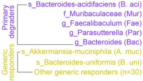
We divided the entire gut microbiota into three eco-groups: 5 primary degraders of inulin; 32 generic responders to inulin intervention; and non-responders. Primary degraders and their competitions are key drivers of baseline-dependent ecological dynamics of microbiota response to dietary fibers.

fiber degraders and responders

SCFA concentrations cannot be maintained at its peak, and drop by 35%-40% even under continuous inulin intake until four weeks. 90%-95% SCFAs produced in colonic lumen are absorbed by gut mucosa. The declining phase of SCFAs in our study may be explained by reduced production rate, increased absorption rate, or both.

Our study confirms findings in the literature and advances understanding of effects of dietary fibers on the gut microbiome at the system level:

  1. The small number of fiber degraders (five for inulin and two for resistant starch) suggested that fiber-induced bacterial shifts are very selective and occur to a restricted number of taxa.
  2. Absolute abundance of many fiber-degrading bacteria, such as taxa related to genus Bifidobacterium, failed to expand in both fibers. This indicates that fiber-induced bacterial enrichment cannot be simply predicted from in vitro growth, and suggests that dietary response of a gut bacterial taxa depends on the ecological context.
  3. Personalized fiber-induced response of gut microbiota were largely determined by baseline abundance of fiber degraders and ecological interactions among these degraders.”

https://www.biorxiv.org/content/10.1101/2021.08.20.457175v1.full “Ecological dynamics of the gut microbiome in response to dietary fiber”


PXL_20210915_133443137

Gut microbiota and critical development periods

This 2021 rodent study focused on global histone acetylation as a model to understand roles of microbially produced short-chain fatty acids in liver function:

“Despite the utility of germ-free mice in probing complex interactions between gut microbiota and host physiology, germ-free mice are developmentally, physiologically, and metabolically unique when compared with their conventionally housed counterparts. We sought to determine whether antibiotic-mediated microbiota depletion would affect global hepatic histone acetylation states through SCFA-dependent mechanisms, as previously observed in germ-free mice.

The inability of antibiotic-mediated microbiota depletion to recapitulate findings observed in germ-free mice suggests that the transition from a germ-free to a colonized mouse leads to resilient alterations in hepatic histone acetylation states that cannot be altered by further modulating the microbial environment. This finding is distinct from other germ-free phenotypes that are considered to be partially reversible, with clear alterations in their function observed after antibiotic treatment.

histone acetylation

Comparing antibiotic-treated and untreated mice that both received CCl4 at 24 and 48 hours after injury, there were almost no histone acetylation differences. This demonstrates that hepatic injury leads to a global shift in histone acetylation that is primarily independent of gut microbiota.

Major chromatin reorganization driven by histone acetylation leads to markers of differentiation, and addition of targeted differentiation signals induces events to stabilize these histone acetylation patterns – a key feature of embryonic development and terminal cellular differentiation. Differences in histone acetylation patterns seen between germ-free and conventionally raised mice may be a developmental-like effect of hepatocytes not yet exposed to microbial by-products.

Results suggest that microbial and dietary modifications to the gut microbiome in conventionally raised mice are not a means to modulate global hepatic histone acetylation. Microbiota-dependent landscaping of the hepatic epigenome appears static in nature, while the hepatic transcriptome is responsive to alterations in the gut microbiota, yet independent of global histone acetylation.

Findings underscore significant differences between these model systems that should be taken into account when considering their relevance to human biology.”

https://aasldpubs.onlinelibrary.wiley.com/doi/10.1002/hep.32043 “Global Microbiota-Dependent Histone Acetylation Patterns Are Irreversible and Independent of Short Chain Fatty Acids” (not freely available) Thanks to Dr. Elliot S. Friedman for providing a copy.


1. By describing “a key feature of embryonic development,” this study provided a gut microbiota-liver analogy of critical periods. If developmental events don’t happen when they are required, it’s probable that their window is missed, and won’t reopen later for a second chance at normalizing.

2. Many studies used a germ-free animal model, such as:

This study provided evidence for a limitation of this model, especially when extrapolating germ-free animal results to humans without similarly testing humans.


PXL_20210914_165000840_exported_1399

Eat oat avenanthramides for your gut microbiota

This 2021 paper covered a 2016 human clinical trial, and several in vitro and rodent follow-up studies:

“Oat has been widely accepted as a key food for human health. It is becoming increasingly evident that individual differences in metabolism determine how different individuals benefit from diet. Both host genetics and gut microbiota play important roles on metabolism and function of dietary compounds.

Results:

  • Avenanthramides (AVAs), the signature bioactive polyphenols of whole-grain (WG) oat, were not metabolized into their dihydro forms, dihydro-AVAs (DH-AVAs), by both human and mouse S9 fractions.
  • DH-AVAs were detected in colon and distal regions, but not in proximal and middle regions of the perfused mouse intestine, and were in specific pathogen–free (SPF) mice but not in germ-free (GF) mice.
  • A kinetic study of humans fed oat bran showed that DH-AVAs reached their maximal concentrations at much later time points than their corresponding AVAs (10.0–15.0 hours vs. 4.0–4.5 hours, respectively).
  • We observed interindividual variations in metabolism of AVAs to DH-AVAs in humans.
  • Faecalibacterium prausnitzii was identified as the individual bacterium to metabolize AVAs to DH-AVAs by 16S rRNA sequencing analysis.
  • Moreover, as opposed to GF mice, F. prausnitzii–monocolonized mice were able to metabolize AVAs to DH-AVAs.

AVA metabolizers

These findings demonstrate that intestinal F. prausnitzii is indispensable for proper metabolism of AVAs in both humans and mice. We propose that abundance of F. prausnitzii can be used to subcategorize individuals into AVA metabolizers and nonmetabolizers after WG oat intake.

Our findings pave the way to use AVAs and DH-AVAs as exposure biomarkers to reflect WG oat intake, which could more accurately record WG oat intake. Whether production of DH-AVAs is part of the beneficial effect of oats on human health will require further investigation.”

https://academic.oup.com/jn/article/151/6/1426/6165027 “Avenanthramide Metabotype from Whole-Grain Oat Intake is Influenced by Faecalibacterium prausnitzii in Healthy Adults”

Commentary at Faecalibacterium prausnitzii Abundance in Mouse and Human Gut Can Predict Metabolism of Oat Avenanthramides.


This study advanced an understanding of inter-individual variability, rather than usual practices that try to sweep individual differences under a statistical rug. Study designs such as four mentioned in Part 2 of Switch on your Nrf2 signaling pathway could have benefited from a similar approach to their research areas.

Not sure why it took over five years to get this paper published after its clinical trial’s January 21, 2016 completion. Meanwhile, science marched on to study effects of specific F. prausnitzii strains, providing results such as three human studies curated in Gut microbiota strains:

  • The third 2018 study found:

    “Only a small number of bacteria with genetic capacity for producing SCFAs were able to take advantage of this new resource and become dominant positive responders. The response, however, was strain specific: only one of the six strains of Faecalibacterium prausnitzii was promoted.”

  • The second 2021 study investigated 135 known strains of F. prausnitzii; and
  • The first 2021 study found beneficial F. prausnitzii strains not yet covered in genomic databases.

Resistant starch therapy recommended de-emphasizing relative gut microbiota abundance measurements, because:

“Relative abundances of smaller keystone communities (e.g. primary degraders) may increase, but appear to decrease simply because cross-feeders [like F. prausnitzii] increase in relative abundance to a greater extent. These limitations illustrate the necessity of sufficiently powering resistant starch interventions where microbiome composition is the primary endpoint, collecting critical baseline data and employing appropriate statistical techniques.”


Four humpback whales successively diving for lunch

PXL_20210914_164307307_exported_16255

PXL_20210914_164307307_exported_26282

PXL_20210914_164307307_exported_41871

PXL_20210914_164307307_exported_50365

Natural products vs. neurodegenerative diseases

I was recently asked about taking rapamycin for its effects on mTOR. I replied that diet could do the same thing. Here’s a 2021 review outlining such effects:

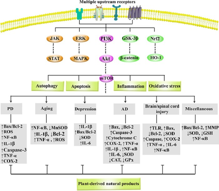
“As common, progressive, and chronic causes of disability and death, neurodegenerative diseases (NDDs) significantly threaten human health, while no effective treatment is available. Recent studies have revealed the role of phosphoinositide 3-kinase (PI3K)/Akt (Protein kinase B)/mammalian target of rapamycin (mTOR) in some diseases and natural products with therapeutic potentials.

Growing evidence highlights the dysregulated PI3K/Akt/mTOR pathway and interconnected mediators in pathogenesis of NDDs. Side effects and drug-resistance of conventional neuroprotective agents urge the need for providing alternative therapies.

1-s2.0-S0944711321002075-ga1_lrg

Polyphenols, alkaloids, carotenoids, and terpenoids have shown to be capable of a great modulation of PI3K/Akt/mTOR in NDDs. Natural products potentially target various important oxidative/inflammatory/apoptotic/autophagic molecules/mediators, such as Bax, Bcl-2, p53, caspase-3, caspase-9, NF-κB, TNF-α, GSH, SOD, MAPK, GSK-3β, Nrf2/HO-1, JAK/STAT, CREB/BDNF, ERK1/2, and LC3 towards neuroprotection.

This is the first systematic and comprehensive review with a simultaneous focus on the critical role of PI3K/Akt/mTOR in NDDs and associated targeting by natural products.”

https://www.sciencedirect.com/science/article/abs/pii/S0944711321002075 “Natural products attenuate PI3K/Akt/mTOR signaling pathway: A promising strategy in regulating neurodegeneration” (not freely available) Thanks to Dr. Sajad Fakhri for providing a copy.


Natural products mentioned in this review that I eat in everyday foods are listed below. The most effective ones are broccoli and red cabbage sprouts, and oats and oat sprouts:

  • Artichokes – luteolin;
  • Blackberries – anthocyanins;
  • Blueberries – anthocyanins, gallic acid, pterostilbene;
  • Broccoli and red cabbage sprouts – anthocyanins, kaempferol, luteolin, quercetin, sulforaphane;
  • Carrots – carotenoids;
  • Celery – apigenin, luteolin;
  • Green tea – epigallocatechin gallate;
  • Oats and oat sprouts – avenanthramides;
  • Strawberries – anthocyanins, fisetin;
  • Tomatoes – fisetin.

Four humpback whales

PXL_20210914_170732350_exported_43137

Screenshot_20210914-121800