Immune memory of pregnancies

This 2018 Israeli human study subject was natural killer cell epigenetic memory of pregnancies:

“Natural killer (NK) cells were first discovered for their ability to kill tumor cells, and later found to also kill pathogen-infected cells.

Different tissue-resident subpopulations of human NK cells exist throughout the body, displaying unique phenotypic and functional properties. One of the most fascinating tissue-resident subsets of NK cells, termed decidual NK cells, is found at the maternal fetal interface (decidua) in direct contact with the placenta.

We discovered a population found in repeated pregnancies, which has a unique transcriptome and epigenetic signature..have open chromatin around the enhancers of [growth factor genes] IFNG [essential for angiogenesis] and VEGFA [supporting vascular formation].

The pregnancy-related NK memory cells identified here might represent the first example of improved function of NK cells that occurs under healthy physiological conditions.”

One source for the experiments was:

“Decidual samples from healthy women who underwent elective first trimester terminations of normal pregnancies.”

https://www.sciencedirect.com/science/article/pii/S1074761318301286 “Trained Memory of Human Uterine NK Cells Enhances Their Function in Subsequent Pregnancies” (not freely available)

Immune memory in the brain

This 2018 German rodent study was a proof-of-principle for immune epigenetic memory in the brain:

“Innate immune memory is a vital mechanism of myeloid [bone marrow] cell plasticity that occurs in response to environmental stimuli and alters subsequent immune responses.

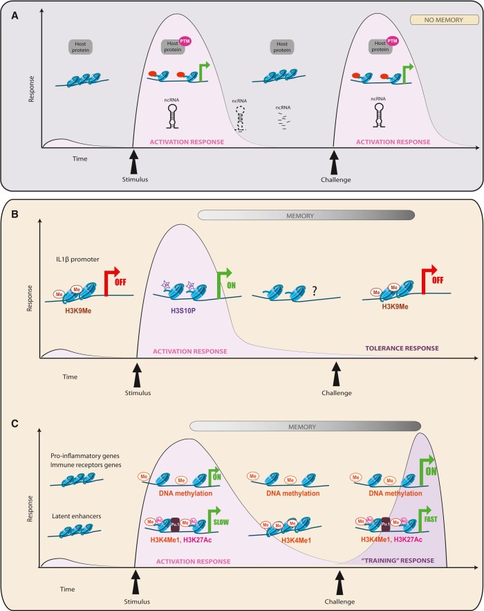
Two types of immunological imprinting can be distinguished – training and tolerance. These are epigenetically mediated and enhance or suppress subsequent inflammation, respectively.

Certain immune stimuli train blood monocytes to generate enhanced immune responses to subsequent immune insults. By contrast, other stimuli induce immune tolerance – suppression of inflammatory responses to subsequent stimuli.

Microglia (brain-resident macrophages) are very long-lived cells. This makes them particularly interesting for studying immune memory, as virtually permanent modification of their molecular profile appears possible. Immune memory in the brain is predominantly mediated by microglia.

In a mouse model of Alzheimer’s pathology, immune training exacerbates cerebral β-amyloidosis and immune tolerance alleviates it; similarly, peripheral immune stimulation modifies pathological features after stroke. Our results identify immune memory in the brain as an important modifier of neuropathology.

Immune memory in the brain could conceivably affect the severity of any neurological disease that presents with an inflammatory component, but this will need to be studied for each individual condition.”


The researchers performed multiple experiments to test different hypotheses about how immune-response experiences are remembered. Modifications to histone methylation and acetylation were targeted.

The stimulus dosage needed to produce immune tolerance – “suppression of inflammatory responses to subsequent stimuli” – was usually four times the dosage used for immune training – “enhanced immune responses to subsequent immune insults.”

https://www.nature.com/articles/s41586-018-0023-4 “Innate immune memory in the brain shapes neurological disease hallmarks” (not freely available)

An example of researchers changing their field’s paradigms

This 2018 German review subject was retroviruses:

“Initial indications that retroviruses are connected to neoplastic transformation were seen more than a century ago. 43% of the human genome is made up of such elements and 8% of the genome is comprised of retroviruses that infected human ancestors, entering cells of the germ line or proliferating thereafter by retrotransposition.

Endogenized retroviruses (ERVs) are abundantly expressed in many transformed cells. In healthy cells, ERV expression is commonly prevented by DNA methylation and other epigenetic control mechanisms.

A recent string of papers has described favorable outcomes of increasing human ERV (HERV) RNA and DNA abundance by treatment of cancer cells with methyltransferase inhibitors. Analogous to an infecting agent, the ERV-derived nucleic acids are sensed in the cytoplasm and activate innate immune responses that drive the tumor cell into apoptosis.”


Some researchers weren’t satisfied with the status quo of this century-old field:

“Chiappinelli et al. (2015) and Roulois et al. (2015) demonstrated a link between DNMTi-induced activation of HERV expression and innate sensing of transcribed viral RNAs and activation of innate immunity signaling pathways leading to an inhibition of tumor cell growth. These results represent a paradigm shift in our comprehension of the antitumor activity of demethylating agents.”

There are opportunities for any researcher whose field can be related to epigenetics to update the way studies are done. Why should researchers settle for mediocrity when they can make a difference?

https://www.ncbi.nlm.nih.gov/pmc/articles/PMC5816757/pdf/fmicb-09-00178.pdf “HERVs New Role in Cancer: From Accused Perpetrators to Cheerful Protectors”

Viruses target epigenetic processes

This 2018 Colorado review subject was general and specific ways viruses target epigenetic processes:

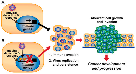
“We describe viral mechanisms and virus-host interactions by which DNA tumor viruses regulate host DNA methylation to evade antiviral immunity.

It is well known that most endogenous retroviruses and retrotransposons in the human genome are inactivated by DNA hypermethylation. In addition to endogenous retroviruses, the genomes of DNA viruses, such as human papillomavirus (HPV), herpes simplex virus 1 (HSV-1), adenovirus, and hepatitis B virus (HBV), are also frequently methylated and silenced in infected cells.

A recently described mechanism for viruses to epigenetically subvert host immunity is repression of immune-related gene expression by induction of DNA hypermethylation. Some host genes are not silenced simply through promoter hypermethylation or histone deacetylation alone, and therefore, viruses may have evolved mechanisms to ensure host gene downregulation through multiple epigenetic modifications.”

http://www.mdpi.com/1999-4915/10/2/82/htm “DNA Tumor Virus Regulation of Host DNA Methylation and Its Implications for Immune Evasion and Oncogenesis”


A second 2018 New York study focused on the Zika virus and DNA methylation:

“We studied the impact of ZIKV infection on the DNA methylation pattern across the entire genome in selected neural cell types. The virus unexpectedly alters the DNA methylome of neural progenitors, astrocytes, and differentiated neurons at genes that have been implicated in the pathogenesis of a number of brain disorders.

It remains open, however, whether the methylation changes come first or whether the viral infection dysregulates epigenetic regulatory genes prior to any epigenetic shift.”

http://msystems.asm.org/content/3/1/e00219-17 “Zika Virus Alters DNA Methylation of Neural Genes in an Organoid Model of the Developing Human Brain”

DNA methylation and childhood adversity

This 2017 Georgia human review covered:

“Recent studies, primarily focused on the findings from human studies, to indicate the role of DNA methylation in the associations between childhood adversity and cardiometabolic disease in adulthood. In particular, we focused on DNA methylation modifications in genes regulating the hypothalamus-pituitary-adrenal axis as well as the immune system.”

Recommendations in the review’s Epigenetics inheritance and preadaptation theory section included:

“Twin studies offer another promising design to explore the mediation effect of DNA methylation between child adversity and cardiometabolic outcomes..which could rule out heterogeneity due to genetic and familia[l]r environmental confounding.”

As it so happened, the below 2018 study provided some evidence.

http://www.sciencedirect.com/science/article/pii/S0167527317352762 “The role of DNA methylation in the association between childhood adversity and cardiometabolic disease” (not freely available) Thanks to lead author Dr. Guang Hao for providing the full study.


This 2018 UK human study:

“Tested the hypothesis that victimization is associated with DNA methylation in the Environmental Risk (E-Risk) Longitudinal Study, a nationally representative 1994-1995 birth cohort of 2,232 twins born in England and Wales and assessed at ages 5, 7, 10, 12, and 18 years. Multiple forms of victimization were ascertained in childhood and adolescence (including physical, sexual, and emotional abuse; neglect; exposure to intimate-partner violence; bullying; cyber-victimization; and crime).

Hypothesis-driven analyses of six candidate genes in the stress response (

  1. NR3C1 [glucocorticoid receptor],
  2. FKBP5 [a regulator of the stress hormone system],
  3. BDNF [brain-derived neurotrophic factor],
  4. AVP [arginine vasopressin],
  5. CRHR1 [corticotropin-releasing hormone receptor 1],
  6. SLC6A4 [serotonin transporter]

) did not reveal predicted associations with DNA methylation.

Epigenetic epidemiology is not yet well matched to experimental, nonhuman models in uncovering the biological embedding of stress.”

One of the sad findings was that as the types of trauma inflicted by other people on the subjects increased, so did the percentage of subjects who hurt themselves by smoking. Two-thirds of teens who reported three or more of the seven adolescent trauma types also smoked by age 18:

Polyvictimization

Self-harming behaviors other than smoking weren’t considered.

Another somber finding was:

“Childhood sexual victimization is associated with stable DNA methylation differences in whole blood in young adulthood. These associations were not observed in relation to sexual victimization in adolescence.”

The researchers guided future studies regarding the proxy measurements of peripheral blood DNA methylation:

“The vast majority of subsequent human studies, including the present one, have relied on peripheral blood. This choice is expedient, but also scientifically reasonable given the aim of detecting effects on stress-related physical health systems that include peripheral circulating processes (immune, neuroendocrine).

But whole blood is heterogeneous, and although cell-type composition can be evaluated and controlled, as in the present study, it does raise the question of whether peripheral blood is a problematic surrogate tissue for research on the epigenetics of stress.

Comparisons of methylomic variation across blood and brain suggest that blood-based EWAS may yield limited information relating to underlying pathological processes for disorders where brain is the primary tissue of interest.”


1. The comment on “epigenetic epidemiology” overstated the study’s findings because the epigenetic analysis, although thorough, was limited to peripheral blood DNA methylation. Other consequential epigenetic effects weren’t investigated, such as histone modifications and microRNA expression.

2. An unstated limitation was that the DNA methylation analyses were constrained by budgets. Studies like The primary causes of individual differences in DNA methylation are environmental factors point out restrictions in the methodology:

“A main limitation with studies using the Illumina 450 K array is that the platform only covers ~1.5 % of overall genomic CpGs, which are biased towards promoters and strongly underrepresented in distal regulatory elements, i.e., enhancers.

WGBS [whole-genome bisulfite sequencing] offers single-site resolution CpG methylation interrogation at full genomic coverage.

Another advantage of WGBS is its ability to access patterns of non-CpG methylation.”

I’d expect that in the future, researchers with larger budgets would reanalyze the study samples using other techniques.

3. The researchers started and ended the study presenting their view of human “embedding of stress” as a fact rather than a paradigm. Epigenetic effects of early life stress exposure compared and contrasted this with another substantiated view.

4. The study focused on the children’s intergenerational epigenetic effects. An outstanding opportunity to advance science was missed regarding transgenerational epigenetic inheritance:

  • Wouldn’t the parents’ blood samples and histories – derived from administering the same questionnaires their twins answered at age 18 – likely provide distant causal evidence for some of the children’s observed effects?
  • And lay the groundwork for hypotheses about aspects of future generations’ physiologies and behaviors?

https://ajp.psychiatryonline.org/doi/full/10.1176/appi.ajp.2017.17060693 “Analysis of DNA Methylation in Young People: Limited Evidence for an Association Between Victimization Stress and Epigenetic Variation in Blood” (not freely available) Thanks to coauthor Dr. Helen Fisher for providing the full study.

Can researchers make a difference in their fields?

The purpose and finding of this 2017 UK meta-analysis of human epigenetics and cognitive abilities was:

“A meta-analysis of the relationship between blood-based DNA methylation and cognitive function.

We identified [two] methylation sites that are linked to an aspect of executive function and global cognitive ability. The latter finding relied on a relatively crude cognitive test..which is commonly used to identify individuals at risk of dementia.

One of the two CpG sites identified was under modest genetic control..there are relatively modest methylation signatures for cognitive function.”

The review’s stated limitations included:

“It is, of course, possible that a reliable blood-based epigenetic marker of cognitive function may be several degrees of separation away from the biological processes that drive cognitive skills.

There are additional limitations of this study:

  • A varying number of participants with cognitive data available for each test;
  • Heterogeneity in relation to the ethnicity and geographical location of the participants across cohorts; and
  • Relating a blood-based methylation signature to a brain-based outcome.

A 6-year window [between ages 70 and 76] is possibly too narrow to observe substantial changes in the CpG levels.”

All of these limitations were known before the meta-analysis was planned and performed. Other “possible” limitations already known by the 47 coauthors include those from Genetic statistics don’t necessarily predict the effects of an individual’s genes.

The paper referenced studies to justify the efforts, such as one (cited twice) coauthored by the lead author of A problematic study of DNA methylation in frontal cortex development and schizophrenia:

“Epigenome-wide studies of other brain-related outcomes, such as schizophrenia, have identified putative blood-based methylation signatures.”


Was this weak-sauce meta-analysis done just to plump up 47 CVs? Why can’t researchers investigate conditions that could make a difference in their fields?

Was this meta-analysis done mainly because the funding was available? I’ve heard that the primary reason there are papers like the doubly-cited one above is that the US NIMH funds few other types of research outside of their biomarker dogma.

The opportunity costs of this genre of research are staggering. Were there no more productive topics that these 47 scientists could have investigated?

Here are a few more-promising research areas where epigenetic effects can be observed in human behavior and physiology:

I hope that the researchers value their professions enough to make a difference with these or other areas of their expertise. And that sponsors won’t thwart researchers’ desires for difference-making science by putting them into endless funding queues.

https://www.nature.com/articles/s41380-017-0008-y “Meta-analysis of epigenome-wide association studies of cognitive abilities”

Transgenerational pathological traits induced by prenatal immune activation

The third paper of Transgenerational epigenetic inheritance week was a 2016 Swiss rodent study of immune system epigenetic effects:

“Our study demonstrates for, we believe, the first time that prenatal immune activation can negatively affect brain and behavioral functions in multiple generations. These findings thus highlight a novel pathological aspect of this early-life adversity in shaping disease risk across generations.”

The epigenetic effects noted in the initial round of experiments included:

  • F1 child and F2 grandchild impaired sociability;
  • F1 and F2 abnormal fear expression;
  • F1 but not F2 sensorimotor gating deficiencies; and
  • F2 but not F1 behavioral despair associated with depressive-like behavior.

These transgenerational effects emerged in both male and female offspring. The prenatal immune activation timing corresponded to the middle of the first trimester of human pregnancy.

The effects were found to be mediated by the paternal but not maternal lineage. The researchers didn’t develop a maternal lineage F3 great-grandchild generation.

The next round of experiments done with the paternal lineage F3 great-grandchildren noted these epigenetic effects:

  • The F3 great-grandchildren had impaired sociability, abnormal fear expression and behavioral despair; and
  • The F3 great-grandchildren had normal sensorimotor gating.

Since the first round of tests didn’t show sex-dependent effects, the F3 great-grandchildren were male-only to minimize the number of animals.

Samples of only the amygdalar complex were taken to develop findings of transcriptomic effects of prenatal immune activation.

Items in the Discussion section included:

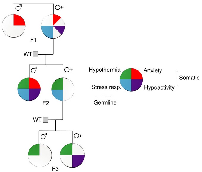
  1. The F2 grandchild and F3 great-grandchild generations’ phenotype of impaired sociability, abnormal fear expression and behavioral despair demonstrated that prenatal immune activation likely altered epigenetic marks in the germ line of the F1 children which resisted erasure and epigenetic reestablishment during germ cell development.
  2. Abnormal F1 child sensorimotor gating followed by normal F2 grandchild and F3 great-grandchild sensorimotor gating demonstrated that prenatal immune activation may also modify somatic but not germ cells.
  3. Non-significant F1 child behavioral despair followed by F2 grandchild and F3 great-grandchild behavioral despair demonstrated that prenatal immune activation may modify F1 germ cells sufficiently to develop a transgenerational phenotype, but unlike item 1 above, somatic cells were insufficiently modified, and the phenotype skipped the F1 children.
  4. Studies were cited that prenatal immune activation later in the gestational process may produce different effects.

The initial round of experiments wasn’t definitive for the maternal lineage. As argued in Transgenerational effects of early environmental insults on aging and disease and A review of epigenetic transgenerational inheritance of reproductive disease, testing of maternal lineage F3 great-grandchildren was needed to control for the variable of direct F2 grandchild germ-line exposure.

Also, effects that didn’t reach statistical significance in the maternal lineage F1 children and F2 grandchildren may have been different in the F3 great-grandchildren. The researchers indirectly acknowledged this lack by noting that these and other effects of immune challenges in a maternal lineage weren’t excluded by the study.

https://www.nature.com/mp/journal/v22/n1/pdf/mp201641a.pdf “Transgenerational transmission and modification of pathological traits induced by prenatal immune activation” (not freely available)


The study’s lead researcher authored a freely-available 2017 review that placed this study in context and provided further details from other studies:

http://www.nature.com/tp/journal/v7/n5/full/tp201778a.html “Epigenetic and transgenerational mechanisms in infection-mediated neurodevelopmental disorders”

It’s transgenerational epigenetic inheritance week!

Transgenerational epigenetic inheritance is a subject whose time has come. This week I sequentially curated two 2017 reviews and two 2016 studies of the subject, and ended with a meta-analysis of human preventive treatments:


It’s the opposite of advancing science for those in the funding chain to give lip service to the subject, and then create an atmosphere where proposals to extend experiments to subsequent generations to study possible transgenerational epigenetic effects are neither encouraged nor funded.

A limited study of parental transmission of anxiety/stress-reactive traits

BehavioralTraitsThis 2016 New York rodent study found:

“Parental behavioural traits can be transmitted by non-genetic mechanisms to the offspring.

We show that four anxiety/stress-reactive traits are transmitted via independent iterative-somatic and gametic epigenetic mechanisms across multiple generations.

As the individual traits/pathways each have their own generation-dependent penetrance and gender specificity, the resulting cumulative phenotype is pleiotropic. In the context of genetic diseases, it is typically assumed that this phenomenon arises from individual differences in vulnerability to the various effects of the causative gene. However, the work presented here reveals that pleiotropy can be produced by the variable distribution and segregated transmission of behavioural traits.”


A primary focus was how anxiety was transmitted from parents to offspring:

“The iterative propagation of the male-specific anxiety-like behaviour is most compatible with a model in which proinflammatory state is propagated from H [serotonin1A receptor heterozygote] F0 to F1 [children] females and in which the proinflammatory state is acquired by F1 males from their H mothers, and then by F2 [grandchildren] males from their F1 mothers.

We propose that increased levels of gestational MIP-1β [macrophage inflammatory protein 1β] in H and F1 mothers, together with additional proinflammatory cytokines and bioactive proteins, are required to produce immune system activation in their newborn offspring, which in turn promotes the development of the anxiety-like phenotype in males.

In particular, increase in the number of monocytes and their transmigration to the brain parenchyma in F1 and F2 males could be central to the development of anxiety.”


The researchers studied transmission of behavioral traits and epigenetic changes. Due to my quick take on the study title – “Behavioural traits propagate across generations..” – I had expectations of this study that weren’t born out. What could the researchers have done versus what they did?

The study design removed prenatal and postnatal parental behavioral transmission of behavioral traits and epigenetic changes as each generation’s embryos were implanted into foster wild-type (WT) mothers.

The study design substituted the foster mothers’ prenatal and postnatal parental environments for the biological parents’ environments. So we didn’t find out, for example:

  • To what extents the overly stress-reactive F1 female children’s prenatal environments and postnatal behaviors induced behaviors and/or epigenetic changes in their children; and
  • Whether the F2 grandchildren’s parental behaviors subsequently induced behaviors and/or epigenetic changes in the F3 great-grandchildren.

How did the study meet the overall goal of rodent studies: to help humans?

    1. Only a minority of humans experienced an early-life environment that included primary caregivers other than our biological parents.
    2. Very, very few of us experienced a prenatal environment other than our biological mothers.
    3. The study’s thorough removal of parental behavior was an outstanding methodology to confirm by falsifiability whether parental behavior was both an intergenerational and transgenerational epigenetic inheritance mechanism.
    4. Maybe the researchers filled in some gaps in previous rodent studies, such as determining what is or isn’t a “true transgenerational mechanism.”

As an example of a rodent study that more closely approximated human conditions, the behavior of a mother whose DNA was epigenetically changed by stress induced the same epigenetic changes to her child’s DNA when her child was stressed per One way that mothers cause fear and emotional trauma in their infants:

“Our results provide clues to understanding transmission of specific fears across generations and its dependence upon maternal induction of pups’ stress response paired with the cue to induce amygdala-dependent learning plasticity.”


How did parental behavioral transmission of behavioral traits and epigenetic changes become a subject not worth investigating? These traits and effects can be seen everyday in real-life human interactions, and in every human’s physiology.

But when investigating human correlates with behavioral epigenetic changes of rodents in the laboratory, parental behavioral transmission of behavioral traits is often treated the way this study treated it: as a confounder.

I doubt that people who have reached some degree of honesty about their early lives and concomitant empathy for others would agree with this prioritization. The papers of Transgenerational epigenetic inheritance week show the spectrum of opportunities to advance science that were intentionally missed.

http://www.nature.com/ncomms/2016/160513/ncomms11492/full/ncomms11492.html “Behavioural traits propagate across generations via segregated iterative-somatic and gametic epigenetic mechanisms”

Enduring epigenetic memories? Or continuous toxic stimulation?

This 2016 French review subject was bacterial infections that produce long-lasting host memories:

“Virulence factors modify the epigenomic landscape through targeting of host signaling cascades, or chromatin complexes directly. Additionally, some bacterial factors have intrinsic catalytic activity enabling them to directly modify chromatin.

Virus, fungi, and parasites also induce similar processes.

Epigenomic changes are not the only possible marks contributing to epigenetic memory. Every inducible change that is not rapidly reversed has the potential to maintain a lasting effect.

Most studies in this field have been performed in vitro with fully terminally differentiated cells such as epithelial cells. Since in such cell types cell fate is already established and a short lifespan often occurs in vivo, this raises the question of whether such memory would be relevant for these cells. The same can be applied to differentiated innate immune cells, which also have a short lifetime.

Looking at the response of undifferentiated cells such as stem cells appears much more appropriate to further explore the concept of innate immune memory.

figureFINAL

The lasting potential of chromatin marks depends not only on the kinetics of the epigenome, but also on the stimulus itself. For example, in contrast to LPS [lipopolysaccharide, the major constituent of the cell wall of Gram-negative bacteria], which is rapidly cleared from the organism, BCG and the anthrax toxin may persist in the host organism.

The lasting epigenomic effect would not be due to memory, but continuous stimulation by persistent pathogens or persistent components.”


The last point emphasized the principle that damaging sources should be addressed. Enduring epigenetic effects may be symptoms rather than causes when toxic conditions persist.

Therapies that attempt to reverse epigenetic changes may not be effective when these changes aren’t the only factors.

https://www.cell.com/cell-host-microbe/fulltext/S1931-3128(16)30148-2 “A Lasting Impression: Epigenetic Memory of Bacterial Infections?”

Epigenetic remodeling creates immune system memory

This 2016 German review subject was memory characteristics of immune cells:

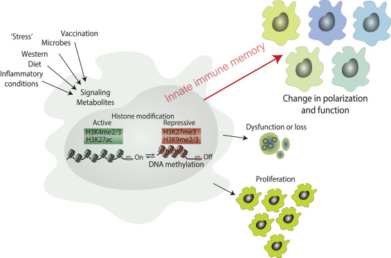
“Innate immune memory has likely evolved as an ancient mechanism to protect against pathogens. However, dysregulated processes of immunological imprinting mediated by trained innate immunity may also be detrimental under certain conditions.

Evidence is rapidly accumulating that innate immune cells can adopt a persistent pro-inflammatory phenotype after brief exposure to a variety of stimuli, a phenomenon that has been termed ‘trained innate immunity.’ The epigenome of myeloid (progenitor) cells is presumably modified for prolonged periods of time, which, in turn, could evoke a condition of continuous immune cell over-activation.”

These reviewers focused on an example of atherosclerosis, although other examples were discussed of epigenetic remodeling to acquire immune memory:

“In the last ten years, several novel non-traditional risk factors for atherosclerosis have been identified that are all associated with activation of the immune system. These include chronic inflammatory diseases such as:

  • Rheumatoid arthritis,
  • Gout,
  • Psoriatic arthritis, and
  • Ankylosing spondylitis,

as well as infections with bacteria or viruses.”

Innate immune memory

http://www.sciencedirect.com/science/article/pii/S1044532316300185 “Long-term activation of the innate immune system in atherosclerosis”


Diets were discussed, mainly regarding their various negative effects. I was interested to see a study that referenced a common dietary supplement:

“Pathway analysis of promoters that were potentiated by β-glucan identified several innate immune and signaling pathways upregulated in trained cells that are responsible for induction of trained immunity.”

Other curated research into epigenetic remodeling of immune system memory includes:

Lack of oxygen’s epigenetic effects

This 2016 Finnish review subject was epigenetic effects of hypoxia:

“Ever since the Cambrian period, oxygen availability has been in the center of energy metabolism. Hypoxia stabilizes expression of hypoxia-inducible transcription factor-1α (HIF-1α), which controls expression of hundreds of survival genes related to enhanced energy metabolism and autophagy.

There are several other signals, mostly related to stresses, which can increase expression of HIF factors and thus improve cellular survival. However, a chronic activation of HIF factors can have detrimental effects, e.g. stimulate cellular senescence and tissue fibrosis commonly enhanced in age-related diseases.

Stabilization of HIF-1α increases expression of histone lysine demethylases (KDM). Hypoxia-inducible KDMs support locally the gene transcription induced by HIF-1α, although they can also control genome-wide chromatin landscape, especially KDMs which demethylate H3K9 and H3K27 sites (repressive epigenetic marks).”

Gene areas where HIF-1α is involved include:

  • “angiogenesis
  • autophagy
  • glucose uptake
  • glycolytic enzymes
  • immune responses
  • embryonic development
  • tumorigenesis
  • generation of miRNAs.”

HIF-1a signaling

Figure 1 above was instructive in that the reviewers pointed out the lack of a feedback mechanism in HIF-1α signaling. A natural lack of feedback to the HIF-1α signaling source contributed to diseases such as:

  • “age-related macular degeneration
  • cancer progression
  • chronic kidney disease
  • cardiomyopathies
  • adipose tissue fibrosis
  • inflammation
  • detrimental effects which are linked to epigenetic changes.”

The point was similar to a study referenced in The PRice “equation” for individually evolving: Which equation describes your life? that:

“Evolution may preferentially mitigate damage to a biological system than reduce the source of this damage.”


This review subject has many interdependencies and timings within a complex network. Contexts are important:

“Cross-talk between NF-κB [nuclear factor kappa B] and HIF-1α in inflammation might be organized in cell type and context-dependent manner.

It seems that ROS [reactive oxygen species] affect HIF-1α signaling in a context-dependent manner.

Hypoxia stimulated expression of KDM3A and KDM4B genes in different cellular contexts. Given that KDM3A and KDM4B are the major histone demethylases which remove repressive H3K9 sites, their role as transcriptional cofactors seems to be important in activation of HIF-1α signaling. Members of KDM4 subfamily have a crucial role in DNA repair systems, although responses seem to be enzyme-specific and appear in a context-dependent manner.

Acute hypoxia can stimulate cell-cycle arrest but does not provoke cellular senescence in all contexts.”

It wasn’t mentioned that hypoxia evokes cellular Adaptations to stress encourage mutations in a DNA area that causes diseases.

The review was tailored for the publishing journal Aging and Disease, and the subject was best summed up by:

“HIF-1α can control cellular fate in adult animals, either stimulating proliferation or triggering cellular senescence, by regulating the expression of different KDMs in a context-dependent manner.”


This review covered hypoxic conditions during human development that are clearly origins of many immediate and later-life diseases. However, cited remedies only addressed symptoms.

That these distant causes can no longer be addressed is a hidden assumption of research and treatment of effects of health problems. Aren’t such assumptions testable here in the current year?

http://www.aginganddisease.org/article/2016/2152-5250/ad-7-2-180.shtml “Hypoxia-Inducible Histone Lysine Demethylases: Impact on the Aging Process and Age-Related Diseases”

Gene therapy by DNA methylation using CRISPR-Cas9

This 2016 Croatian human cell study was a proof-of-concept to induce specific DNA methylation of two genes:

“In this work we have created and characterized a novel CRISPR-Cas9-based epigenome editing tool, the dCas9-DNMT3A, which enabled targeted and specific CpG methylation at the promoter of two loci, the BACH2 and the IL6ST.

We have demonstrated the ability of the dCas9-DNMT3A construct to silence gene expression.

The BACH2 and IL6ST loci were previously associated with IgG glycosylation and inflammatory as well as autoimmune diseases.”

A few limitations:

“CpG methylation achieved using the active dCas9-DNMT3A construct was not stable in cultured cells. We found a ‘window’ of high methylation activity between days 5 and 15.

The relatively higher number of sgRNA [short complementary single guide RNA] targets in the BACH2 promoter compared to the IL6ST promoter (8 versus 4, respectively) might account for the higher statistical significance of gene silencing with inactive construct in the case of BACH2.”

http://nar.oxfordjournals.org/content/early/2016/03/10/nar.gkw159.full “Repurposing the CRISPR-Cas9 system for targeted DNA methylation”

Epigenetic regulation of natural killer cells

This 2016 German review focused on how epigenetic processes affected the natural killer cell part of the immune system:

“Natural killer (NK) cells recognize and eliminate tumor- and virus-infected cells, parasites as well as certain types of bacteria. NK cell activity is related to a complex interaction of activating and inhibiting receptors on the NK cell surface.

During the development of HPCs [hemopoietic progenitor cells] to mature NK cells, the DNA demethylation of KIR [killer cell immunoglobin-like receptors] genes leads to KIR expression. But DNA methylation does not just determine which KIR gene is expressed, it also determines which allele expresses the KIR gene. KIR genes are also regulated by microRNA.

KIR genes exhibit highly similar histone acetylation signatures, which are typically found in expressed genes. This fact puts the KIR genes into a state of readiness for transcription which is depending on the DNA methylation as critical epigenetic modification in the regulation of KIR gene expression.

Epigenetic modifications have been reported to be involved in the expression of NKG2D, which is one the most important activating NK cell receptor.”

The reviewers included a section on NK cell activity and external stimuli. They summarized:

“The significance of the described findings is limited by study designs. Although human NK cells were frequently used, in most cases treatment took place in ex vivo experiments.”

The reviewers also provided a good three-paragraph explanation of general epigenetic mechanisms.

http://www.mdpi.com/1422-0067/17/3/326/htm “Natural Killer Cells—An Epigenetic Perspective of Development and Regulation”

Telomerase activity outside of telomere maintenance

This 2016 Singapore review was on the role of telomerase in cancers. From its background section:

“Telomeres are conserved, repetitive sequences located at the ends of eukaryotic chromosomes which protect the integrity of genomic DNA. DNA polymerase is unable to replicate the 5′ [carbon number] ends of chromosomes, hence, cells require a RNA dependent DNA polymerase called telomerase to synthesize DNA on the lagging strand. Telomerase activity is tightly regulated and seen mainly in germ cells, stem cells and some immune cell types which have high proliferative needs.

In contrast, somatic cells do not display detectable telomerase activity. As a result, the chromosomes of normal somatic cells shorten 50–200 bp [base pair] each replication at the telomeres due to the problem of end replication. Thus, somatic cells are eventually burdened with DNA damage, replication crisis, cellular senescence or apoptosis and can divide only limited number of times, whereas cells that have active telomerase possess unlimited proliferative potential.”

The main section of the review described the details of how:

“Reactivation of telomerase has been considered as a strategy for telomere maintenance and is a major hallmark of cancer. Although the major function of telomerase is thought to be telomere elongation, accumulating evidence has suggested that it can modulate expression of various genes which affect cancer progression and tumorigenesis.”

http://link.springer.com/article/10.1007/s00018-016-2146-9/fulltext.html “Reactivation of telomerase in cancer”