Prenatal programming of human HPA axis development

This 2017 UC Irvine human review subject provided details of how fetal hypothalamic-pituitary-adrenal components and systems develop, and how they are epigenetically changed by the mother’s environment:

“The developmental origins of disease or fetal programming model predicts that intrauterine exposures have life-long consequences for physical and psychological health. Prenatal programming of the fetal hypothalamic-pituitary-adrenal (HPA) axis is proposed as a primary mechanism by which early experiences are linked to later disease risk.

Development of the fetal HPA axis is determined by an intricately timed cascade of endocrine events during gestation and is regulated by an integrated maternal-placental-fetal steroidogenic unit. Mechanisms by which stress-induced elevations in hormones of maternal, fetal, or placental origin influence the structure and function of the emerging fetal HPA axis are discussed.

Human gestational physiology and fetal HPA axis development differ even from that of closely related nonhuman primates, thereby limiting the generalizability of animal models. This review will focus solely on studies of prenatal stress and fetal HPA axis development in humans.”


1. Every time I read a prenatal study I’m in awe of all that has to go right – and at the appropriate times and sequences – for a fetus to be undamaged. Add in what needs to happen at birth, during infancy, and throughout early childhood, and it seems impossible for any human to escape epigenetic damage.

2. The reviewers referenced animal studies and human research performed with postnatal subjects, despite the disclaimer:

This review will focus solely on studies of prenatal stress and fetal HPA axis development in humans.”

This led to blurring of what had been studied or not with human fetuses regarding the subject.

3. These reviewers uncritically listed many dubious human studies that had both stated and undisclosed severe limitations on their findings. Other reviewers offer informed analysis of cited studies, as Sex-specific impacts of childhood trauma summarized with cortisol:

“Findings are dependent upon variance in extenuating factors, including but not limited to, different measurements of:

  • early adversity,
  • age of onset,
  • basal cortisol levels, as well as
  • trauma forms and subtypes, and
  • presence and severity of psychopathology symptomology.”

4. The paper would have been better had it stayed on topic with its title “Developmental origins of the human hypothalamic-pituitary-adrenal axis.” Let other reviews cover animals, post-natal humans, and questionable evidence.

5. I asked the reviewers to provide a searchable file to facilitate using their work as a reference.

https://www.researchgate.net/publication/318469661_Developmental_origins_of_the_human_hypothalamic-pituitary-adrenal_axis “Developmental origins of the human hypothalamic-pituitary-adrenal axis” (registration required)

Hidden hypotheses of epigenetic studies

This 2018 UK review discussed three pre-existing conditions of epigenetic genome-wide association studies:

“Genome-wide technology has facilitated epigenome-wide association studies (EWAS), permitting ‘hypothesis-free’ examinations in relation to adversity and/or mental health problems. Results of EWAS are in fact conditional on several a priori hypotheses:

  1. EWAS coverage is sufficient for complex psychiatric problems;
  2. Peripheral tissue is meaningful for mental health problems; and
  3. The assumption that biology can be informative to the phenotype.

1. CpG sites were chosen as potentially biologically informative based on consultation with a consortium of DNA methylation experts. Selection was, in part, based on data from a number of phenotypes (some medical in nature such as cancer), and thus is not specifically targeted to brain-based, stress-related complex mental health phenotypes.

2. The assumption is often that distinct peripheral tissues are interchangeable and equally suited for biomarker detection, when in fact it is highly probable that peripheral tissues themselves correspond differently to environmental adversity and/or disease state.

3. Analyses result in general statements such as ‘neurodevelopment’ or the ‘immune system’ being involved in the aetiology of a given phenotype. Whether these broad categories play indeed a substantial role in the aetiology of the mental health problem is often hard to determine given the post hoc nature of the interpretation.”


The reviewers mentioned in item #2 the statistical flaw of assuming that measured entities are interchangeable with one another. They didn’t mention that the problem also affected item #1 methodologies of averaging CpG methylation measurements in fixed genomic bins or over defined genomic regions, as discussed in:

The reviewers offered suggestions for reducing the impacts of these three hypotheses. But will doing more of the same, only better, advance science?

Was it too much to ask of researchers whose paychecks and reputations depended on a framework’s paradigm – such as the “biomarker” mentioned a dozen and a half times – to admit the uselessness of gathering data when the framework in which the data operated wasn’t viable? They already knew or should have known this.

Changing an individual’s future behavior even before they’re born provided one example of what the GWAS/EWAS framework missed:

“When phenotypic variation results from alleles that modify phenotypic variance rather than the mean, this link between genotype and phenotype will not be detected.”

DNA methylation and childhood adversity concluded that:

“Blood-based EWAS may yield limited information relating to underlying pathological processes for disorders where brain is the primary tissue of interest.”

The truth about complex traits and GWAS added another example of how this framework and many of its paradigms haven’t produced effective explanations of “the aetiology of the mental health problem”

“The most investigated candidate gene hypotheses of schizophrenia are not well supported by genome-wide association studies, and it is likely that this will be the case for other complex traits as well.”

Researchers need to reevaluate their framework if they want to make a difference in their fields. Recasting GWAS as EWAS won’t make it more effective.

https://www.sciencedirect.com/science/article/pii/S2352250X18300940 “Hidden hypotheses in ‘hypothesis-free’ genome-wide epigenetic associations”

Restoration of a “normal” epigenetic landscape

This 2018 Texas human review subject was prostate cancer epigenetics:

“We comprehensively review the up-to-date roles of epigenetics in the development and progression of prostate cancer. We especially focus on three epigenetic mechanisms: DNA methylation, histone modifications, and noncoding RNAs. We elaborate on current models/theories that explain the necessity of these epigenetic programs in driving the malignant phenotypes of prostate cancer cells.

It is now generally accepted that epigenetics contributes to the development of nearly every stage of PCa [prostate cancer]. Considering the highly heterogeneous nature of PCa, it is quite likely that [the] effect of a particular epigenetic pattern on growth of cancer cells varies from case to case and [is] context specific.

Restoration of a “normal” epigenetic landscape holds promise as a cure for prostate cancer.”

The review’s Epigenetic Therapy section explained much of what’s going on in the above graphic. Its Table 3 was instructive for up-to-date clinical trial information on epigenetic treatments of prostate cancer.


“Restoration of a “normal” epigenetic landscape” won’t guarantee a healthy outcome once diseases start. Prevention seems desirable to avoid the situation where:

“Numerous epigenetic alterations reinforce the establishment of a context-specific transcriptional profile that favors self-renewal, survival, and invasion of PCa cells.”

http://www.ajandrology.com/article.asp?issn=1008-682X;year=2019;volume=21;issue=3;spage=279;epage=290;aulast=Liao “Epigenetic regulation of prostate cancer: the theories and the clinical implications”

Measuring epigenetic changes at a single-cell level

This 2018 Canadian cell study described the development of a single-cell protocol to:

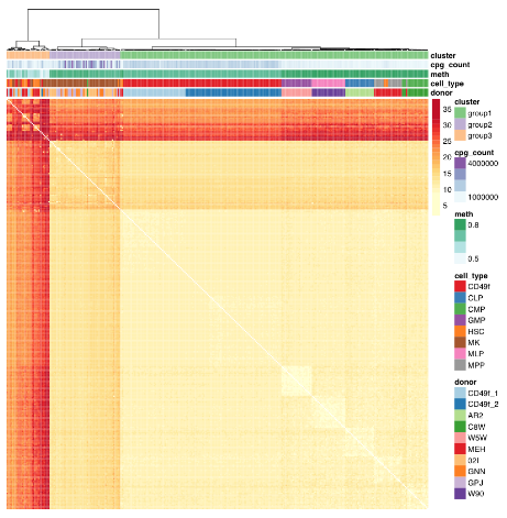
“Profile primitive hematopoietic cells of mouse and human origin to identify epigenetically distinct subpopulations. Deep sampling of the CpG content of individual HSCs allowed for the near complete reconstitution of regulatory states from epigenetically defined subpopulations of HSCs and revealed a high level of redundancy of CpG methylation states within these phenotypically defined hematopoietic cell types.

Hematopoietic stem cells (HSCs) are functionally defined cells that display evidence of extensive self-renewal of their ability to generate mature blood cells for the lifetime of the organism and following transplantation into myelosuppressed permissive hosts. Most of the epigenetic measurements underpinning these observations represent consensus values experimentally derived from thousands of cells partially enriched in HSCs or their progeny, thus failing to discern distinct epigenetic states within HSCs.

Current analytical strategies for single-cell DNA methylation measurements average DNA methylation in fixed genomic bins or over defined genomic regions.

However, inference across cells (as well as sequence context) assumes homogeneity across cells, which is at cross-purposes with the generation of single-cell molecular measurements through the potential to mask rare subpopulations.

We identified donor as a significant source of consistent epigenetic heterogeneity, which was reduced but not eliminated by correcting for personal genetic variants. This observation is consistent with previous reports that showed genetic diversity as related to but not accountable for all DNA methylation differences and suggests that in utero environmental differences may be encoded within the HSC compartment.”


The study advanced science not only by measuring CpG methylation within each HSC, but also by producing another data point “that in utero environmental differences may be encoded within the HSC compartment.”

The paragraph with “assumes homogeneity across cells” bold text provided another example of the statistical analysis flaw that gives individually inapplicable results per Group statistics don’t necessarily describe an individual. The above graphic of human hematopoietic phenotypes demonstrated that the researchers have potentially solved this problem by measuring individual cells.

The researchers discussed another aspect of the study that’s similar to the epigenetic clock methodology:

“Phenotype-specific methylation signatures are characterized by extensive redundancy such that distinct epigenetic states can be accurately described by only a small fraction of single-CpG methylation states. In support of such a notion, the unique components of a DNA methylation “age” signature are contained in ∼353 CpGs sites, presumably representing a random sample of a total age signature that involves many more sites not detected using the reduced representation strategies from which these signatures have been derived.”

Also, in The epigenetic clock theory of aging the originator of the epigenetic clock characterized HSCs as an effective intervention against epigenetic aging:

“In vivo, haematopoietic stem cell therapy resets the epigenetic age of blood of the recipient to that of the donor.”

https://www.cell.com/stem-cell-reports/article/S2213-6711(18)30308-4/fulltext “High-Resolution Single-Cell DNA Methylation Measurements Reveal Epigenetically Distinct Hematopoietic Stem Cell Subpopulations”

Epigenetic effects of breast cancer treatments

This 2018 UC San Diego review subject was the interplay between breast cancer treatments and their effects on aging:

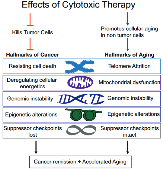
“Although current breast cancer treatments are largely successful in producing cancer remission and extending lifespan, there is concern that these treatments may have long lasting detrimental effects on cancer survivors, in part, through their impact on non-tumor cells. It is unclear whether breast cancer and/or its treatments are associated with an accelerated aging phenotype.

In this review, we have highlighted five of nine previously described cellular hallmarks of aging that have been described in the context of cytotoxic breast cancer treatments:

  1. Telomere attrition;
  2. Mitochondrial dysfunction;
  3. Genomic instability;
  4. Epigenetic alterations; and
  5. Cellular senescence.”


The review was full of caveats weakening the above graphic’s associations:

  1. “Telomere attrition – Blood TL [telomere length] was not associated with chemotherapy in three out of four studies;
  2. Mitochondrial dysfunction – How cancer therapies affect cellular energetics as they relate to rate of aging is unclear;
  3. Genomic instability – Potentially contributing to accelerated aging;
  4. Epigenetic alterations – Although some of the key regulators of these processes have begun to be identified, including DNA and histone methylases and demethylases, histone acetylases and de-acetylases and chromatin remodelers, how they regulate the changes in aging through alteration of global transcriptional programs, remains to be elucidated; and
  5. Cellular senescence – Dysregulated pathways can be targeted by cytotoxic chemotherapies, resulting in preferential cell death of tumor cells, but how these treatments also affect normal cells with intact pathways is unclear.”

To their credit, these reviewers at least presented some of the contrary evidence, and didn’t continue on with a directed narrative as other reviewers are prone to do.

https://www.sciencedirect.com/science/article/pii/S1879406818301176 “Breast cancer treatment and its effects on aging” (not freely available)


The originator of the epigenetic clock methodology was a coauthor of the review. Only one of his works was cited in the Epigenetic alterations subsection:

https://link.springer.com/article/10.1007%2Fs10549-017-4218-4 “DNA methylation age is elevated in breast tissue of healthy women”

This freely-available 2017 study quoted below highlighted that epigenetic clock measurements as originally designed were tissue-specific:

“To our knowledge, this is the first study to demonstrate that breast tissue epigenetic age exceeds that of blood tissue in healthy female donors. In addition to validating our earlier finding of age elevation in breast tissue, we further demonstrate that the magnitude of the difference between epigenetic age of breast and blood is highest in the youngest women in our study (age 20–30 years) and gradually diminishes with advancing age. As women approach the age of the menopausal transition, we found that the epigenetic of age of blood approaches that of the breast.”

Additional caution was justified in both interpreting age measurements and extending them into “cellular hallmarks” when the tissue contained varying cell types:

“Our studies were performed on whole breast tissue. Diverse types of cells make up whole breast tissue, with the majority of cells being adipocytes. Other types of cells include epithelial cells, cuboidal cells, myoepithelial cells, fibroblasts, inflammatory cells, vascular endothelial cells, preadipocytes, and adipose tissue macrophages.

This raises the possibility that the magnitude of the effects we observe, of breast tissue DNAm age being greater than other tissues, might be an underestimation, since it is possible that not all of the cells of the heterogenous sample have experienced this effect. Since it is difficult to extract DNA from adipose tissue, we suspect that the majority of DNA extracted from our whole breast tissues was from epithelial and myoepithelial cells.”

Allergies and epigenetic histone modifications

This 2018 German review provided short summaries of 44 studies on the contribution of histone modifications to allergies. An overall summary of their search results was:

“There are at least two levels at which the role of histone modifications is manifested.

  • One is the regulation of cells that contribute to the allergic inflammation (T cells and macrophages) and those that participate in airway remodeling.
  • The other is the direct association between histone modifications and allergic phenotypes.

Inhibitors of histone-modifying enzymes may potentially be used as anti-allergic drugs. Furthermore, epigenetic patterns may provide novel tools in the diagnosis of allergic disorders.”


This type of search is what’s expected of researchers who will perform either:

  • A meta-analysis of studies selected from the search results; or
  • Their own study.

These reviewers didn’t indicate that they were proceeding along either path.

The review was fine for the purpose of presenting current studies of the subject. But this was just the preparatory stage of research.

https://aacijournal.biomedcentral.com/articles/10.1186/s13223-018-0259-4 “Histone modifications and their role in epigenetics of atopy and allergic diseases”

A study of our evolutionary remnants

This 2018 Michigan human cell study subject was factors affecting the expression of human endogenous retroviruses:

“We provide a comprehensive genomic and epigenomic map of the more than 500,000 endogenous retroviruses (ERVs) and fragments that populate the intergenic regions of the human genome.

The repressive epigenetic marks associated with the ERVs, particularly long terminal repeats (LTRs), show a remarkable switch in silencing mechanisms, depending on the evolutionary age of the LTRs:

  • Young LTRs tend to be CpG-rich and are mainly suppressed by DNA methylation, whereas
  • Intermediate age LTRs are associated predominantly with histone modifications, particularly histone H3 lysine 9 (H3K9) methylation.
  • The evolutionarily old LTRs are more likely inactivated by the accumulation of loss-of-function genetic mutations.

Because the expression of ERVs is potentially dangerous to the host cell, understanding the repressive mechanisms is important. Earlier studies have implicated the aberrant expression of ERVs in autoimmune disease pathogenesis. However, this “enemy within” may also play a beneficial role in cancer therapy.

The same kinds of chromatin dynamics appear to be used both by LTRs and genes.”


I wasn’t going to curate this study before I saw the above graphic of our Boreoeutherian ancestor. Evolutionary subjects seem very abstract until an artist reconstructs the data visually.

https://genome.cshlp.org/content/early/2018/07/03/gr.234229.118.full.pdf “Switching roles for DNA and histone methylation depend on evolutionary ages of human endogenous retroviruses” (not freely available)


This post has somehow become a target for spammers, and I’ve disabled comments. Readers can comment on other posts and indicate that they want their comment to apply here, and I’ll re-enable comments.

Starving awakens ancient parasite DNA within us

This 2018 Italian human cell study conducted a series of experiments on the effects of nutrient deprivation:

“Reduced food intake, and in particular protein or amino acid (AA) restriction, extends lifespan and healthspan.

We have previously shown that, in mammalian cells, deprivation of essential AAs (methionine/cysteine or tyrosine) leads to the transcriptional reactivation of integrated silenced transgenes by a process involving epigenetic chromatic remodeling and histone acetylation.

Here we show that the deprivation of methionine/cysteine also leads to the transcriptional upregulation of endogenous retroviruses [ERVs], suggesting that essential AA starvation affects the expression not only of exogenous non-native DNA sequences, but also of endogenous anciently-integrated and silenced parasitic elements of the genome.

ERVs, comprising 8% of the human genome, represent the remnants of past infections of germ cells by exogenous retroviruses, and are mostly unable to retrotranspose in the human genome. However, they can reactivate during physiological development, or in pathological conditions like cancer, and regulate the expression of nearby genes by their LTR elements, leading to general transcriptional reprogramming.

Dissection of the underlying mechanism ruled out a role for the main AA-deficiency sensor GCN2 and pointed to the ribosome as the possible master controller.”

http://journals.plos.org/plosone/article?id=10.1371/journal.pone.0200783 “Amino acid deprivation triggers a novel GCN2-independent response leading to the transcriptional reactivation of non-native DNA sequences”


The study found that reality is sometimes stranger than what fiction writers dream up. 🙂

The authors cited a 2016 Danish review I hadn’t previously curated:

https://www.nature.com/articles/nrendo.2016.87 “The role of diet and exercise in the transgenerational epigenetic landscape of T2DM” (not freely available)

Contrary to what’s implied by its title, though, and as I noted in How to hijack science: Ignore its intent and focus on the 0.0001%, those reviewers didn’t cite any human studies that adequately demonstrated transgenerational epigenetic inheritance causes and effects. They admitted:

“Direct evidence that epigenetic factors drive the inheritance of T2DM [type 2 diabetes mellitus] in humans is lacking.”

The Danish reviewers then continued on as if proof of human transgenerational epigenetic inheritance was a foregone conclusion! It didn’t serve any valid scientific purpose to assume such evidence into existence.

A dietary supplement that trains the innate immune system

This 2018 Netherlands review topic was long-term epigenetic programming of the innate immune system:

“Immunological memory has been classically described for the adaptive immune system, in which naive B and T lymphocytes develop antigen-specific, long-lasting memory cells after encountering a new antigen.

Immunological memory is not an exclusive trait of lymphocytes. The function of cells from the innate immune system, such as monocytes, macrophages, dendritic cells, and NK cells, is also influenced by contact with different stimuli, undergoing functional reprogramming.

β-glucan, the prototypical trained immunity-inducing agonist:

  • Modulates hematopoietic stem and progenitor cells, influencing behavior and responsiveness of peripheral myeloid cells;
  • Leads to a shift of cellular metabolism from oxidative phosphorylation toward aerobic glycolysis.

Analysis of transcriptional data from macrophages stimulated with β-glucan revealed that the cholesterol synthesis pathway is highly up-regulated in trained immunity. A follow-up of this study showed that activation of the cholesterol synthesis pathway, but not its synthesis itself, is crucial for innate memory. In agreement with this, inhibition of cholesterol synthesis in mice reduced induction of trained immunity by β-glucan.

β-glucan-induced changes in trimethylation of histone 3 lysine 4 (H3K4me3) and acetylation of histone 3 lysine 27 (H3K27ac) in human monocytes 7 days after the first stimulation in vitro were associated with a switch to glycolysis, suggesting a deep, long lasting reprogramming of cells.

Inducers of cellular reprogramming such as β-glucan have shown potential as a treatment or adjuvant for osteosarcoma, influenza, or skin lesions, among others.”

https://jlb.onlinelibrary.wiley.com/doi/pdf/10.1002/JLB.MR0318-104R “Long-term reprogramming of the innate immune system”

A mid-year selection of epigenetic topics

Here are the most popular of the 65 posts I’ve made so far in 2018, starting from the earliest:

The pain societies instill into children

DNA methylation and childhood adversity

Epigenetic mechanisms of muscle memory

Sex-specific impacts of childhood trauma

Sleep and adult brain neurogenesis

This dietary supplement is better for depression symptoms than placebo

The epigenetic clock theory of aging

A flying human tethered to a monkey

Immune memory in the brain

The lack of oxygen’s epigenetic effects on a fetus

Addictive behavior and epigenetic DNA methylation

This 2018 McGill paper reviewed findings from animal and human studies on the relationships between drug-seeking behavior and epigenetic DNA methylation:

“Although there is an increasing line of evidence from preclinical models of addiction, there are only a few human studies that systematically assessed DNA methylation in addiction. Most of the studies were done on small cohorts and focused on one or a few candidate genes, except in the case of alcohol use where larger studies have been carried out.

A long line of evidence suggests that abnormal patterns of gene expression occur in brain regions related to drug addiction such as the nucleus accumbens, prefrontal cortex, amygdala, and the ventral tegmental area.

Using the “incubation of craving” model in rats trained to self-administer cocaine, and treated with either SAM or RG108, the genome-wide DNA methylation and gene expression landscape in the nucleus accumbens after short (1 day) and long (30 days) abstinence periods and the effects of epigenetic treatments were delineated. The main findings are:

  • A long incubation period results in robust changes in methylation;
  • Direct accumbal infusion of SAM that is paired with a “cue” after long incubation times increases drug-seeking behavior,
  • Whereas a single treatment with RG108 decreases this behavior.

Importantly, the effects of these single administrations of a DNA methylation inhibitor remain stable for 30 more days. These data suggest that DNA methylation might be mediating the impact of “incubation” on the craving phenotype and that this phenotype could be reprogrammed by a DNA demethylation agent.”


The subject has a large scope, and a narrow aspect was presented in this paper. Rodent research by one of the coauthors that was cited, Chronic pain causes epigenetic changes in the brain and immune system, provided some relevant details.

The review covered neither human dimensions of the impacts of unfulfilled needs nor investigations of exactly what pain may impel human drug-seeking behavior. The “Implications for Diagnostic and Therapeutics” were largely at the molecular level.

https://www.sciencedirect.com/science/article/pii/S1877117318300164 “The Role of DNA Methylation in Drug Addiction: Implications for Diagnostic and Therapeutics” (not freely available)

A protein involved in fasting’s epigenetic effects

This 2018 Illinois rodent study conducted a series of experiments on a protein that’s activated by fasting:

“Jumonji D3 (JMJD3) histone demethylase epigenetically regulates development and differentiation, immunity, and tumorigenesis by demethylating a gene repression histone mark, H3K27-me3. JMJD3 has what we believe to be a novel metabolic role and epigenetically regulates mitochondrial β-oxidation.

Epigenetic modifications play a critical role in linking environmental signals, such as changes in nutrient and hormonal levels and the circadian rhythm, to regulate genes to maintain homeostasis. Epigenetics is particularly relevant to metabolic regulation.

In response to fasting, the interaction of JMJD3 with both SIRT1 and PPARα is induced, which leads to epigenetic activation of their own genes and of β-oxidation network genes. Downregulation of hepatic JMJD3 leads to intrinsic defects in β-oxidation, which results in liver steatosis as well as glucose and insulin intolerance.

JMJD3 was required for the beneficial effects mediated by expression of SIRT1 in obese mice and vice versa. Restoration of JMJD3 to normal levels in HFD [high-fat diet]-fed obese mice leads to improved fatty acid β-oxidation and ameliorates metabolic symptoms of obesity and these beneficial effects are largely dependent on SIRT1.”


Have to hand it to the researchers who named this protein to coincidentally rhyme with a children’s book and movie. It certainly provokes more interest than other ways of naming discoveries, such as after what it resembles and/or the discoverer’s name.

https://www.jci.org/articles/view/97736 “Fasting-induced JMJD3 histone demethylase epigenetically activates mitochondrial fatty acid β-oxidation”

A disturbance in the paradigm of child abuse

The principal way science advances is through a principle Einstein expressed as:

“No amount of experimentation can ever prove me right; a single experiment can prove me wrong.”

The scientific community and public should be satisfied that the scientific process is working well when hypotheses are discarded due to nonconfirming evidence. Researchers should strive to develop evidence that rejects paradigms, and be lauded for their efforts.

The opposite took place with this 2018 commentary on two studies where evidence didn’t confirm current biases. I curated one of these studies in DNA methylation and childhood adversity.

Commentators’ dismissive tone was set in the opening paragraph:

“Is early exposure to adversity associated with a genetic or an epigenetic signature? At first glance, two articles in this issue -..and the other from Marzi et al., who measured genome-wide DNA methylation in a prospective twin cohort assessed at age 18 – appear to say that it is not.”

Commentators – one of whom was a coauthor of Manufacturing PTSD evidence with machine learning, – went on to protect their territory. Nevermind these two studies’ advancement of science that didn’t coincide with commentators’ vested interests.


My main concern with the curated study was that although child subjects had been studied at ages 5, 7, 10, 12, and 18, parents had never been similarly evaluated! Those researchers passed up an opportunity to develop parents as a F0 generation for understanding possible human transgenerational inherited epigenetic causes and effects.

That study focused on the children’s intergenerational epigenetic effects. However, animal studies have often demonstrated transgenerational effects that skip over F1 generation children! For example:

  1. Transgenerational pathological traits induced by prenatal immune activation found a F2 grandchild and F3 great-grandchild phenotype of impaired sociability, abnormal fear expression and behavioral despair – effects that weren’t present in F1 children;
  2. A self-referencing study of transgenerational epigenetic inheritance found histone modifications in the F3 generation that weren’t found in F1 and F2 generations; and
  3. A study not cited in – but completely appropriate for – The lack of oxygen’s epigenetic effects on a fetus found heart disease effects in the F1 generation that were different from the heart disease effects found in F2 and F3 generations.

https://ajp.psychiatryonline.org/doi/pdf/10.1176/appi.ajp.2018.18020156 “Considering the Genetic and Epigenetic Signature of Early Adversity Within a Biopsychosocial Framework” (not freely available)

An evolutionary view of stress and cancer

This 2018 Michigan review subject was cancer evolution:

“Based on the fact that cancer typically represents a complex adaptive system, where there is no linear relationship between lower-level agents (such as each individual gene mutation) and emergent properties (such as cancer phenotypes), we call for a new strategy based on the evolutionary mechanism of aneuploidy [abnormal number of chromosomes] in cancer, rather than continuous analysis of various individual molecular mechanisms.

Cancer evolution can be understood by the dynamic interaction among four key components:

  1. Internal and external stress;
  2. Elevated genetic and non-genetic variations (either necessary for cellular adaptation or resulting from cellular damages under stress);
  3. Genome-based macro-cellular evolution (genome replacement, emergent as new systems); and
  4. Multiple levels of system constraint which prevent/slow down cancer evolution (from tissue/organ organization to the immune system interaction).

Since the sources of stress are unlimited and unavoidable (as they are required by all living systems), there are large numbers of gene mutations / epigenetic events / chromosomal aberrations, such as aneuploidy, that can be linked to stress-mediated genomic variants. Furthermore, as environmental constraints are constantly changing, even identical instances of aneuploidy will have completely different outcomes in the context of cancer evolution, as the results of each independent run of evolution will most likely differ.

Most current research efforts are focusing on molecular profiles based on an average population, and outliers are eliminated or ignored, either by the methods used or statistical tools. The traditional view of biological research is to identify patterns from “noise,” without the realization that the so-called “noise” in fact is heterogeneity, which represents a key feature of cancer evolution by functioning as the evolutionary potential.

Understanding the molecular mechanism (both cause and effect) of aneuploidy is far from enough. A better strategy is to monitor the evolutionary process by measuring evolutionary potential. For example, the overall degree of CIN [chromosome instability] is more predictive than individual gene mutation profile.”


Although I read many abstracts of cancer research papers every week, I usually don’t curate them. I curated this paper because the reviewers emphasized several themes of this blog, including:

  • Further examples of how stress may shape one’s life.
  • How researchers miss information when they ignore or process away variation:

    Studies have demonstrated the importance of outliers in cancer evolution, as cancer is an evolutionary game of outliers. While this phenomenon can provide a potential advantage for cellular adaptation, it can also, paradoxically, generate non-specific system stress, which can further produce more genetic and non-genetic variants which favor the disease condition.”

Epigenetics researchers may benefit from evolutionary viewpoints that incorporate the interactions of stress and “genetic and non-genetic variants.”

Since epigenetic changes require inheritance in order to persist, it would be a step forward to see researchers start “measuring evolutionary potential” of these inheritance processes.

https://molecularcytogenetics.biomedcentral.com/articles/10.1186/s13039-018-0376-2 “Understanding aneuploidy in cancer through the lens of system inheritance, fuzzy inheritance and emergence of new genome systems”

Common features of autoimmune diseases

This 2018 French review subject was mechanisms of autoimmunity:

“Autoimmune diseases (AIDs) encompass more than 80 distinct chronic disorders characterized by inflammatory reactions that can either be systemic or organ specific. In all cases, the disease development is the consequence of the effects of environmental factors in predisposed individuals.

Most of the genes identified by genome-wide association studies (GWAS) on AIDs are related to immunity. However, functional immune parameters that are commonly dysregulated in AIDs do not necessarily stem from these genetic variants. Rather than performing even larger GWAS, understanding complex traits, such as human diseases, may require meticulous analysis or cell-specific gene networks and take into account not only core genes but also seemingly irrelevant genes that may overall have an impact on the disease.

Treg cell defects have been considered a primary cause of AIDs. However, one could ask whether the Treg cell dysfunction exists before the onset of the disease or is provoked by the inflammatory event induced by the triggering components. The defect of Treg cells generally coexists with the inflammatory processes, suggesting several hypotheses:

  1. The inflammation might develop because of a poor regulation of the immune system,
  2. The Treg cells could become inefficient because of the inflammatory environment, or
  3. A common factor concomitantly leads to both effects.

It is likely that autoimmunity results from a chronic imbalance involving both environmental and intrinsic factors. It is now clear that polygenic explanations did not fulfill expectations and that more efforts are needed to understand how the interplay of environmental clues may have a phenotypic impact.”

https://nyaspubs.onlinelibrary.wiley.com/doi/full/10.1111/nyas.13560 “Pathophysiological mechanisms of autoimmunity” (not freely available) Thanks to Dr. Julien Verdier for providing a copy.