This 2018 UC San Diego review subject was the interplay between breast cancer treatments and their effects on aging:
“Although current breast cancer treatments are largely successful in producing cancer remission and extending lifespan, there is concern that these treatments may have long lasting detrimental effects on cancer survivors, in part, through their impact on non-tumor cells. It is unclear whether breast cancer and/or its treatments are associated with an accelerated aging phenotype.
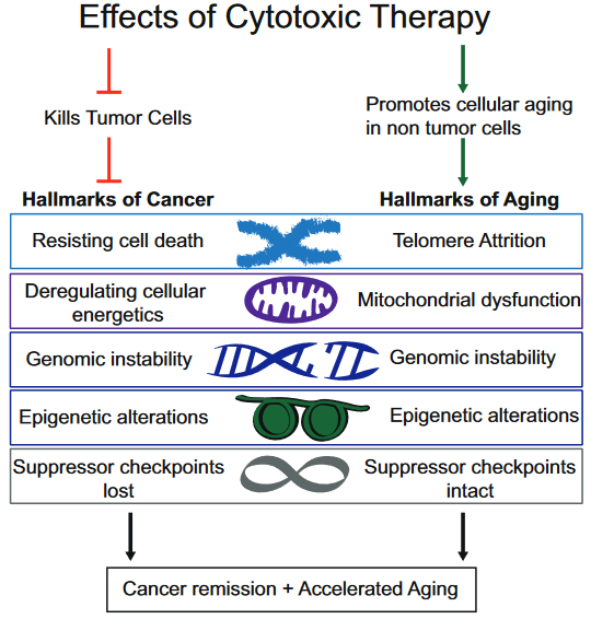
In this review, we have highlighted five of nine previously described cellular hallmarks of aging that have been described in the context of cytotoxic breast cancer treatments:
- Telomere attrition;
- Mitochondrial dysfunction;
- Genomic instability;
- Epigenetic alterations; and
- Cellular senescence.”
The review was full of caveats weakening the above graphic’s associations:
- “Telomere attrition – Blood TL [telomere length] was not associated with chemotherapy in three out of four studies;
- Mitochondrial dysfunction – How cancer therapies affect cellular energetics as they relate to rate of aging is unclear;
- Genomic instability – Potentially contributing to accelerated aging;
- Epigenetic alterations – Although some of the key regulators of these processes have begun to be identified, including DNA and histone methylases and demethylases, histone acetylases and de-acetylases and chromatin remodelers, how they regulate the changes in aging through alteration of global transcriptional programs, remains to be elucidated; and
- Cellular senescence – Dysregulated pathways can be targeted by cytotoxic chemotherapies, resulting in preferential cell death of tumor cells, but how these treatments also affect normal cells with intact pathways is unclear.”
To their credit, these reviewers at least presented some of the contrary evidence, and didn’t continue on with a directed narrative as other reviewers are prone to do.
https://www.sciencedirect.com/science/article/pii/S1879406818301176 “Breast cancer treatment and its effects on aging” (not freely available)
The originator of the epigenetic clock methodology was a coauthor of the review. Only one of his works was cited in the Epigenetic alterations subsection:
https://link.springer.com/article/10.1007%2Fs10549-017-4218-4 “DNA methylation age is elevated in breast tissue of healthy women”
This freely-available 2017 study quoted below highlighted that epigenetic clock measurements as originally designed were tissue-specific:
“To our knowledge, this is the first study to demonstrate that breast tissue epigenetic age exceeds that of blood tissue in healthy female donors. In addition to validating our earlier finding of age elevation in breast tissue, we further demonstrate that the magnitude of the difference between epigenetic age of breast and blood is highest in the youngest women in our study (age 20–30 years) and gradually diminishes with advancing age. As women approach the age of the menopausal transition, we found that the epigenetic of age of blood approaches that of the breast.”
Additional caution was justified in both interpreting age measurements and extending them into “cellular hallmarks” when the tissue contained varying cell types:
“Our studies were performed on whole breast tissue. Diverse types of cells make up whole breast tissue, with the majority of cells being adipocytes. Other types of cells include epithelial cells, cuboidal cells, myoepithelial cells, fibroblasts, inflammatory cells, vascular endothelial cells, preadipocytes, and adipose tissue macrophages.
This raises the possibility that the magnitude of the effects we observe, of breast tissue DNAm age being greater than other tissues, might be an underestimation, since it is possible that not all of the cells of the heterogenous sample have experienced this effect. Since it is difficult to extract DNA from adipose tissue, we suspect that the majority of DNA extracted from our whole breast tissues was from epithelial and myoepithelial cells.”
