Epigenetic clocks so far in 2022

2022’s busiest researcher took time out this month to update progress on epigenetic clocks. If I curated every study he’s contributed to, it would require at least three blog posts a week. I’ll link to a few he’s posted in August 2022 that are more appreciated in the researcher community.

“In my lab, we are looking for clocks that apply to multiple species at the same time, for example, universal pan-mammalian clocks. It’s all about enhancing translation.

If you have an intervention that rejuvenates a mouse, a rat, a dog, and a cat according to the same clock, then chances are high that it will also work in humans. Naked Mole-Rat Hyaluronan Synthase 2 Promotes Longevity and Enhances Healthspan in Mice

Several groups, including mine, are working on single cell methylation clocks. Researchers are building clocks that respond to lifestyle interventions, such as exercise.

Moving away from methylation, it would be nice to build similar clocks for other ‘omics’ data. Many researchers build clocks on the basis of other omics data, such as for chromatin, proteomics, and gene expression.

There are different platforms, but they all attempt to measure the same thing: biological age. LINE-1 RNA causes heterochromatin erosion and is a target for amelioration of senescent phenotypes in progeroid syndromes

Epigenetic clocks are ‘life course clocks.’ I don’t know any other biomarkers of aging that applies to fetal tissues as well, because most other biomarkers measure organ dysfunction. Epigenetic profiling and incidence of disrupted development point to gastrulation as aging ground zero in Xenopus laevis

There’s this company called Intervene Immune, founded by Greg Fahy, and they are using GrimAge and other epigenetic clocks in clinical trials. They are doing a Phase II clinical trial. By the way, I’m one of the participants.

I could name several other groups who are using epigenetic clocks in clinical trials. It would be interesting if more people would measure epigenetic age in clinical trials in humans, at least as a secondary outcome, because there’s always an opportunity to make a discovery.

If you compare GrimAge to other biomarkers, such as cholesterol or glucose levels, you will see similar noise levels there. Epigenetic clocks are remarkably robust compared to what else is used in the clinic. I would say that the issue with technical noise in epigenetic clocks has been solved.

I’m really glad that different companies and researchers pursue different avenues, since it diversifies our risk. If one of these approaches works, it will change the world.”

https://www.lifespan.io/news/steve-horvath-on-the-present-and-future-of-epigenetic-clocks/ “Steve Horvath on the Present and Future of Epigenetic Clocks”


PXL_20220819_095802653

If you lose mobility, you lose cognitive function

This 2022 human study used four epigenetic clocks to assess aging:

“This cohort study was a secondary analysis of 3 Women’s Health Initiative (WHI) ancillary studies among 1813 women eligible to survive to age 90 years by end of study period. The study found that increased epigenetic age acceleration (EAA) as measured by 4 epigenetic clocks was associated with lower odds of survival to age 90 years with intact mobility; results were similar when including intact cognitive functioning.

This study benefited from a large, racially and ethnically diverse sample of women who were followed up to at least age 90 years with detailed longitudinal data on a host of lifestyle and health history factors. This study is generalizable to WHI women owing to use of IPW weights, and may be generalizable to a large range of women in the United States.

zoi220662t1_1658260078.05222

Among 1813 women, there were:

  • 464 women who survived to age 90 years with intact mobility and cognitive functioning;
  • 420 women who survived to age 90 years without intact mobility and cognitive functioning; and
  • 929 women who did not survive to age 90 years.

Only 29 women were reclassified from the healthy longevity group to surviving to age 90 years without intact mobility and cognitive functioning. Although it was of great interest to investigate the association between EAA and survival to age 90 years with intact cognitive function independently, this study population did not have sufficient numbers of women who experienced loss of cognitive function (without loss of mobility) to do so.”

https://jamanetwork.com/journals/jamanetworkopen/fullarticle/2794706 “Analysis of Epigenetic Age Acceleration and Healthy Longevity Among Older US Women”


Early humans who lost mobility in our African savanna ancestral environment during the Pleistocene Epoch (approximately 2.6M to 12K years ago) were prey. I highly doubt that immobile individuals successfully became our ancestors.

I downgraded this study because these researchers misguidedly soiled worthwhile findings with BMI and education level non-causal associations. They intentionally did this, as several of them were coauthors of the execrable Epigenome-wide meta-analysis of BMI in nine cohorts: examining the utility of epigenetic BMI in predicting metabolic health.

See Findings, or fun with numbers? and Does a societal mandate cause DNA methylation? for opposing research.


PXL_20220813_102515183

Broccoli sprouts and your brain

A 2022 review of Nrf2 signaling hilariously avoided mentioning sulforaphane, although of ~4,000 sulforaphane published articles, two were cited. I’ll curate it anyway to highlight referenced brain effects.

“A good stability of NRF2 activity is crucial to maintain redox balance and therefore brain homeostasis. In this review, we have gathered recent data about the contribution of the NRF2 pathway in the healthy brain as well as during metabolic diseases, ageing, and ageing-related neurodegenerative diseases.

A functional NRF2 system is important to regulate both neuroinflammation, i.e., activation of microglia and astrocytes, and oxidative stress in the brain. NRF2 and NF-κB transcription factors regulate cellular responses to inflammation and oxidative stress in order to maintain brain homeostasis. Both pathways have been described to inhibit each other.

Nrf2 brain aging

Future challenges will be to establish novel therapies to:

  • Increase NRF2 activation in specific cell types and/or brain regions; and
  • Modulate NRF2 pathway in senescent cells.

Modulation of NRF2 signalling pathway by using specific food products [like unmentioned broccoli sprouts] and phytochemicals [like unmentioned sulforaphane], dietary supplements [like unmentioned Vitamin D3], drugs, and epigenetic modifiers, alone or in combination, will help to limit inflammatory diseases, ageing process, and subsequently ageing-related diseases.”

https://www.mdpi.com/2076-3921/11/8/1426/htm “Normal and Pathological NRF2 Signalling in the Central Nervous System”


PXL_20220808_095334058

Trained immunity epigenetics

Two papers on trained immunity, starting with a 2022 review:

“Live attenuated vaccines such as the Bacillus Calmette–Guérin, measles-containing vaccines, and the oral polio vaccine have been shown to reduce overall mortality beyond their effects attributable to the targeted diseases.

After an encounter with a primary stimulus, epigenetic and metabolic reprogramming of bone marrow progenitor cells and functional changes of tissue immune cell populations result in augmented immune responses against a secondary challenge. This process has been termed trained immunity.

Main epigenetic events during induction of trained immunity are:

  1. Chromosomal reorganization on the level of topologically associated domains;
  2. Induction of long noncoding RNA activity;
  3. Histone modifications and chromatin accessibility; and
  4. DNA (de)methylation.

trained immunity mechanisms

An epigenetic enzyme belonging to the lysine methyltransferase family, Set7, possesses vital function in β-glucan training of monocytes. When inhibited, trained immunity phenotype is diminished, while Set7 deficient mice cannot establish innate immune memory.

β-glucan is recognized by Dectin-1, and has been known to lead to a shift from oxidative phosphorylation (OXPHOS) to glycolysis as an ATP source. However, a more recent study reported an increase in both glycolysis and oxygen consumption following training, which signals a higher rate of OXPHOS. This discrepancy is explained by the difference in concentration of β-glucan used in the experiments.

Stopping vaccination with measles and polio once the pathogens are eradicated, or replacing live attenuated polio with inactivated polio, should be done with caution, as it may have a substantial impact on childhood mortality. Trained immunity may also represent an important new approach to improve current vaccines, or to develop novel vaccines that combine induction of classical adaptive immune memory and innate immune memory.”

https://www.sciencedirect.com/science/article/pii/S0952791522000371 “Trained immunity: implications for vaccination”


Reference 34 was a 2020 study by two of the same coauthors that provided details on the above discrepancy:

“Findings presented by the current study suggest that the disparity in terms of the role of OXPHOS arises from the stimulatory dose of β-glucan [by intraperitoneal injection]. A β-glucan concentration of 1 μg/mL induces both glycolysis and OXPHOS, whereas a concentration of 10 μg/mL induces glycolysis but inhibits OXPHOS.”

https://www.cell.com/cell-reports/fulltext/S2211-1247(20)30458-7 “The Set7 Lysine Methyltransferase Regulates Plasticity in Oxidative Phosphorylation Necessary for Trained Immunity Induced by β-Glucan”


PXL_20220728_094358955.NIGHT

The goddess of rainbows

Two 2022 papers, starting with a review of irisin:

“This article is an overview of irisin generation, secretion, and tissue distribution. Its targeting of tissues or organs for prevention and treatment of chronic diseases is systematically summarized, with discussion of underlying molecular mechanisms.

Irisin is an exercise-induced myokine expressed as a bioactive peptide in multiple tissues and organs. Exercise and cold exposure are major inducers for its secretion.

Mechanistic studies confirm that irisin is closely correlated with lipid metabolism, insulin resistance, inflammation, ROS, endocrine, neurotrophic factors, cell regeneration and repairing, and central nervous system regulation. Irisin decreases with age, and is closely associated with a wide range of aging-related diseases.

A number of studies in elderly humans and animal models have shown that exercise can promote the body’s circulation and increase irisin levels in some tissues and organs. Resistance, aerobic, or combined exercise seem to play a positive role. However, exercise could not change serum irisin in some reported studies.

irisin human studies

There are large individual differences in exercise training in the elderly population. Since the half-life of irisin in the body is less than 1 h, it is necessary to pay attention to the time of blood sampling after a single exercise intervention. Some factors that impede detection of irisin levels in vivo include the half-life of irisin protein, sampling time, different tissues, and different health statuses before and after intervention.

It is worth noting that high-intensity exercise shows higher irisin levels even with the same energy expenditure during exercise. Precision studies of irisin in elderly subjects following exercise intervention need to be further clarified.”

https://www.sciencedirect.com/science/article/pii/S1568163722001222 “Irisin, An Exercise-induced Bioactive Peptide Beneficial for Health Promotion During Aging Process” (not freely available) Thanks to Dr. Ning Chen for providing a copy.


A second paper was a human study too recent to be cited by the first paper. I’ll highlight its irisin findings:

“We investigated the complex relationship among DNAm based biomarkers of aging, including DNAmFitAge, a variety of physiological functioning variables, blood serum measures including cholesterol, irisin level, and redox balance, and the microbiome on 303 healthy individuals aged between 33 and 88 years with a diverse level of physical fitness. Regular exercise was associated with younger biological age, better memory, and more protective blood serum levels.

Our research intends to show that regular physical exercise is related to microbiota and methylation differences which are both beneficial to aging and measurable. Our research provides the first investigation between microbiome derived metabolic pathways and DNAm based aging biomarkers.

Irisin levels decrease with age (0.23 average decrease for every 1 year older). We found age-related decreases in irisin levels were attenuated by exercise training. The link between irisin to GrimAge Acceleration and FitAge Acceleration is a novel observation.

HDL is positively associated with irisin. HDL and irisin have complex roles in physiology, and the positive relationship we observe between physical exercise and HDL and irisin align with protective effects seen between HDL and irisin with glucose homeostasis.

This work further supports the biological importance of irisin to the aging process. It is possible our research motivates interventions to boost irisin, like through physical exercise, as possible anti-aging therapies.”

https://www.medrxiv.org/content/10.1101/2022.07.22.22277842v1 “DNA methylation clock DNAmFitAge shows regular exercise is associated with slower aging and systemic adaptation


PXL_20220725_095201761

Variable aging measurements

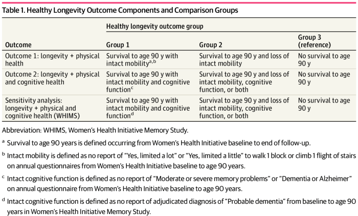
Two papers on aging measurements, starting with a 2022 human study:

“We collected longitudinally across the adult age range a comprehensive list of phenotypes within four domains (body composition, energetics, homeostatic mechanisms and neurodegeneration / neuroplasticity) and functional outcomes. We integrated individual deviations from population trajectories into a global longitudinal phenotypic metric of aging.

blsa participant ages

We demonstrate that accelerated longitudinal phenotypic aging is associated with faster physical and cognitive decline, faster accumulation of multimorbidity, and shorter survival.”

https://www.nature.com/articles/s43587-022-00243-7 “Longitudinal phenotypic aging metrics in the Baltimore Longitudinal Study of Aging”


I disagree with this study’s methodology.

1. Although it acknowledged individual variability, nothing was done to positively adjust to those facts. What could have been done per A review of biological variability was:

“Obtain a measurement of variability that is independent of the mean to ensure to not confound changes in variability with shifts in mean.”

2. A usual research practice is to take at least three measurements, and use their average as representative. That wasn’t done here, maybe because of time and expense considerations?

3. An important measurement for physical function was the time to finish a 400 meter walk. I walk more than ten times that almost every day. I use the first 400 meters as a warmup period while getting to the beach to walk eastward and enjoy the predawn light and water animal activity. I concentrate on gait speed during the last third while walking westward on a straightaway bike path.

This study would measure my gait speed as a sometimes old person during the first 400 meters, rather than a gait speed that usually approaches a young person’s during the last 400 meters. Even if I tried to walk my fastest right out of the gate, I wouldn’t be surprised to find a decade or two difference by this study’s measurements between a morning walk’s first and last 400 meter gait speeds.

4. An important cognitive function measurement was the Digital Symbol Substitution Test, apparently taken during subjects’ fasted state? Sometimes after exercising, I’m okay cognitively when starting work in a fasted state at 6:30 a.m., and other times I’m tired.

Two days ago during the last hour of work 1:30-2:30 p.m., I did outstanding work, four hours after eating whole oats for breakfast, and after drinking two coffees and three teas. I took time to put together pieces of puzzles into proper contexts for management’s attention. My bosses weren’t too pleased with the story it told, but it is what it is.

5. Are measurements of how you start what matters? Or is it how you finish, as is common in competitive sports?

This study would measure my cognitive function as a sometimes old person, rather than performance that approaches a young person’s later in the workday. For both physical and cognitive function, my abilities to ramp up and come close to young people’s capabilities are features that I work on, not random, inconvenient measurement variability.

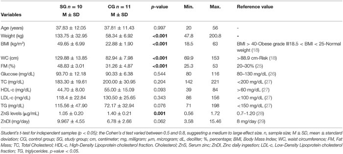
6. Blood measurements were downgraded as having “limited coverage of the four phenotypic domains.” These were taken to fit into specific paradigms and epigenetic clocks. They predictably failed to show causality, as acknowledged with:

“Our analysis showed strong associations between global longitudinal phenotypic score and changes in physical and cognitive function. We did not have sufficient observations to fully separate these two dimensions over time, which would have strengthened the assumption of causality.”

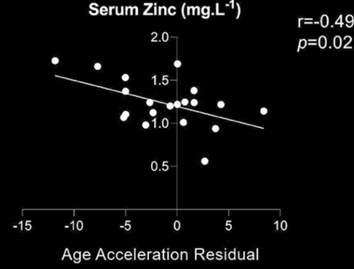
Nowhere in this study was it hinted that all measurements were downstream effects of unmeasured causes. A follow-on study could reanalyze these subjects’ blood samples, MRI, and other measurements for originating upstream factors of signaling pathways and cascades per Signaling pathways and aging and An environmental signaling paradigm of aging.


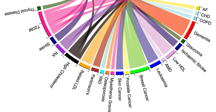
Reference 35 of this first study was a 2021 human and rodent study that was tossed in as a limitation with:

“We might not have all of the relevant phenotypic measures (for example, more detailed immune profiles) for all participants.”

Its findings included:

“From the blood immunome of 1,001 individuals aged 8–96 years, we developed a deep-learning method based on patterns of systemic age-related inflammation. The resulting inflammatory clock of aging (iAge) tracked with multimorbidity, immunosenescence, frailty and cardiovascular aging, and is also associated with exceptional longevity in centenarians.

Canonical markers of acute infection such as IL-6 and tumor necrosis factor-α were not major contributors to iAge, indicating that, except for IL-1β, infection-driven inflammatory markers of the acute inflammatory response do not contribute to age-related chronic inflammation.

We conducted a follow-up study in an independent cohort of 97 extremely healthy adults (aged 25–90 years) matched for cardiovascular risk factors (including conserved levels of high-sensitivity C-reactive protein), selected from a total of 151 recruited participants using strict selection criteria. In this healthy cohort, inflammation markers were measured using a 48-plex cytokine panel. Only 6 circulating immune proteins were significantly correlated with age, with CXCL9 again the largest contributor to age-related inflammation.

CXCL9 is a T-cell chemoattractant induced by IFN-γ and is mostly produced by neutrophils, macrophages and endothelial cells (ECs). We find that CXCL9 is mainly produced by aged endothelium and predicts subclinical levels of cardiovascular aging in nominally healthy individuals.

We did not find any significant correlation between known disease risk factors reported in the study (BMI, smoking, dyslipidemia) and levels of CXCL9 gene or protein expression. We hypothesize that one root cause of CXCL9 overproduction is cellular aging per se, which can trigger metabolic dysfunction.

As ECs but not cardiomyocytes expressed the CXCL9 receptor, CXCR3, we hypothesize that this chemokine acts both in a paracrine fashion (when it is produced by macrophages to attract T cells to the site of injury) and in an autocrine fashion (when it is produced by the endothelium) creating a positive feedback loop. In this model, increasing doses of CXCL9 and expression of its receptor in these cells leads to cumulative deterioration of endothelial function in aging.

IFN-γ did not increase in expression in our cellular aging RNA-seq experiment, suggesting that there are triggers of CXCL9 (other than IFN-γ) that play a role in cellular senescence in the endothelium that are currently unknown. However, in our 1KIP study, IFN-γ was in fact the second-most important negative contributor to iAge, which could be explained by the cell-priming effect of cytokines, where the effect of a first cytokine alters the response to a different one.

iAge derived from immunological cytokines gives us an insight into the salient cytokines that are related to aging and disease. A notable difference compared to other clocks is that iAge is clearly actionable as shown by our experiments in CXCL9 where we can reverse aging phenotypes. More practical approaches range from altering a person’s exposomes (lifestyle) and/or the use of interventions to target CXCL9 and other biomarkers described here.

Our immune metric for human health can identify within healthy older adults with no clinical or laboratory evidence of cardiovascular disease, those at risk for early cardiovascular aging. We demonstrate that CXCL9 is a master regulator of vascular function and cellular senescence, which indicates that therapies targeting CXCL9 could be used to prevent age-related deterioration of the vascular system and other physiological systems as well.”

https://www.nature.com/articles/s43587-021-00082-y “An inflammatory aging clock (iAge) based on deep learning tracks multimorbidity, immunosenescence, frailty and cardiovascular aging”


PXL_20220721_093128925.NIGHT

Non-CpG methylation

Three 2022 papers on methylation epigenetic modifiers, starting with a human study focused on mitochondrial DNA non-CpG methylation involving nucleobases other than guanine (arginine, cytosine, or thymine):

“We collected brain tissue in the nucleus accumbens and prefrontal cortex from deceased individuals without (n = 39) and with (n = 14) drug use, and used whole-genome bisulfite sequencing to cover cytosine sites in the mitochondrial genome. Epigenetic clocks in illicit drug users, especially in ketamine users, were accelerated in both brain regions by comparison with nonusers.

Unlike the predominance of CpG over non-CpG methylation in the nuclear genome, the average CpG and non-CpG methylation levels in the mitochondrial genome were almost equal. The utility of non-CpG methylation was further illustrated by the three indices constructed in this study with non-CpG sites having better distinction between brain areas, age groups, and the presence or absence of drug use than indices consisting of CpG sites only. Results of previous studies on the mitochondrial genome that were solely based on CpG sites should be interpreted cautiously.

The epigenetic clock made up of age-related cytosine sites in mtDNA of the control group was consistently replicated in these two brain regions. One possibility for the correlation is the cycle theory that involves mitochondrial activity, mitochondrial DNA methylation, and alpha-ketoglutarate.

As mitochondrial activity fades with aging, mitochondria gradually lose the ability to eliminate methylation on cytosines through alpha-ketoglutarate. Further investigation of the underlying mechanisms is warranted.

To our knowledge, this is the first report that ketamine might change the mitochondrial epigenetic clock in human brain tissues. We believe this is the first report to elucidate comprehensively the importance of mitochondrial DNA methylation in human brain.”

https://clinicalepigeneticsjournal.biomedcentral.com/articles/10.1186/s13148-022-01300-z “Mitochondrial DNA methylation profiling of the human prefrontal cortex and nucleus accumbens: correlations with aging and drug use”


A second rodent study focused on RNA methylation:

“We investigated the role of RNA N6-methyladenosine (m6A) in improved resilience against chronic restraint stress. A combination of molecular, behavioral, and in vivo recording data demonstrates exercise-mediated restoration of m6A in the mouse medial prefrontal cortex, whose activity is potentiated to exert anxiolytic effects. To provide molecular explanations, it is worth noting that epigenetic regulation, such as histone modification, microRNA, and DNA methylation all participate in mental and cognitive rehabilitation following exercise.

To generalize these rodent data to humans, we recruited a small group of patients with major depressive disorder with prominent anxiety disorders. Compared to age- and sex-matched healthy individuals, patients displayed decreased circulating methyl donor S-adenosyl methionine (SAM) levels. Serum SAM levels were found to be inversely correlated with the Hamilton Anxiety Scale, suggesting the potential value of SAM as a biomarker for depression or anxiety disorders.

Hepatic biosynthesis of methyl donors is necessary for exercise to improve brain RNA m6A to counteract environmental stress. The dependence on hepatic-brain axis suggests the ineffectiveness of exercise training on people with hepatic dysfunctions.

This novel liver-brain axis provides an explanation for brain network changes upon exercise training, and provides new insights into diagnosis and treatment of anxiety disorders. Exercise-induced anxiolysis might be potentiated by further replenishment of RNA methylation donors, providing a strategy of exercise plus diet supplement in preventing anxiety disorders.”

https://onlinelibrary.wiley.com/doi/10.1002/advs.202105731 “Physical Exercise Prevented Stress-Induced Anxiety via Improving Brain RNA Methylation”


A third paper was a review of mitochondrial-to-nuclear epigenetic regulation. I’ll highlight one mitochondrial metabolite, alpha-ketoglutarate (α-KG):

“Apart from established roles in bioenergetics and biosynthesis, mitochondria are signaling organelles that communicate their fitness to the nucleus, triggering transcriptional programs to adapt homeostasis stress that is essential for organismal health and aging. Emerging studies revealed that mitochondrial-to-nuclear communication via altered levels of mitochondrial metabolites or stress signals causes various epigenetic changes, facilitating efforts to maintain homeostasis and affect aging.

Metabolites generated by the tricarboxylic acid (TCA) cycle, the electron transport chain (ETC), or one-carbon cycle within mitochondria can act as substrates or cofactors to control epigenetic modification, especially histone acetylation and methylation and DNA methylation. α-KG produced in the TCA cycle serves as an essential cofactor for the chromatin-modifying Jumonji C (JmjC) domain-containing lysine demethylases (JMJDs) and ten-eleven translocation (TETs) DNA demethylases. Changes in α-KG levels are capable of driving nuclear gene expression by affecting DNA and histone methylation profiles.

1-s2.0-S0968000422000676-gr2_lrg

α-KG deficiency in progenitor stem cells increases with age. For example, the level of α-KG is reduced in follicle fluids of aged humans, and supplementation with α-KG preserves ovarian function in mice.

α-KG extends lifespan in Drosophila by activating AMPK signaling and inhibiting the mTOR pathway. Supplementing α-KG in the form of a calcium salt promoted a longer and healthier life associated with decreased levels of inflammatory cytokines in old mice.

A human study showed a nearly 8-year reversal in DNA methylation clock biological ages of 42 individuals taking an α-KG based formulation for 4–10 months. α-KG supplementation leads to both demethylation and hypermethylation of some CpG sites in the genome, suggesting that α-KG may have a broader effect on methylation-based aging, such as metabolic functions.

Outstanding questions:

  1. How is production of mitochondrial metabolites regulated both spatially and temporally to elicit epigenetic changes in response to mitochondrial dysfunction?
  2. What are specific epigenetic factors involved in mitochondrial-to-nuclear communications, and how do they cooperate with transcription factors in response to various external and internal stimuli?
  3. Do various mitochondrial metabolites act alone or in concert on the epigenome to regulate the aging process?
  4. Are some organs or tissues more at risk than others in maintaining mitochondrial-to-nuclear communication during aging?
  5. Can intervention of mitochondrial-to-nuclear communications mimic beneficial epigenetic changes to delay aging or alleviate age-onset diseases?”

https://www.sciencedirect.com/science/article/pii/S0968000422000676 “Mitochondrial-to-nuclear communication in aging: an epigenetic perspective”


PXL_20220706_093129304

Betaine and diabetes

Two 2022 papers on betaine’s effects, starting with a review:

“Rodent studies provide evidence that betaine effectively limits many diabetes-related disturbances.

  • Betaine therapy improves glucose tolerance and insulin action, which is strongly associated with changes in insulin-sensitive tissues, such as skeletal muscle, adipose tissue, and liver.
  • Betaine supplementation positively affects multiple genes, which expression is dysregulated in diabetes.
  • AMP-activated protein kinase is thought to play a central role in the mechanism underlying anti-diabetic betaine action.
  • Studies with animal models of type 2 diabetes have shown that betaine exerts anti-inflammatory and anti-oxidant effects, and also alleviates endoplasmic reticulum stress.

1-s2.0-S0753332222003353-gr2_lrg

These changes contribute to improved insulin sensitivity and better blood glucose clearance. Results of animal studies encourage exploration of therapeutic betaine efficacy in humans with type 2 diabetes.”

https://www.sciencedirect.com/science/article/pii/S0753332222003353 “The anti-diabetic potential of betaine. Mechanisms of action in rodent models of type 2 diabetes”


Reference 31 was a human study:

“Few studies on humans have comprehensively evaluated intake composition of methyl-donor nutrients choline, betaine, and folate in relation to visceral obesity (VOB)-related hepatic steatosis (HS), the hallmark of non-alcoholic fatty liver diseases.

  • Total choline intake was the most significant dietary determinant of HS in patients with VOB.
  • Combined high intake of choline and betaine, but not folate, was associated with an 81% reduction in VOB-related HS.
  • High betaine supplementation could substitute for choline and folate to normalize homocysteine levels under methyl donor methionine-restriction conditions.
  • Preformed betaine intake from whole-grain foods and vegetables can lower obesity-increased choline and folate requirements by sparing choline oxidation for betaine synthesis and folate for methyl donor conversion in one-carbon metabolism.

Our data suggest that combined dietary intake of choline and betaine reduces the VOB-related HS risk in a threshold-dependent manner.”

https://www.mdpi.com/2072-6643/14/2/261/htm “Optimal Dietary Intake Composition of Choline and Betaine Is Associated with Minimized Visceral Obesity-Related Hepatic Steatosis in a Case-Control Study”

Increasing betaine intake to lower choline and folate requirements was similar to an idea in Treating psychopathological symptoms will somehow resolve causes? that:

“Such positive effects of taurine on glutathione levels may be explained by the fact that cysteine is the essential precursor to both metabolites, whereby taurine supplementation may drive metabolism of cysteine towards GSH synthesis.”


I came across this first paper by it citing All about the betaine, Part 2:

“This review focuses on biological and beneficial effects of dietary betaine (trimethylglycine), a naturally occurring and crucial methyl donor that restores methionine homeostasis in cells. Betaine is endogenously synthesized through metabolism of choline, or exogenously consumed through dietary intake.

Human intervention studies showed no adverse effects with 4 g/day supplemental administration of betaine in healthy subjects. However, overweight subjects with metabolic syndrome showed a significant increase in total and LDL-cholesterol concentrations. These effects were not observed with 3 g/day of betaine administration.

Betaine exerts significant therapeutic and biological effects that are potentially beneficial for alleviating a diverse number of human diseases and conditions.”

https://www.mdpi.com/2079-7737/10/6/456/htm “Beneficial Effects of Betaine: A Comprehensive Review”


PXL_20220518_094703198

Brain changes

This 2022 human study investigated healthy young adult brain changes using MRI and epigenetic clock technologies:

“We aimed to characterize the association of epigenetic age (i.e. estimated DNA methylation age) and its acceleration with surface area, cortical thickness, and volume in healthy young adults. It is largely unknown how accelerated epigenetic age affects multiple cortical features among young adults from 19 to 49 years. Prior findings imply not only that these dynamic changes reveal different aspects of cortical aging, but also that chronological age itself is not a reliable factor to understand the process of cortical aging.

accelerated epigenetic age vs brain features

Seventy-nine young healthy individuals participated in this study. Findings of our study should be interpreted within the context of relatively small sample size, without older adults, and with epigenetic age assessed from saliva.

Additional and unique regional changes due to advanced and accelerated epigenetic age, compared to chronological age-related changes, suggest that epigenetic age could be a viable biomarker of cortical aging. Longitudinal and cross-sectional studies with a larger sample and wider age range are necessary to characterize ongoing effects of epigenetic cortical aging, not only for healthy but also for pathological aging.”

https://doi.org/10.1093/cercor/bhac043 “The effects of epigenetic age and its acceleration on surface area, cortical thickness, and volume in young adults” (not freely available) Thanks to Dr. Yong Jeon Cheong for providing a copy.

Thyroid function

This 2022 review subject was thyroid function changes:

“Circulating concentrations of thyrotropin (TSH) and thyroxine (T4) are tightly regulated. Each individual has setpoints for TSH and free T4 which are genetically determined, and subject to environmental and epigenetic influence.

What is normal for one individual may not be normal for another, even within conventional definitions of euthyroidism. Notably, circulating TSH exists in several different isoforms with varying degrees of glycosylation, sialylation, and sulfonation which affect tissue availability and bioactivity. This is not reflected in immunoreactive TSH concentrations determined by routine laboratory assays.

enm-2022-1463f2

TSH and free T4 relationship analyzed by age in 120,403 patients who were not taking thyroxine treatment. Median TSH for each free T4 integer value (in pmol/ L) was calculated, then plotted as 20-year age bands in adults. Dotted horizontal and vertical lines mark the TSH reference range (0.4 to 4.0 mU/L) and free T4 reference range (10 to 20 pmol/L), respectively.

Mild TSH elevation in older people does not predict adverse health outcomes. In fact, higher TSH is associated with greater life expectancy, including extreme longevity.

In older people, TSH increases with aging without an accompanying fall in free T4. Clinical guidelines now recommend against routine levothyroxine treatment in older people with mild subclinical hypothyroidism.”

https://e-enm.org/journal/view.php?doi=10.3803/EnM.2022.1463 “Thyroid Function across the Lifespan: Do Age-Related Changes Matter?”


PXL_20220427_190457415

Vitamin D and pain

This 2022 human study investigated epigenetic clock associations:

“We assessed the potential relationship of Vitamin D’s effects on pain intensity and disability through associations in epigenetic aging in individuals with and without knee osteoarthritis (KOA). We hypothesized that associations between Vitamin D levels with pain intensity and interference in persons with KOA would be significantly mediated by epigenetic aging.

As a whole, the sample had a mean Vitamin D serum level of 26.7 ng/mL (± 12.8 ng/mL). The mean AgeAccelGrim was 2.4 years (± 5.6 years). There were no significant differences in Vitamin D levels between sex, race, and study site categories.

There was a significant difference in Vitamin D levels between the pain groups, with individuals in the High Impact Pain group showing significantly lower mean levels of Vitamin D (24.01 ng/mL) compared to the Low Impact Pain (28.30 ng/mL) and No Pain (27.30 ng/mL) groups.

vitamin d and pain

Data from this study highlight the important role that Vitamin D plays within the genomic environment, as well as in relation to health outcomes including pain intensity and disability.”

https://link.springer.com/article/10.1007/s12603-022-1758-z “Accelerated Epigenetic Aging Mediates the Association between Vitamin D Levels and Knee Pain in Community-Dwelling Individuals” (not freely available)


It’s good to see a study relating biological age to nutrition status. I didn’t see much discussion of other obvious factors involved in either pain or biological age in their limitations paragraph.

Subjects’ Vitamin D 26.7 ng/mL ± 12.8 ng/mL status indicated that most didn’t spend a few cents every day for their own one precious life. And Vitamin D supplementation wasn’t an exclusion criterion.

The local fire and rescue squad came last Friday to take away a younger neighbor’s body who died overnight. Last I talked with them, they were at least 50 pounds overweight and never exercised. Expressed condolences to their spouse, but wasn’t shocked.

I don’t live in a community-dwelling situation (old people who live on their own as opposed to those taken care of in nursing homes) like this study’s subjects. My youngest neighbors are in their twenties.

Nature hasn’t cared about our lives after our early teens, because we survived long enough to reproduce. What happens in our lives after puberty is largely up to each individual.

PXL_20220502_215338364

Signaling pathways and aging

This 2022 study investigated biological mechanisms of aging:

“Age-related multimorbidity, the presence of more than one age-related disease (ARD) in an individual, poses a major and increasing challenge. Open questions are whether mechanisms of aging can explain ARD co-occurrence in patients, and whether intervention into these mechanisms could prevent or treat multiple ARDs simultaneously.

Five signaling pathways/ cascades were significantly enriched across protein lists for all nine aging hallmarks. These pathways are likely to play a key role in the etiology of ARDs.

Among these five signaling pathways, three were involved in the innate and/ or adaptive immune response. Underlying genes were derived from ARDs comprising metabolic syndrome disorders, autoimmune disorders, and cancers, highlighting the immune response across multiple ARDs.

The ‘intrinsic apoptotic signaling pathway in response to DNA damage by a p53 class mediator’ was also significantly enriched across all aging hallmark protein lists. Underlying genes were derived from multiple cancers and metabolic syndrome disorders.

The ERK1/2 pathway regulates many processes including cell survival, metabolism, and inflammation and was significantly enriched across all aging hallmark protein lists. Underlying genes were derived from 22 aging hallmark-associated ARDs.

erk1-2 pathway

Our study provides evidence for the role of aging hallmarks in the etiology of human ARD multimorbidities and ARDs with incompletely understood pathogenesis. We also raise the possibility that multiple ARDs may be prevented by targeting common signaling pathways.”

https://onlinelibrary.wiley.com/doi/10.1111/acel.13524 “Biological mechanisms of aging predict age-related disease co-occurrence in patients”


I’ll assume that this study finding the importance of innate and adaptive immunity, intrinsic apoptotic, and ERK1/2 signaling pathways in aging was incorporated into A rejuvenation therapy and sulforaphane treatment. Its lead laboratory researcher Dr. Harold Katcher said in interviews that the treatment was formulated from existing research findings.

Its first follow-on lifespan study is going well (4/30/2022 update). 7 6 of 8 treated subjects are alive, compared with 5 4 of 8 control group subjects.

Subjects’ age at the follow-on study’s February 2021 start was 24 months. They are 38-months-old now, and rat maximum lifespan is 45 months, so there should be preliminary results in 2022.

Regarding healthspan, grip strength in treated subjects after a fourth dose was recently measured at 2.6 times control subjects.

Grip+Strength

Longevity+study+(02.09.2021)-modificado-2

Other health measurements are body weight, and TNF-α and IL-6 cytokines.

A second follow-on study uses 18-month-old subjects of both sexes. The initial study was all males, and the first follow-on study is all females.

This second follow-on treatment group will be dosed at 45-day intervals vs. 90-day intervals of the first two studies. Human equivalent doses would be once every 4 years vs. every 8 years.

The treatment works per Beginning of the cure for aging and Reinvigorated. This second follow-on study is research and development to approximate optimal treatment times by age and possibly sex. The idea per Week 37 of Changing to a youthful phenotype with broccoli sprouts is that “by the second rejuvenation you’re already starting at ‘young’.”

Update #2 6/17/2022

Update #3 2/8/2023

Are blood epigenetic clock measurements optimal?

This 2022 human study investigated tissue-specific epigenetic clock measurements:

“We used DNA methylation data representing 11 human tissues (adipose, blood, bone marrow, heart, kidney, liver, lung, lymph node, muscle, spleen, and pituitary gland) to quantify the extent to which epigenetic age acceleration (EAA) in one tissue correlates with EAA in another tissue.

Epigenetic age was moderately correlated across tissues:

  • Blood had the greatest number and degree of correlation, most notably with spleen and bone marrow. Blood did not correlate with epigenetic age of liver.
  • EAA in liver was weakly correlated with EAA in kidney, adipose, lung, and bone marrow.
  • Hypertension was associated with EAA in several tissues, consistent with multiorgan impacts of this illness.
  • HIV infection was associated with positive age acceleration in kidney and spleen.
  • Men were found to exhibit higher EAA than women across all tissues when analyzed together. Significant results were also observed in individual tissues (muscle, spleen, and lymph nodes).

men age faster

Blood alone will often fail to detect EAA in other tissues. It will be advisable to profile several sources of DNA (including blood, buccal cells, adipose, and skin) to get a comprehensive picture of the epigenetic aging state of an individual.”

https://link.springer.com/article/10.1007/s11357-022-00560-0 “HIV, pathology and epigenetic age acceleration in different human tissues”


PXL_20220415_184720157

Biological age and zinc

This 2022 human study investigated zinc’s influence in modulating DNA methylation patterns:

“The purpose of this study was to identify epigenetic variables related to serum Zn (ZnS) levels and Zn daily ingestion (ZnDI) in a case-control cohort. Individuals were selected and classified according to their body mass index into two groups: control group of 11 women without obesity, and study group composed of 10 women with obesity. Inclusion criteria were women aged 18–50 years with stable body weight for at least 6 months.

Novel Zinc-Related Differentially Methylated Regions in Leukocyt

A negative correlation of ZnS with epigenetic age acceleration residual suggested that the higher the ZnS levels, the lower the aging rate:

serum zinc

Our results regarding Zn homeostasis in women with obesity suggested regulation by other mechanisms besides ingestion:

  • Zn-associated differentially methylated regions may exert downstream effects on inflammation, macronutrient metabolism, and DNA/cellular process repair.
  • Hypomethylation of the PM20D1 gene could interconnect DNA methylation and nutritional status.”

https://www.frontiersin.org/articles/10.3389/fnut.2022.785281/full “Novel Zinc-Related Differentially Methylated Regions in Leukocytes of Women With and Without Obesity”


This study emphasized that nutrients aren’t the whole story on health. We also have to be in metabolic zones where our diet and nutrient choices can achieve desired effects.

Subjects’ selection criteria (BMI) was more than double the control group’s. Sometimes people’s lives show others what not to do with their own.