Stress consequences on gut bacteria, behavior, immune system, and neurologic function

This 2015 Canadian rodent study found:

“Chronic social defeat induced behavioral changes that were associated with reduced richness and diversity of the gut microbial community.

The degree of deficits in social, but not exploratory behavior, was correlated with group differences between the microbial community profile.

Defeated mice also exhibited reduced abundance of pathways involved in biosynthesis and metabolism of tyrosine and tryptophan: molecules that serve as precursors for synthesis of dopamine, norepinephrine, serotonin, and melatonin, respectively.

This study indicates that stress-induced disruptions in neurologic function are associated with altered immunoregulatory responses.”

These researchers had an extensive Discussion section where they placed study findings in contexts with other rodent and human studies. For example:

“Our analyses also predicted reduced frequency of fatty acid biosynthesis and metabolism pathways, including that of propanoate and butanoate – byproducts of dietary carbohydrate fermentation by intestinal microorganisms.

Butyrate is a potent histone deacetylase (HDAC) inhibitor that exerts antidepressant-like effects by increasing histone acetylation in the frontal cortex and hippocampus, and consequentially, raising BDNF transcript levels.

Although it was previously unclear whether systemic levels of these metabolites achieved in vivo were sufficient to produce behavioral changes, progress has been made by discovering their presence in cerebrospinal fluid and the brain, and demonstrating that colon-derived SCFAs [short chain fatty acids] cross the blood–brain barrier and preferentially accumulate in the hypothalamus, where they can affect CNS activity.”

http://www.psyneuen-journal.com/article/S0306-4530%2815%2900934-8/fulltext “Structural & functional consequences of chronic psychosocial stress on the microbiome & host”
Testing the null hypothesis of oxytocin’s effects in humans

“There are so many reports of relationships between oxytocin and social behaviors. It is impossible that not a single one of these effects is real.

Isn’t it?

When running a battery of three tasks for every subject who underwent oxytocin treatment..finding false effects becomes almost guaranteed – over 90%.”

http://theneuroeconomist.com/2016/01/the-self-justification-molecule-how-have-we-accumulated-a-vast-behavioral-oxytocin-literature-for-over-a-decade/ “The self-justification molecule: how have we accumulated a vast behavioral oxytocin literature for over a decade”


From one of the references, Why Most Published Research Findings Are False:

“For many current scientific fields, claimed research findings may often be simply accurate measures of the prevailing bias.”


Also see the researcher’s response on their blog post Does oxytocin increase trust in humans? Frequently asked questions:

“Scientists publish only positive findings and not negative ones, and I cannot think of a single study in the vast human oxytocin literature that was replicated by an independent research group.”

Epigenetic consequences of early-life trauma: What are we waiting for?

This 2015 UK human review discussed:

“The progress that has been made by studies that have investigated the relationship between depression, early trauma, the HPA axis and the NR3C1 [glucocorticoid receptor] (GR) gene.

Gene linkage studies for depression, as well as for other common complex disorders, have been perceived by some to be of only limited success; hence the focus on GWAS [genome-wide association studies]. However, even for simple traits, genetic variants identified by GWAS are rarely shown to account for more than 20% of the heritability.

Epigenetic changes are potentially reversible and therefore amenable to intervention, as has been seen in cancer, cardiovascular disease and neurological disorders.”


Five of the review’s references included FKBP5 (a gene that produces a protein that dampens glucocorticoid receptor sensitivity) in their titles, but it wasn’t mentioned in the review itself. A search on FKBP5 also showed human studies such as the 2014 Placental FKBP5 Genetic and Epigenetic Variation Is Associated with Infant Neurobehavioral Outcomes in the RICHS Cohort that found:

“Adverse maternal environments can lead to increased fetal exposure to maternal cortisol, which can cause infant neurobehavioral deficits. The placenta regulates fetal cortisol exposure and response, and placental DNA methylation can influence this function.

Placental FKBP5 methylation reduces expression in a genotype specific fashion, and genetic variation supersedes this effect. These genetic and epigenetic differences in expression may alter the placenta’s ability to modulate cortisol response and exposure, leading to altered neurobehavioral outcomes.”


The authors listed seven human studies conducted 2008-2015 “investigating interactions between methylation of NR3C1, depression and early adversity”:

“Newborn offspring exposed to maternal depression in utero had increased methylation at [a GR CpG site] as well as adverse neurobehavioural outcomes.

Unlike the majority of animal studies examining NR3C1 methylation, many types of potential stressors, sometimes at different developmental stages, have been used to represent early human adversity.

Substantial differences can be expected in the nature of stresses prenatally compared with postnatally, as well as their developmental consequences.”

Seven human studies over the past eight years was a very small number considering both the topic’s importance and the number of relevant animal studies during the period.

Is the topic too offensive for human studies? What makes people pretend that adverse prenatal and perinatal environments have no lasting consequences to the child?

“Many more studies will be needed before effects directly attributable to early life trauma can be separated from those relating to tissue type.

Although investigators have amassed a considerable amount of evidence for an association between differential methylation and HPA axis function in humans, a causal relationship still needs to be fully established.”

Factors that disrupt neurodevelopment may be the largest originators of epigenetic changes that are sustained throughout an individual’s entire lifespan.

Are the multitude of agendas that have resources thrown at them more important than ensuring the well-being of a human before and after they are born?

https://www.researchgate.net/publication/282048312_Early_life_trauma_depression_and_the_glucocorticoid_receptor_gene_-_an_epigenetic_perspective “Early life trauma, depression and the glucocorticoid receptor gene–an epigenetic perspective”

Brain-region-specific energy metabolism affected the social competitiveness of highly-anxious rats

This 2015 Swiss rodent study found:

Mitochondrial function in the nucleus accumbens, a brain region relevant for motivation and depression, is a critical mediating factor in the subordinate status displayed by high-anxious rats.

Treatment with nicotinamide, an amide form of vitamin B3 that boosts mitochondrial respiration, into the NAc [nucleus accumbens] of high-anxious rats at a time point before the social encounter and at a dose that increased accumbal mitochondrial respiration, abolished the disadvantage of high-anxious animals to become dominant against low-anxious animals.

Our findings highlight a key role for brain energy metabolism in social behavior and point to mitochondrial function in the nucleus accumbens as a potential marker and avenue of treatment for anxiety-related social disorders.”

The researchers handled individual differences of the outbred subjects by separating them into high-, intermediate-, and low-anxiety categories according to their responses on two tests. The high- and low-anxiety subjects were matched by weight, age, and social experience.

Here are a few examples of the researchers thoroughly ruling out confounding factors:

“Differences in social competitiveness are not related to overall differences in social motivation or sociability.


Although social competition did significantly increase corticosterone compared with baseline levels, there were no significant differences between anxiety groups at either time point.


Microinfusion of either ROT, MA, or 3NP [mitochondrial respiration inhibitors] reduced the success of treated animals to win the social contest.

Importantly, these treatments did not induce side effects on social investigation or auto-grooming during social competition, or alter locomotor activity, anxiety, or sociability in additional experiments.

Furthermore, these inhibitor treatments did not produce neurotoxic effects, as the drugs were infused at low doses and we confirmed the absence of lesion and neuronal death.

The effects of complex I or complex II inhibition on social competition were specific for the NAc, as infusions of the same inhibitors into the BLA [basolateral amygdala] had no effect on social dominance and did not affect general locomotor activity.

We further showed that, unlike infusion of muscimol [a GABA receptor agonist] in the BLA that interferes with BLA-dependent auditory fear conditioning, 3NP did not affect conditioning in this task, discarding that neuronal inactivation could be a general mechanism whereby impairing mitochondrial function would affect putative functions from the affected brain region.


The impact of mitochondrial function in social competition described here is not mediated by oxidative stress.”

http://www.pnas.org/content/112/50/15486.full “Mitochondrial function in the brain links anxiety with social subordination”

Increased epigenetic brain capacity is an evolved human characteristic

This 2015 George Washington study compared human and chimpanzee brain attributes to find:

“The morphology of the human cerebral cortex is substantially less genetically heritable than in chimpanzees and therefore is more responsive to molding by environmental influences.”

From the news coverage:

“We found that the anatomy of the chimpanzee brain is more strongly controlled by genes than that of human brains, suggesting that the human brain is extensively shaped by its environment no matter its genetics.

Though our findings suggest that the increased plasticity found in human brains has many benefits for adaptation, it is also possible that it makes our brain more vulnerable to many human-specific neurodegenerative and neurodevelopment disorders.”

The study demonstrated an aspect of how natural selection of species leading to Homo sapiens – after humans and chimpanzees shared a common ancestor – favored our increased capacity to adapt to our environments.

http://www.pnas.org/content/112/48/14799.full “Relaxed genetic control of cortical organization in human brains compared with chimpanzees”

Neural pathways for forgetting bad smells

This 2015 New York fruit fly study found:

“Forgetting is regulated by multiple neural pathways that impinge upon a memory center.

Forgetting over time and the acute forgetting of conflicting memory during reversal learning rely on separable neural circuits.

Inactivating these neurons inhibits memory decay without altering learning, whereas activating them promotes forgetting. These neurons [include] a cluster of dopaminergic neurons and a pair of glutamatergic neurons.

Although activity of these neurons is required for memory decay over time, they are not required for acute forgetting during reversal learning. Our results thus not only establish the presence of multiple neural pathways for forgetting in Drosophila but also suggest the existence of diverse circuit mechanisms of forgetting in different contexts.”

Here’s a 3D view of the glutamatergic neurons:

http://movie-usa.glencoesoftware.com/video/10.1073/pnas.1512792112/video-2

http://www.pnas.org/content/112/48/E6663.full “Dissecting neural pathways for forgetting in Drosophila olfactory aversive memory”

Familiar stress opens up an epigenetic window of neural plasticity

This 2015 Italian rodent study found:

“There is a window of plasticity that allows familiar and novel experiences to alter anxiety– and depressive-like behaviors, reflected also in electrophysiological changes in the dentate gyrus (DG).

A consistent biomarker of mood-related behaviors in DG is reduced type 2 metabotropic glutamate (mGlu2), which regulates the release of glutamate. Within this window, familiar stress rapidly and epigenetically up-regulates mGlu2..and improves mood behaviors.

These hippocampal responses reveal a window of epigenetic plasticity that may be useful for treatment of disorders in which glutamatergic transmission is dysregulated.”

The current study included two of the authors of A common dietary supplement that has rapid and lasting antidepressant effects.

The supplementary material showed the:

“Light–dark test as a screening method allowed identification of clusters of animals with a different baseline anxiety profile”

for the BDNF Val66Met subjects. This research methodology better handled the individual differences that often confound studies.

The study’s press release provided further details such as:

“Here again, in experiments relevant to humans, we saw the same window of plasticity, with the same up-then-down fluctuations in mGlu2 and P300 in the hippocampus, Nasca says. This result suggests we can take advantage of these windows of plasticity through treatments, including the next generation of drugs, such as acetyl-L-carnitine, that target mGlu2—not to ‘roll back the clock’ but rather to change the trajectory of such brain plasticity toward more positive directions.”


I disagree with the authoring researchers’ extrapolation of these rodent findings to humans, which seemed to favor chemical intervention. Causes of human stress should be removed or otherwise addressed.

I hope that the study’s “familiar stress” findings won’t be use to attempt to justify potentially harmful practices such as Critical Incident Stress Debriefing, which mandatorily guides people to process recent trauma. Instead, An interview with Dr. Rachel Yehuda on biological and conscious responses to stress made a point about “windows of plasticity” that’s relevant to who we are as feeling human beings:

“What I hear from trauma survivors — what I’m always struck with is how upsetting it is when other people don’t help, or don’t acknowledge, or respond very poorly to needs or distress.”

http://www.pnas.org/content/112/48/14960.full “Stress dynamically regulates behavior and glutamatergic gene expression in hippocampus by opening a window of epigenetic plasticity”

An interview with Dr. Rachel Yehuda on biological and conscious responses to stress

How Trauma and Resilience Cross Generations

“The purpose of epigenetic changes, I think, is simply to increase the repertoire of possible responses.

So let’s say, for some reason, your parents transmitted to you biologic changes that are very appropriate to starvation, but you don’t live in a culture where food is not plentiful.

You’re just not optimized, but I think that if we develop an awareness of what the biologic changes from stress and trauma are meant to do, then I think we can develop a better way of explaining to ourselves what our true capabilities and potentials are.


What I hear from trauma survivors — what I’m always struck with is how upsetting it is when other people don’t help, or don’t acknowledge, or respond very poorly to needs or distress.


Feel it instead of running to someone to give you a sleeping pill.”

Transcript: http://www.onbeing.org/program/rachel-yehuda-how-trauma-and-resilience-cross-generations/transcript/7791

A study of methylation’s mechanical effects on DNA molecules

This 2015 Italian study investigated effects of DNA methylation on mechanical properties of single DNA molecules:

As a consequence of cytosine methylation, the binding of proteins that are implicated in transcription to gene promoters is severely hindered, which results in gene regulation and, eventually, gene silencing. To date, the mechanisms by which methylation biases the binding affinities of proteins to DNA are not fully understood; however, it has been proposed that changes in double-strand conformations, such as stretching, bending, and over-twisting, as well as local variations in DNA stiffness/flexibility may play a role.

We observe that methylation induces no relevant variations in DNA contour lengths, but produces measurable incremental changes in persistence lengths [stiffness/flexibility].

The results reported herein support the claim that the biological consequences of the methylation process, specifically difficulties in protein-DNA binding, are at least partially due to DNA conformation modifications.”

http://www.sciencedirect.com/science/article/pii/S0304416515002706 “Effects of cytosine methylation on DNA morphology: An atomic force microscopy study”

A molecular study of the epigenetic regulation of memory

This 2015 Norwegian rodent study provided:

“New insights into the molecular underpinnings of synaptic plasticity.

We report the first global transcriptome [all RNA found in specific cells] analysis of in vivo synaptic plasticity, using the well-established model of LTP [long-term potentiation, an increase in synaptic strength that underlies memory] in the rat dentate gyrus [a region of the hippocampus where neurogenesis occurs].

We have identified a number of novel lncRNAs [long (more than 200 nucleotides) noncoding (non-protein coding) RNA] that are dynamically regulated in response to LTP. In addition, we also observed an altered expression of multiple classes of repeat elements [mobile DNA sequences often involved in mutations] including retrotransposons [a repeat element type formed by copy-and-paste mechanisms].

The results presented here reveal a vast extension of mRNAs [messenger RNA, a large RNA that carries codes for protein production] previously not associated with neuronal plasticity; the discovery of extensive, dynamic regulation of lncRNAs, repeat elements, and tRNA [transfer RNA that links mRNA and amino acids during protein production] following LTP induction in the adult rat brain.

These findings provide a broader foundation for elucidating the transcriptional and epigenetic regulation of synaptic plasticity.”

Regarding lncRNA:

“We annotate a total of 10,256 novel lncRNAs in the rat transcriptome.

To infer possible functions of lncRNAs, we correlated [71] differentially expressed lncRNAs with regulated protein coding genes.

There are no established rules for predicting the function of lncRNAs.”

Regarding repeat elements:

“It is intriguing to consider that expression of repeat elements during LTP is the first step toward retrotransposition and reshaping of the neuronal genome. A hypothetical mechanism for how these repeat elements could be linked to memory, would be that a certain stimuli, whether it is stress or a learning task (here LTP), deregulate the repression of repeat elements which are then rapidly and transiently transcribed. These elements reinsert themselves back into the genome of stimulated neurons where they influence the expression of neighboring genes.

The present work supports the intriguing hypothesis that dynamic retrotransposition may act as a molecular means to reprogram the neuronal genome as part of long-term synaptic plasticity and memory formation.”

See RNA as a proxy signal for context-specific biological activity for more about lncRNA.

http://journal.frontiersin.org/article/10.3389/fnins.2015.00351/full “Dynamic expression of long noncoding RNAs and repeat elements in synaptic plasticity”

Transgenerational epigenetic programming with stress and microRNA

This 2015 Pennsylvania rodent study found:

“Sperm miRs [microRNAs, a small non-coding RNA that has a role in gene expression] function to reduce maternal mRNA [messenger RNA, a large RNA that carries codes for protein production] stores in early zygotes, ultimately reprogramming gene expression in the offspring hypothalamus and recapitulating the offspring stress dysregulation phenotype.”

These researchers caused stress-induced changes at an early stage of embryonic development with microRNA injections. Resultant adverse effects weren’t observed until subjects were adults!

Most news coverage focused on it being a male’s stress, not a female’s, that affected a developing embryo. Either or both sexes can epigenetically disadvantage a fetus – okay.

Demonstrating how a damaging influence can begin immediately after conception, but symptoms didn’t present until adulthood made this study newsworthy.


Although the term “transgenerational” was used in the study’s title, abstract, and elsewhere, studied epigenetic effects were intergenerational rather than transgenerational. Per A review of epigenetic transgenerational inheritance of reproductive disease, for the term to apply, researchers need to provide evidence in at least the next 2 male or non-gestating female generations and/or 3 gestating female generations of:

“Altered epigenetic information between generations in the absence of continued environmental exposure.”


From a press release, a study coauthor who also coauthored How to make a child less capable even before they are born: stress the pregnant mother-to-be stated:

“Bale suspects that when a male experiences stress it may trigger the release of miRs contained in exosomes from epithelial cells that line the epididymis, the storage and maturation site for sperm between the testes and the vas deferens. These miRs may be incorporated into maturing sperm and influence development at fertilization.”

Not all stress-related gene expression in pituitary and adrenal glands differed.

http://www.pnas.org/content/112/44/13699.full “Transgenerational epigenetic programming via sperm microRNA recapitulates effects of paternal stress”

A review of the epigenetic basis for mental illness

This 2015 New York combined animal and human review of epigenetic studies noted:

“While genetic factors are important in the etiology of most mental disorders, the relatively high rates of discordance among identical twins, particularly for depression and other stress-related syndromes, clearly indicate the importance of additional mechanisms.

Environmental factors such as stress are known to play a role in the onset of these illnesses.

Exposure to such environmental insults induces stable changes in gene expression, neural circuit function, and ultimately behavior, and these maladaptations appear distinct between developmental versus adult exposures.

Increasing evidence indicates that these sustained abnormalities are maintained by epigenetic modifications in specific brain regions.”

Placing the “maladaptations” and “sustained abnormalities” phrases into their contexts:

  • A fetus biologically adapted to their environment – however toxic it was – in order to best survive.
  • These adaptations for survival were subsequently viewed as Disrupted Neurodevelopment and “maladaptations” from the perspectives of normal development and environments.
  • The “sustained abnormalities” caused within the earlier environments “are maintained by epigenetic modifications.” An improved environment wasn’t impetus enough to change developmental “maladaptations.”

Per the below link, it’s been a month since this review was published. Why has there been ZERO news coverage of it?

One reason may be that the Friedman Brain Institute, Icahn School of Medicine at Mount Sinai, didn’t issue a press release or otherwise publicize it. Another reason may be the groups that are opposed to its findings:

  • Parents who provided harmful environments for their children, beginning at conception;
  • People who feel threatened when scientific causal evidence resonates with what happened in their own lives, and in response, limit their empathetic understanding of others’ problems;
  • Social workers, psychologists, and others in industries whose paychecks depend on efforts that aren’t directed towards ameliorating the causes for these later-life effects;
  • Psychiatrists and medical personnel whose livelihoods depend on pharmaceutical and other treatments that only alleviate symptoms;
  • Researchers whose funding depends on producing non-etiologic findings.

Despite resistance to this review’s findings, a large number of people would benefit from publicizing evidence for:

“These sustained abnormalities are maintained by epigenetic modifications in specific brain regions.”

http://nro.sagepub.com/content/early/2015/09/24/1073858415608147 “Epigenetic Basis of Mental Illness”

Stress-induced epigenetic DNA modifications may be inherited

This 2015 Australian plant summary study made several points:

“Non-transmission of epigenetic marks through meiosis may be regarded as an epigenetic modification in itself. We should understand the implications for plant evolution in the context of both selection for and selection against transgenerational epigenetic memory.

Both epigenetic inheritance and resetting are mechanistically directed and targeted. Stress-induced epigenetic modifications may buffer against DNA sequence-based evolution to maintain plasticity, or may form part of plasticity’s adaptive potential.

In some cases the signature of the stress experience remains in the epigenome after relief from the stress, providing a “memory.” If this memory conditions the response to stress during subsequent development, the organism is said to be epigenetically primed. If the memory of the stress experienced by a parent conditions the response of its progeny, this epigenetic priming may be transgenerational.

Epigenetic and genetic variation co-evolve. Epigenetic plasticity does not completely buffer evolvability and reduce the correlation between fitness and genotype, slowing selection.”

One of the summarized studies found that a transgenerational epigenetic change eventually silenced itself after the 40th copy!

The Are stress-induced epigenetic changes to DNA inherited across generations? study was cited, although it argued for the opposing viewpoint.

http://journal.frontiersin.org/article/10.3389/fpls.2015.00699/full “Transgenerational inheritance or resetting of stress-induced epigenetic modifications: two sides of the same coin”

Genetic causes for epigenetic symptoms

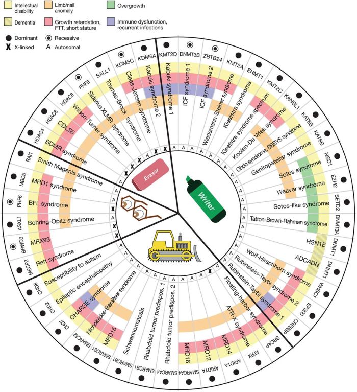
This 2015 human summary study was of 44 genetic disorders that disrupt the maintenance of epigenetic modifications:

“..making them likely to have significant downstream epigenetic consequences. Interestingly, these patients often demonstrate neurological dysfunction, suggesting that precise epigenetic regulation may be critical for neuronal homeostasis. However, at the same time, it is important to keep in mind that many of these proteins have additional non-epigenetic roles.

Mutations in many of these components have now been linked to a number of well-known causes of intellectual disability. Intellectual disability is generally defined as deficits of intellectual function and adaptive behavior that occur during the developmental period.

Given the opposing activity of many of the components of the epigenetic machinery, the pathogenic sequence in these disorders involves an imbalance of chromatin states. Keeping a subset of genes under “pressure” from two opposing systems may allow the cellular system to rapidly respond to environmental stimuli.

These disorders, on average, have unusual phenotypic breadth. Similarly, there is a shift in distribution toward a higher number of organ systems affected.

In addition to developmental phenotypes (multiple congenital anomalies), in some cases there appear to be ongoing defects that remain consequential in post-natal life. An example of the latter is the hippocampal memory defects seen in many of the mouse models.

This raises the question whether cells undergoing neurogenesis and synaptogenesis are particularly sensitive to subtle defects of the epigenetic machinery and downstream epigenetic abnormalities. A major remaining question is whether neurogenesis defects and/or abnormalities of synaptic plasticity are a unifying pathophysiological process.”

The researchers represented the 44 genetic disorders on a wheel graph:

F1.large

I look forward to further research that includes non-genetic disruptors of epigenetic modifications.

http://genome.cshlp.org/content/25/10/1473.full “The Mendelian disorders of the epigenetic machinery”

Leaky gates, anxiety, and grocery store trips without buying list items

An interview with Jeff Link, the editor of Dr. Arthur Janov’s 2011 book “Life Before Birth: The Hidden Script that Rules Our Lives” with Ken Rose:

“Even further confirmation for some of the views of Janov, that maybe weren’t widely accepted for a time, it’s new research now being done into memory and what a lot of scientist are seeing, a lot of different studies is that memory reactivates the same neuroimpulses that were initially firing off when the event happened.

So a traumatic event when you remember it, the act of remembering it is actually creating a neuromirror of what went on initially.

In a lot of ways that is what Primal Therapy is attempting to do; is to go back to that place and reconnect, or as it’s sometimes referred to, reconsolidate the brain state so that real healing can take place.”

Transcript (part 4 of 6): http://cigognenews.blogspot.com/2015/09/ken-rose-on-life-before-birth-part-46.html

MP3: http://www.pantedmonkey.org/podcastgen/download.php?filename=2011-12-15_1300_what_now_jeff_link.mp3