People will forgive you for being wrong, but they will never forgive you for being right – especially if events prove you right while proving them wrong. Thomas Sowell
Starting the fifth year of this blog with a 2018 presentation by the founder of the epigenetic clock method describing the state of the art up through July 2018. The webinar was given on the release day of The epigenetic clock now includes skin study.
Segments before the half-hour mark provide an introduction to the method and several details about the concurrently-released study. The Q&A section starts a little before the hour mark.
This 2018 Austrian human study subject was various associations of prenatal testosterone levels to fetal development:
“The available evidence suggests, albeit not conclusively, that prenatal testosterone levels may be one cause for the association of sexual orientation with handedness. Associations among women were consistent with predictions of the Geschwind–Galaburda theory (GGT), whereas those among men were consistent with predictions of the callosal hypothesis. However, research on the associations between sexual orientation and handedness appears to be compromised by various methodological and interpretational problems which need to be overcome to arrive at a clearer picture.
The GGT posits that high prenatal testosterone levels cause a delay in the fetal development of the left cerebral hemisphere which results in a right-hemisphere dominance and hence in a tendency for left-handedness. According to the GGT, high prenatal testosterone levels entail not only a masculinization of the female fetus, but also a feminization of the male fetus (contrary to neurohormonal theory). Overall, the male fetus is subjected to higher levels of intrauterine testosterone than the female fetus. The GGT is thus consistent with the higher prevalence of left-handedness among men than among women.
The callosal hypothesis applies to men only and assumes, in line with neurohormonal theory, that low prenatal testosterone levels are associated with later homosexuality. According to the CH, high prenatal testosterone enhances processes of cerebral lateralization through mechanisms of axonal pruning, thereby resulting in stronger left-hemisphere dominance and a smaller corpus callosum. Consistent with this, women have a larger corpus callosum than men.”
The study’s Limitations section included the following:
“Limitations of the current study pertain to the self-report nature of our data. Behavioral data may provide differing results from those obtained here.
Assessment of sexual orientation relied on a single-item measure. Utilization of rating scales (e.g., the Kinsey Sexual Orientation Scale) or of multi-item scales, and assessing different components of sexual orientation, would have allowed for a more fine-grained analysis and for a cross-validation of sexual orientation ratings with sexual attraction.
Albeit both our samples were large, the proportions of bisexual and homosexual individuals were, expectedly, only small, as were effects of lateral preferences. Thus, in analysis we could not differentiate bisexual from homosexual individuals. Bisexual and homosexual individuals may differ with regard to the distribution of lateral preferences.
Some effect tests in this study have been underpowered. Independent replications with even larger samples are still needed.”
The largest unstated limitation was no fetal measurements. When a fetus’ epigenetic responses and adaptations aren’t considered, not only can the two competing hypotheses not be adequately compared, but causes for the studied phenotypicprogramming and other later-life effects will also be missed.
The poor substitutes for evidence in this 2018 US study guaranteed that human transgenerational epigenetically inherited effects wouldn’t be found in the generations that followed after prenatal diethylstilbestrol (DES) exposure:
“A synthetic, nonsteroidal estrogen, DES was administered to pregnant women under the mistaken belief it would reduce pregnancy complications and losses. From the late 1930s through the early 1970s, DES was given to nearly two million pregnant women in the US alone.
Use of DES in pregnancy was discontinued after a seminal report showed a strong association with vaginal clear cell adenocarcinoma in prenatally exposed women. A recent analysis of the US National Cancer Institute (NCI) DES Combined Cohort Follow-up Study showed elevated relative risks of twelve adverse health outcomes.
We do not have sufficient data concerning the indication for DES in the grandmother to determine whether adverse pregnancy outcomes in the third generation might resemble those of their grandmothers. Fourth generation effects of prenatal exposures in humans have not been reported.”
https://www.sciencedirect.com/science/article/pii/S0890623818304684 “Reproductive and Hormone-Related Outcomes in Women whose Mothers were Exposed in utero to Diethylstilbestrol (DES): A Report from the US National Cancer Institute DES Third Generation Study” (not freely available)
Its non-statistical data was almost all unverified self-reports by a self-selected sample of the F2 grandchildren, average age 37.
No detailed physical measurements or samples were taken of the F2 grandchildren, or of their F1 parents, or of their F0 grandparents, all of which are required as baselines for any transgenerational epigenetic inheritance findings.
No detailed physical measurements or samples were taken of the F3 great-grandchildren, which is the generation that may provide transgenerational evidence if the previous generations also have detailed physical baselines.
That study’s researchers drew enough participants (360) such that their statistics package allowed them to impute and assume into existence a LOT of data. But the scientific method constrained them to make factual statements of what the evidence actually showed. They admitted:
“In conclusion, we did not find a transgenerational effect of prenatal famine exposure on the health of grandchildren in this study.”
The current study similarly used the faulty methods 1-3 above to produce results such as:
“We do not have sufficient data concerning the indication for DES in the [F0] grandmother to determine whether adverse pregnancy outcomes in the [F2] third generation might resemble those of their grandmothers.
Fourth [F3] generation effects of prenatal exposures in humans have not been reported.“
Zero studies of probably more than 10,000,000 F3 great-grandchildren of DES-exposed women just here in the US?
Who is against funding these studies? Who is afraid of what such studies may find?
The F3 generation can have disease while the F1 and F2 generations do not.
Ancestral exposure to toxicants is a risk factor that must be considered in the molecular etiology of ovarian disease.
For the current study:
What could be expected from a study design that didn’t include F3 women and men, which is the only generation that didn’t have direct DES exposure?
What a nonsensical study design to permit NON-evidence like educational level!
Human studies of possible intergenerational and transgenerational epigenetic inheritance are urgently needed. There will be abundant evidence to discover if researchers will take their fields seriously.
“A single dimension is able to measure a person’s liability to mental disorder, comorbidity among disorders, persistence of disorders over time, and severity of symptoms.”
The coauthors partially based this on:
“Repeated diagnostic interviews carried out over 25 years, when the research participants were 11, 13, 15, 18, 21, 26, 32, and 38 years old, and include information about seven diagnostic groups: anxiety, depression, attention deficit hyperactivity disorder, conduct disorder, substance dependence, bipolar disorder, and schizophrenia.”
“Dunedin and other studies show that most people have at least one episode of mental illness during their lifetime.”
What compels people to manufacture “universal” truths? Aren’t such beliefs poor substitutes for feeling? For understanding historical, factual, personal truths?
What if the price we pay for avoiding and pressuring down our feelings is: A wasted life?
What if the grand hypothesis worth proving is: For one’s life to have meaning, each individual has to regain their feelings?
Let’s start out the new year with a repost of a cautionary reminder:
“Both “predict and “explain” imply that investigators have uncovered a reliable structure to phenomena, the latter involving hypotheses describing unseen mechanisms, leading to a new ability to control events and produce formerly unpredicted/unpredictable outcomes. This is clearly not a fair description of post hoc correlation-fishing.
The current publication system almost forces authors to make causal statements using filler verbs (e.g. to drive, alter, promote) as a form of storytelling (Gomez-Marin, 2017); without such a statement they are often accused of just collecting meaningless facts.”
The originator of the 2013 epigenetic clock improved its coverage with this 2018 UCLA human study:
“We present a new DNA methylation-based biomarker (based on 391 CpGs) that was developed to accurately measure the age of human fibroblasts, keratinocytes, buccal cells, endothelial cells, skin and blood samples. We also observe strong age correlations in sorted neurons, glia, brain, liver, and bone samples.
The skin & blood clock outperforms widely used existing biomarkers when it comes to accurately measuring the age of an individual based on DNA extracted from skin, dermis, epidermis, blood, saliva, buccal swabs, and endothelial cells. Thus, the biomarker can also be used for forensic and biomedical applications involving human specimens.
The biomarker applies to the entire age span starting from newborns, e.g. DNAm of cord blood samples correlates with gestational week.
Furthermore, the skin & blood clock confirms the effect of lifestyle and demographic variables on epigenetic aging. Essentially it highlights a significant trend of accelerated epigenetic aging with sub-clinical indicators of poor health.
Conversely, reduced aging rate is correlated with known health-improving features such as physical exercise, fish consumption, high carotenoid levels. As with the other age predictors, the skin & blood clock is also able to predict time to death.
Collectively, these features show that while the skin & blood clock is clearly superior in its performance on skin cells, it crucially retained all the other features that are common to other existing age estimators.”
An introduction to the study highlighted several items:
“Although the skin-blood clock was derived from significantly less samples (~900) than Horvath’s clock (~8000 samples), it was found to more accurately predict chronological age, not only across fibroblasts and skin, but also across blood, buccal and saliva tissue. A potential factor driving this improved accuracy in blood could be related to the approximate 18-fold increase in genomic coverage afforded by using Illumina 450k/850k beadarrays.
It serves as a roadmap for future clock studies, pointing towards the importance of constructing tissue or cell-type specific epigenetic clocks, to more accurately measure biological aging in the given tissue/cell-type, and therefore with the potential to be more informative of disease-risk or the success of disease interventions in the tissue or cell-type of interest.”
A subset of memory recall–induced neurons in the DG [dentate gyrus] becomes reactivated after memory attenuation,
The degree of fear reduction positively correlates with this reactivation, and
The continued activity of memory recall–induced neurons is critical for remote fear memory attenuation.
Although other brain areas such as the prefrontal cortex and the amygdala are likely to be implicated in remote fear memories and remain to be investigated, these results suggest that fear attenuation at least partially occurs in memory recall–induced ensembles through updating or unlearning of the original memory trace of fear.
These data thereby provide the first evidence at an engram-specific level that fear attenuation may not be driven only by extinction learning, that is, by an inhibitory memory trace different from the original fear trace.
Rather, our findings indicate that during remote fear memory attenuation both mechanisms likely coexist, albeit with the importance of the continued activity of memory recall–induced neurons experimentally documented herein. Such activity may not only represent the capacity for a valence change in DG engram cells but also be a prerequisite for memory reconsolidation, namely, an opportunity for learning inside the original memory trace.
As such, this activity likely constitutes a physiological correlate sine qua non for effective exposure therapies against traumatic memories in humans: the engagement, rather than the suppression, of the original trauma.”
The researchers also provided examples of human trauma:
“We dedicate this work to O.K.’s father, Mohamed Salah El-Dien, and J.G.’s mother, Wilma, who both sadly passed away during its completion.”
So, how can this study help humans? The study had disclosed and undisclosed limitations:
1. Humans aren’t lab rats. We can ourselves individually change our responses to experiential causes of ongoing adverse effects. Standard methodologies can only apply external treatments.
2. It’s a bridge too far to go from neural activity in transgenic mice to expressing unfounded opinions on:
“A physiological correlate sine qua non for effective exposure therapies against traumatic memories in humans.”
Human exposure therapies have many drawbacks, in addition to being applied externally to the patient on someone else’s schedule. A few others were discussed in The role of DNMT3a in fear memories:
“Inability to generalize its efficacy over time,
Potential return of adverse memory in the new/novel contexts,
Context-dependent nature of extinction which is widely viewed as the biological basis of exposure therapy.”
3. Rodent neural activity also doesn’t elevate recall to become an important goal of effective human therapies. Clearly, what the rodents experienced should have been translated into human reliving/re-experiencing, not recall! Terminology used in animal studies preferentially has the same meaning with humans, since the purpose of animal studies is to help humans.
4. The researchers acknowledged that:
“Other brain areas such as the prefrontal cortex and the amygdala are likely to be implicated in remote fear memories and remain to be investigated.”
“The findings imply that in response to traumatic stress, some individuals, instead of activating the glutamate system to store memories, activate the extra-synaptic GABA system and form inaccessible traumatic memories.”
The study I curated yesterday, Organ epigenetic memory, demonstrated organ memory storage. It’s hard to completely rule out that other body areas may also store traumatic memories.
The wide range of epigenetic memory storage vehicles is one reason why effective human therapies need to address the whole person, the whole body, and each individual’s entire history.
This post has somehow become a target for spammers, and I’ve disabled comments. Readers can comment on other posts and indicate that they want their comment to apply here, and I’ll re-enable comments.
This 2018 New York rodent study not only wasted resources but also speciously attempted to extrapolate animal study findings to humans:
“While it is clear that behavioralexperience modulates epigenetic profiles, it is less evident how the nature of that experience influences outcomes and whether epigenetic/genetic “biomarkers” could be extracted to classify different types of behavioral experience.
Male and female mice were subjected to either:
a Fixed Interval (FI) schedule of food reward, or
a single episode of forced swim followed by restraint stress, or
no explicit behavioral experience
after which global expression levels of two activating (H3K9ac and H3K4me3) and two repressive (H3K9me2 and H3k27me3) post-translational histone modifications (PTHMs), were measured in hippocampus (HIPP) and frontal cortex (FC).
A random subset of 5 of the 12 animals from each sex/behavioral experience group were used for these analyses. FC and HIPP were dissected from each of those 5 brains and homogenized for subsequent analyses. Thus, sample size for PTHM expression levels was n = 5 for each region/sex/behavioral treatment group and all PTHM expression level analyses utilized the homogenized tissue.
The specific nature of the behavioral experience differentiated profiles of PTHMs in a sex- and brain region-dependent manner, with all 4 PTHMs changing in parallel in response to different behavioral experiences. Global PTHMs may provide a higher-order pattern recognition function.”
The researchers knew or should have known that measuring “global expression levels” in “homogenized tissue” of “n = 5” subjects was flawed, and they did it anyway. They acknowledged some of the numerous study design defects with qualifiers such as:
“Even though these were global levels of histone modifications (and thus not indicative of changes at specific genes or sites on genes)..
As FS-RS behavioral experience was completed before FI behavioral experience, a longer overall post-behavior experience time (approximately 1 week) elapsed for this group, resulting in some differences in overall timing between these experiences and global PTHM assessment. However, extending the duration of the FS-RS experience (i.e., repeated exposures) would also have led to habituation..”
Did they purposely make these mistakes because of the “biomarkers”paradigm?
What would they have found if they had followed their judgments and training to design a better study? Experience-dependent histone modifications that differed by gender and brain region was certainly a promising research opportunity.
As for extrapolating the cited animal study findings to humans? Ummm..NO!
https://www.ncbi.nlm.nih.gov/pmc/articles/PMC6060276/ “Different Behavioral Experiences Produce Distinctive Parallel Changes in, and Correlate With, Frontal Cortex and Hippocampal Global Post-translational Histone Levels”
It’s dawned on me that although links in blog posts are indexed by search engines, links in comments may not be. Here’s a post to elevate links in three comments that may have escaped notice.
“It is my view that all researchers have a narrow focus on what they want to research, without having an over-riding paradigm in which to fit the research and its results. Janovian Primal Therapy and theory, with its focus and understanding of the three different levels of consciousness would provide for a much needed over-arching paradigm, especially in the area of mental health.”
“You are right on. The Norcross survey, in particular, is utter crap. More than half of those “experts” surveyed were CBT therapists who knew nothing about PT and yet deemed themselves confident to judge “primal scream therapy” as “discredited.” I feel the therapy will never be understood for what it is.”
“There is of course, reversibility. Michael Meaney’s baby rats had their epigenetic changes reversed with loving maternal care. There are several compounds in development which have been shown to reverse methylation. This former physician and researcher says, “Epigenetic changes affect the level of activity of our genes. Genetic activity levels affect our emotions, beliefs, and our bodies. Exploring epigenetics and chronic illness may help us understand causes that many of us suspect have played a role in the onset and evolution of our illnesses. Furthermore, these epigenetic changes have been found to be reversible, at least some of the time, even with a seemingly indirect treatment such as psychotherapy.” Epigenetics and Chronic Illness: Why Symptoms May Be Reversible
So what gives? I suspect that your researcher is working with his/her head in the sand, hamstrung by their ideological biases. If CBT can effect epigenetic changes, imagine what primal therapy can do.”
And a seven-year anniversary repost of events that affect me every day:
This 2017 UC Irvine human review subject provided details of how fetalhypothalamic-pituitary-adrenal components and systems develop, and how they are epigenetically changed by the mother’s environment:
“The developmental origins of disease or fetal programming model predicts that intrauterine exposures have life-long consequences for physical and psychological health. Prenatal programming of the fetal hypothalamic-pituitary-adrenal (HPA) axis is proposed as a primary mechanism by which early experiences are linked to later disease risk.
Development of the fetal HPA axis is determined by an intricately timed cascade of endocrine events during gestation and is regulated by an integrated maternal-placental-fetal steroidogenic unit. Mechanisms by which stress-induced elevations in hormones of maternal, fetal, or placental origin influence the structure and function of the emerging fetal HPA axis are discussed.
Human gestational physiology and fetal HPA axis development differ even from that of closely related nonhuman primates, thereby limiting the generalizability of animal models. This review will focus solely on studies of prenatal stress and fetal HPA axis development in humans.”
1. Every time I read a prenatal study I’m in awe of all that has to go right – and at the appropriate times and sequences – for a fetus to be undamaged. Add in what needs to happen at birth, during infancy, and throughout early childhood, and it seems impossible for any human to escape epigenetic damage.
2. The reviewers referenced animal studies and human research performed with postnatal subjects, despite the disclaimer:
This review will focus solely on studies of prenatal stress and fetal HPA axis development in humans.”
This led to blurring of what had been studied or not with human fetuses regarding the subject.
3. These reviewers uncritically listed many dubious human studies that had both stated and undisclosed severe limitations on their findings. Other reviewers offer informed analysis of cited studies, as Sex-specific impacts of childhood trauma summarized with cortisol:
“Findings are dependent upon variance in extenuating factors, including but not limited to, different measurements of:
presence and severity of psychopathology symptomology.”
4. The paper would have been better had it stayed on topic with its title “Developmental origins of the human hypothalamic-pituitary-adrenal axis.” Let other reviews cover animals, post-natal humans, and questionable evidence.
5. I asked the reviewers to provide a searchable file to facilitate using their work as a reference.
This 2018 Chinese study electronically modeled the brain’s circuits to evaluate memory transfer mechanisms:
“During non-rapid-eye-movement (NREM) sleep, thalamo-cortical spindles and hippocampal sharp wave-ripples have been implicated in declarative memory consolidation. Evidence suggests that long-term memory consolidation is coordinated by the generation of:
enabling memory transfer from the hippocampus to the cortex.
Consolidation has also been demonstrated in other brain tasks, such as:
In the acquisition of motor skills, where there is a shift from activity in prefrontal cortex to premotor, posterior parietal, and cerebellar structures; and
In the transfer of conscious to unconscious tasks, where activity in initial unskilled tasks and activity in skilled performance are located in different regions, the so-called ‘scaffolding-storage’ framework.
By separating a neural circuit into a feedforward chain of gating populations and a second chain coupled to the gating chain (graded chain), graded information (i.e. information encoded in firing rate amplitudes) may be faithfully propagated and processed as it flows through the circuit. The neural populations in the gating chain generate pulses, which push populations in the graded chain above threshold, thus allowing information to flow in the graded chain.
In this paper, we will describe how a set of previously learned synapses may in turn be copied to another module with a pulse-gated transmission paradigm that operates internally to the circuit and is independent of the learning process.”
The study had neither been peer-reviewed, nor were the mechanisms tested in living beings.
“Genome-wide technology has facilitated epigenome-wide association studies (EWAS), permitting ‘hypothesis-free’ examinations in relation to adversity and/or mental health problems. Results of EWAS are in fact conditional on several a priori hypotheses:
EWAS coverage is sufficient for complex psychiatric problems;
Peripheral tissue is meaningful for mental health problems; and
The assumption that biology can be informative to the phenotype.
1. CpG sites were chosen as potentially biologically informative based on consultation with a consortium of DNA methylation experts. Selection was, in part, based on data from a number of phenotypes (some medical in nature such as cancer), and thus is not specifically targeted to brain-based, stress-related complex mental health phenotypes.
2. The assumption is often that distinct peripheral tissues are interchangeable and equally suited for biomarker detection, when in fact it is highly probable that peripheral tissues themselves correspond differently to environmental adversity and/or disease state.
3. Analyses result in general statements such as ‘neurodevelopment’ or the ‘immune system’ being involved in the aetiology of a given phenotype. Whether these broad categories play indeed a substantial role in the aetiology of the mental health problem is often hard to determine given the post hoc nature of the interpretation.”
The reviewers mentioned in item #2 the statistical flaw of assuming that measured entities are interchangeable with one another. They didn’t mention that the problem also affected item #1 methodologies of averaging CpG methylation measurements in fixed genomic bins or over defined genomic regions, as discussed in:
The reviewers offered suggestions for reducing the impacts of these three hypotheses. But will doing more of the same, only better, advance science?
Was it too much to ask of researchers whose paychecks and reputations depended on a framework’s paradigm – such as the “biomarker” mentioned a dozen and a half times – to admit the uselessness of gathering data when the framework in which the data operated wasn’t viable? They already knew or should have known this.
“When phenotypic variation results from alleles that modify phenotypic variance rather than the mean, this link between genotype and phenotype will not be detected.”
“Blood-based EWAS may yield limited information relating to underlying pathological processes for disorders where brain is the primary tissue of interest.”
The truth about complex traits and GWAS added another example of how this framework and many of its paradigms haven’t produced effective explanations of “the aetiology of the mental health problem”
“The most investigated candidate gene hypotheses of schizophrenia are not well supported by genome-wide association studies, and it is likely that this will be the case for other complex traits as well.”
Researchers need to reevaluate their framework if they want to make a difference in their fields. Recasting GWAS as EWAS won’t make it more effective.
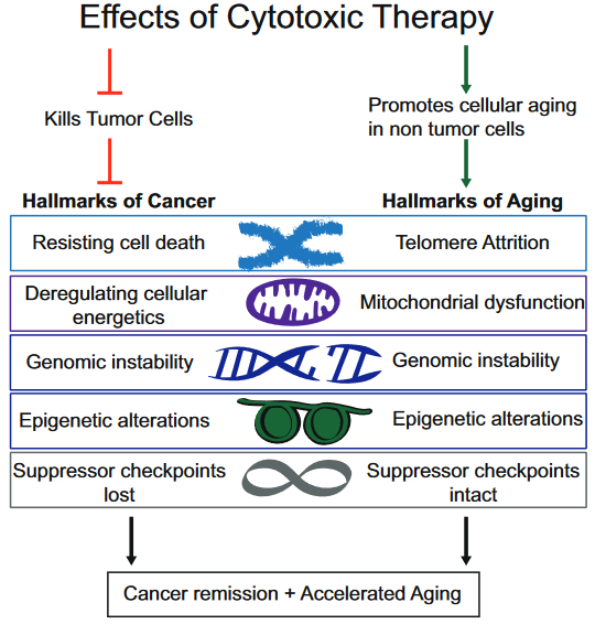
This 2018 UC San Diego review subject was the interplay between breast cancer treatments and their effects on aging:
“Although current breast cancer treatments are largely successful in producing cancer remission and extending lifespan, there is concern that these treatments may have long lasting detrimental effects on cancer survivors, in part, through their impact on non-tumor cells. It is unclear whether breast cancer and/or its treatments are associated with an accelerated aging phenotype.
In this review, we have highlighted five of nine previously described cellular hallmarks of aging that have been described in the context of cytotoxic breast cancer treatments:
The review was full of caveats weakening the above graphic’s associations:
“Telomere attrition – Blood TL [telomere length] was not associated with chemotherapy in three out of four studies;
Mitochondrial dysfunction – How cancer therapies affect cellular energetics as they relate to rate of aging is unclear;
Genomic instability – Potentially contributing to accelerated aging;
Epigenetic alterations – Although some of the key regulators of these processes have begun to be identified, including DNA and histone methylases and demethylases, histone acetylases and de-acetylases and chromatin remodelers, how they regulate the changes in aging through alteration of global transcriptional programs, remains to be elucidated; and
Cellular senescence – Dysregulated pathways can be targeted by cytotoxic chemotherapies, resulting in preferential cell death of tumor cells, but how these treatments also affect normal cells with intact pathways is unclear.”
To their credit, these reviewers at least presented some of the contrary evidence, and didn’t continue on with a directed narrative as other reviewers are prone to do.
The originator of the epigenetic clock methodology was a coauthor of the review. Only one of his works was cited in the Epigenetic alterations subsection:
This freely-available 2017 study quoted below highlighted that epigenetic clock measurements as originally designed were tissue-specific:
“To our knowledge, this is the first study to demonstrate that breast tissue epigenetic age exceeds that of blood tissue in healthy female donors. In addition to validating our earlier finding of age elevation in breast tissue, we further demonstrate that the magnitude of the difference between epigenetic age of breast and blood is highest in the youngest women in our study (age 20–30 years) and gradually diminishes with advancing age. As women approach the age of the menopausal transition, we found that the epigenetic of age of blood approaches that of the breast.”
Additional caution was justified in both interpreting age measurements and extending them into “cellular hallmarks” when the tissue contained varying cell types:
“Our studies were performed on whole breast tissue. Diverse types of cells make up whole breast tissue, with the majority of cells being adipocytes. Other types of cells include epithelial cells, cuboidal cells, myoepithelial cells, fibroblasts, inflammatory cells, vascular endothelial cells, preadipocytes, and adipose tissue macrophages.
This raises the possibility that the magnitude of the effects we observe, of breast tissue DNAm age being greater than other tissues, might be an underestimation, since it is possible that not all of the cells of the heterogenous sample have experienced this effect. Since it is difficult to extract DNA from adipose tissue, we suspect that the majority of DNA extracted from our whole breast tissues was from epithelial and myoepithelial cells.”
This 2018 McGill paper reviewed findings from animal and human studies on the relationships between drug-seeking behavior and epigenetic DNA methylation:
“Although there is an increasing line of evidence from preclinical models of addiction, there are only a few human studies that systematically assessed DNA methylation in addiction. Most of the studies were done on small cohorts and focused on one or a few candidate genes, except in the case of alcohol use where larger studies have been carried out.
A long line of evidence suggests that abnormal patterns of gene expression occur in brain regions related to drug addiction such as the nucleus accumbens, prefrontal cortex, amygdala, and the ventral tegmental area.
Using the “incubation of craving” model in rats trained to self-administer cocaine, and treated with either SAM or RG108, the genome-wide DNA methylation and gene expression landscape in the nucleus accumbens after short (1 day) and long (30 days) abstinence periods and the effects of epigenetic treatments were delineated. The main findings are:
A long incubation period results in robust changes in methylation;
Direct accumbal infusion of SAM that is paired with a “cue” after long incubation times increases drug-seeking behavior,
Whereas a single treatment with RG108 decreases this behavior.
Importantly, the effects of these single administrations of a DNA methylation inhibitor remain stable for 30 more days. These data suggest that DNA methylation might be mediating the impact of “incubation” on the craving phenotype and that this phenotype could be reprogrammed by a DNA demethylation agent.”
The review covered neither human dimensions of the impacts of unfulfilled needs nor investigations of exactly what pain may impel human drug-seeking behavior. The “Implications for Diagnostic and Therapeutics” were largely at the molecular level.