Experiential feeling therapy addressing the pain of the lack of love.
Neurochemicals
The hypothalamus couples with the brainstem to cause migraines
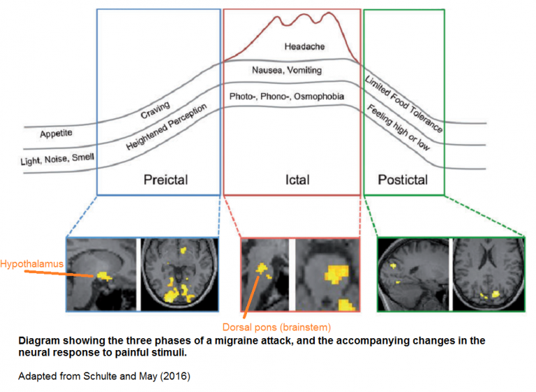
This 2016 German human study with one subject found:
“The hypothalamus to be the primary generator of migraine attacks which, due to specific interactions with specific areas in the higher and lower brainstem, could alter the activity levels of the key regions of migraine pathophysiology.”
The subject underwent daily fMRI scans, and procedures to evoke brain activity. She didn’t take any medications, and suffered three migraine attacks during the 31-day experimental period.
Neuroskeptic commented:
“The dorsal pons has previously been found to be hyperactive during migraine. It’s been dubbed the brain’s ‘migraine generator.’ Schulte and May’s data suggest that this is not entirely true – rather, it looks like the hypothalamus may be the true generator of migraine, while the brainstem could be a downstream mediator of the disorder.
A hypothalamic origin of migraines would help to explain some of the symptoms of the disorder, such as changes in appetite, that often accompany the headaches.”
The above graphic looks to me like the result of feedback mechanisms that either didn’t exist or inadequately handled the triggering event. Other examples of the hypothalamus lacking feedback or being involved in a deviated feedback loop include:
- Lack of feedback to the HIF-1α signaling source mentioned in Lack of oxygen’s epigenetic effects
- Impaired hippocampal glucocorticoid negative feedback mentioned in Treating prenatal stress-related disorders with an oxytocin receptor agonist
There are many unanswered questions with a one-person study, of course. Addressing the cause of this painful condition would find out when, where, and how a person’s hypothalamus became modified to express migraine tendencies.
I’d guess that migraine tendencies may appear as early as the first trimester of pregnancy, given that a highly functional hypothalamus is needed for survival and development in our earliest lives. Gaining as much familial and historical information as possible from the person would be necessary steps in therapies that address migraine causes.
http://blogs.discovermagazine.com/neuroskeptic/2016/05/22/pinpointing-origins-of-migraine/ “Pinpointing the Origins of Migraine in the Brain”
https://academic.oup.com/brain/article/139/7/1987/2464241 “The migraine generator revisited: continuous scanning of the migraine cycle over 30 days and three spontaneous attacks”
As mentioned in How to cure the ultimate causes of migraines? comments are turned off for this post due to it somehow becoming a magnet for spammers. Readers can comment on that post instead.
Genetic imprinting, sleep, and parent-offspring conflict
This 2016 Italian review subject was the interplay of genetic imprinting and sleep regulation:
“Sleep results from the synergism between at least two major processes: a homeostatic regulatory mechanism that depends on the accumulation of the sleep drive during wakefulness, and a circadian self-sustained mechanism that sets the time for sleeping and waking throughout the 24-hour daily cycle.
REM sleep apparently contravenes the restorative aspects of sleep; however, the function of this ‘paradoxical’ state remains unknown. Although REM sleep may serve important functions, a lack of REM sleep has no major consequences for survival in humans; however, severe detrimental effects have been observed in rats.
Opposite imprinting defects at chromosome 15q11–13 are responsible for opposite sleep phenotypes as well as opposite neurodevelopmental abnormalities, namely the Prader-Willi syndrome (PWS) and the Angelman syndrome (AS). Whilst the PWS is due to loss of paternal expression of alleles, the AS is due to loss of maternal expression.
Maternal additions or paternal deletions of alleles at chromosome 15q11–13 are characterized by temperature control abnormalities, excessive sleepiness, and specific sleep architecture changes, particularly REM sleep deficits. Conversely, paternal additions or maternal deletions at chromosome 15q11–13 are characterized by reductions in sleep and frequent and prolonged night wakings.
The ‘genomic imprinting hypothesis of sleep’ remains in its infancy, and several aspects require attention and further investigation.”
http://journals.plos.org/plosgenetics/article?id=10.1371/journal.pgen.1006004 “Genomic Imprinting: A New Epigenetic Perspective of Sleep Regulation”
A commenter to the review referenced a 2014 study Troubled sleep: night waking, breastfeeding, and parent–offspring conflict that received several reactions, including one by the same commenter. Here are a few quotes from the study author’s consolidated response:
“‘Troubled sleep’ had two major purposes. The first was to draw attention to the oppositely perturbed sleep of infants with PWS and AS and explore its evolutionary implications. The involvement of imprinted genes suggests that infant sleep has been subject to antagonistic selection on genes of maternal and paternal origin with genes of maternal origin favoring less disrupted sleep.
My second major purpose was a critique of the idea that children would be happier, healthier and better-adjusted if we could only return to natural methods of child care. This way of thinking is often accompanied by a belief that modern practices put children at risk of irrevocable harm.
The truth of such claims is ultimately an empirical question, but the claims are sometimes presented as if they had the imprimatur of evolutionary biology. This appeal to scientific authority often seems to misrepresent what evolutionary theory predicts: that which evolves is not necessarily that which is healthy.
Why should pregnancy not be more efficient and more robust than other physiological systems, rather than less? Crucial checks, balances and feedback controls are lacking in the shared physiology of the maternal–fetal unit.
Infant sleep may similarly lack the exquisite organization of systems without evolutionary conflict. Postnatal development, like prenatal development, is subject to difficulties of evolutionarily credible communication between mothers and offspring.”
The author addressed comments related to attachment theory:
“Infants are classified as having insecure-resistant attachment if they maintain close proximity to their mother after a brief separation while expressing negative emotions and exhibiting contradictory behaviors that seem to both encourage and resist interaction. By contrast, infants are classified as having insecure-avoidant attachment if they do not express negative emotion and avoid contact with their mother after reunion.
Insecure-avoidant and insecure-resistant behaviors might be considered antithetic accommodations of infants to less responsive mothers; the former associated with reduced demands on maternal attention, the latter with increased demands. A parallel pattern is seen in effects on maternal sleep. Insecure-avoidant infants wake their mothers less frequently, and insecure-resistant infants more frequently, than securely attached infants.
Parent–child interactions are transformed once children can speak. Infants with more fragmented sleep at 6 months had less language at 18 and 30 months.
Infants with AS have unconsolidated sleep and never learn to speak. The absence of language in the absence of expression of one or more MEGs [maternally expressed imprinted genes] is compatible with a hypothesis in which earlier development of language reduces infant demands on mothers.”
Regarding cultural differences:
“China, Taiwan and Hong Kong have both high rates of bed-sharing and high rates of problematic sleep compared with western countries. Within this grouping, however, more children sleep in their own room but parents report fewer sleep problems in Hong Kong than in either China or Taiwan.
Clearly, cultural differences are significant, and the causes of this variation should be investigated, but the differences cannot be summarized simply as ‘west is worst’.
The fitness [genetic rather than physical fitness] gain to mothers of an extra child and the benefits for infants of longer IBIs [interbirth intervals] are substantial. These selective forces are unlikely to be orders of magnitude weaker than the advantages of lactase persistence, yet the selective forces associated with dairying have been sufficient to result in adaptive genetic differentiation among populations.
The possibility of gene–culture coevolution should not be discounted for behaviors associated with infant-care practices.”
Regarding a mismatch between modern and ancestral environments:
“I remain skeptical of a tendency to ascribe most modern woes to incongruence between our evolved nature and western cultural practices. We did not evolve to be happy or healthy but to leave genetic descendants, and an undue emphasis on mismatch risks conflating health and fitness.
McKenna [a commenter] writes ‘It isn’t really nice nor maybe even possible to fool mother nature.’ Here I disagree. Our genetic adaptations often try to fool us into doing things that enhance fitness at costs to our happiness.
Our genes do not care about us and we should have no compunction about fooling them to deliver benefits without serving their ends. Contraception, to take one obvious example, allows those who choose childlessness to enjoy the pleasures of sexual activity without the fitness-enhancing risk of conception.
Night waking evolved in environments in which there were strong fitness costs from short IBIs and in which parents lacked artificial means of birth-spacing. If night waking evolved because it prolonged IBIs, then it may no longer serve the ends for which it evolved.
Nevertheless, optimal infant development might continue to depend on frequent night feeds as part of our ingrained evolutionary heritage.
It could also be argued that when night waking is not reinforced by feeding, and infants sleep through the night, then conflict within their genomes subsides. Infants would then gain the benefit of unfragmented sleep without the pleiotropic costs of intragenomic conflict. Plausible arguments could be presented for either hypothesis and a choice between them must await discriminating evidence.”
Commenters on the 2014 study also said:
“[Crespi] The profound implications of Haig’s insights into the roles of evolutionary conflicts in fetal, infant and maternal health are matched only by the remarkable absence of understanding, appreciation or application of such evolutionary principles among the research and clinical medical communities, or the general public.
[Wilkins] A mutation may be selected for its effect on the trait that is the basis of the conflict, but that mutation also likely affects other traits. In general, we expect that these pleiotropic effects to be deleterious: conflict over one trait can actually drive other traits to be less adapted. Natural selection does not necessarily guarantee positive health outcomes.
[McNamara] Assuming that AS/REM is differentially influenced by genes of paternal origin then both REM properties and REM-associated awakenings can be better explained by mechanisms of genomic conflict than by traditional claims that REM functions as an anti-predator ‘sentinel’ for the sleeping organism.
[Hinde] Given this context of simultaneous coordination and conflict between mother and infant, distinguishing honest signals of infant need from self-interested, care-extracting signals poses a challenge.“
A limited study of parental transmission of anxiety/stress-reactive traits
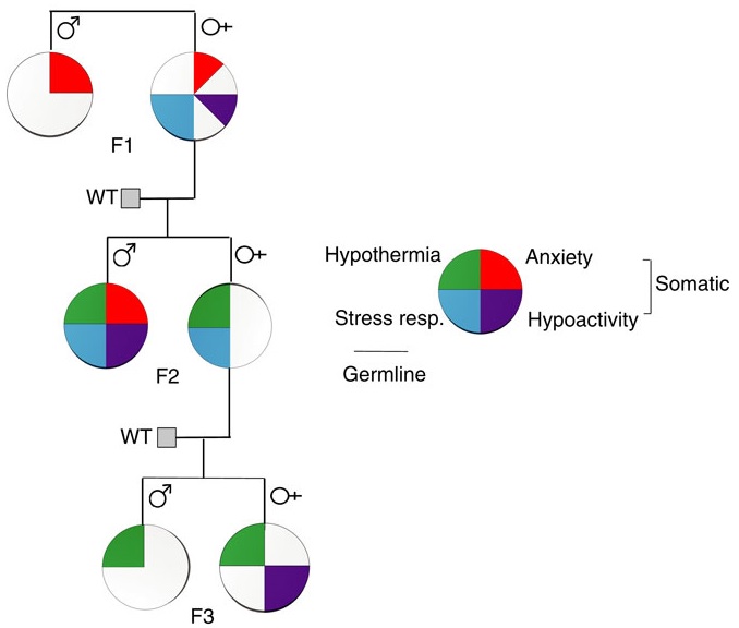 This 2016 New York rodent study found:
“Parental behavioural traits can be transmitted by non-genetic mechanisms to the offspring.
We show that four anxiety/stress-reactive traits are transmitted via independent iterative-somatic and gametic epigenetic mechanisms across multiple generations.
As the individual traits/pathways each have their own generation-dependent penetrance and gender specificity, the resulting cumulative phenotype is pleiotropic. In the context of genetic diseases, it is typically assumed that this phenomenon arises from individual differences in vulnerability to the various effects of the causative gene. However, the work presented here reveals that pleiotropy can be produced by the variable distribution and segregated transmission of behavioural traits.”
A primary focus was how anxiety was transmitted from parents to offspring:
“The iterative propagation of the male-specific anxiety-like behaviour is most compatible with a model in which proinflammatory state is propagated from H [serotonin1A receptor heterozygote] F0 to F1 [children] females and in which the proinflammatory state is acquired by F1 males from their H mothers, and then by F2 [grandchildren] males from their F1 mothers.
We propose that increased levels of gestational MIP-1β [macrophage inflammatory protein 1β] in H and F1 mothers, together with additional proinflammatory cytokines and bioactive proteins, are required to produce immune system activation in their newborn offspring, which in turn promotes the development of the anxiety-like phenotype in males.
In particular, increase in the number of monocytes and their transmigration to the brain parenchyma in F1 and F2 males could be central to the development of anxiety.”
The researchers studied transmission of behavioral traits and epigenetic changes. Due to my quick take on the study title – “Behavioural traits propagate across generations..” – I had expectations of this study that weren’t born out. What could the researchers have done versus what they did?
The study design removed prenatal and postnatal parental behavioral transmission of behavioral traits and epigenetic changes as each generation’s embryos were implanted into foster wild-type (WT) mothers.
The study design substituted the foster mothers’ prenatal and postnatal parental environments for the biological parents’ environments. So we didn’t find out, for example:
- To what extents the overly stress-reactive F1 female children’s prenatal environments and postnatal behaviors induced behaviors and/or epigenetic changes in their children; and
- Whether the F2 grandchildren’s parental behaviors subsequently induced behaviors and/or epigenetic changes in the F3 great-grandchildren.
How did the study meet the overall goal of rodent studies: to help humans?
-
- Only a minority of humans experienced an early-life environment that included primary caregivers other than our biological parents.
- Very, very few of us experienced a prenatal environment other than our biological mothers.
- The study’s thorough removal of parental behavior was an outstanding methodology to confirm by falsifiability whether parental behavior was both an intergenerational and transgenerational epigenetic inheritance mechanism.
- Maybe the researchers filled in some gaps in previous rodent studies, such as determining what is or isn’t a “true transgenerational mechanism.”
As an example of a rodent study that more closely approximated human conditions, the behavior of a mother whose DNA was epigenetically changed by stress induced the same epigenetic changes to her child’s DNA when her child was stressed per One way that mothers cause fear and emotional trauma in their infants:
“Our results provide clues to understanding transmission of specific fears across generations and its dependence upon maternal induction of pups’ stress response paired with the cue to induce amygdala-dependent learning plasticity.”
How did parental behavioral transmission of behavioral traits and epigenetic changes become a subject not worth investigating? These traits and effects can be seen everyday in real-life human interactions, and in every human’s physiology.
But when investigating human correlates with behavioral epigenetic changes of rodents in the laboratory, parental behavioral transmission of behavioral traits is often treated the way this study treated it: as a confounder.
I doubt that people who have reached some degree of honesty about their early lives and concomitant empathy for others would agree with this prioritization. The papers of Transgenerational epigenetic inheritance week show the spectrum of opportunities to advance science that were intentionally missed.
http://www.nature.com/ncomms/2016/160513/ncomms11492/full/ncomms11492.html “Behavioural traits propagate across generations via segregated iterative-somatic and gametic epigenetic mechanisms”
Why drugs aren’t ultimately therapeutic
This 2016 Oregon review’s concept was the inadequacy of drug-based therapies, explored with the specific subject of epilepsy:
“Currently used antiepileptic drugs:
- [aren’t] effective in over 30% of patients
- [don’t] affect the comorbidities of epilepsy
- [don’t] prevent the development and progression of epilepsy (epileptogenesis).
Prevention of epilepsy and its progression [requires] novel conceptual advances.”
The overall concept that current drug-based therapies poorly address evolutionary biological realities was illustrated by a pyramid, with the comment that:
“If the basis of the pyramid depicted in Figure 1 is overlooked, it becomes obvious that a traditional pharmacological top-down treatment approach has limitations.”
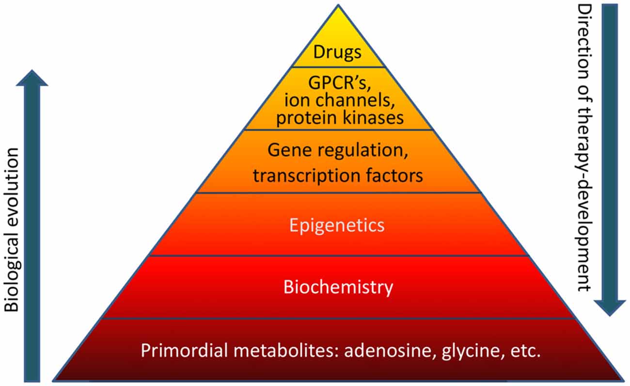
I would have liked the reviewer to further address the “therapeutic reconstruction of the epigenome” point he made in the Abstract:
“New findings based on biochemical manipulation of the DNA methylome suggest that:
- Epigenetic mechanisms play a functional role in epileptogenesis; and
- Therapeutic reconstruction of the epigenome is an effective antiepileptogenic therapy.”
As it was, the reviewer lapsed into the prevalent belief that the causes of and cures for human diseases will always be found on the molecular level – for example, the base of the above pyramid – and never in human experiences. This preconception leads to discounting human elements – notably absent in the above pyramid – that generate epigenetic changes.
A consequence of ignoring experiential causes of diseases is that the potential of experiential therapies to effect “therapeutic reconstruction of the epigenome” isn’t investigated.
http://journal.frontiersin.org/article/10.3389/fnmol.2016.00026/full “The Biochemistry and Epigenetics of Epilepsy: Focus on Adenosine and Glycine”
Contending with epigenetic consequences of violence to women
This 2016 UK review subject was the interplay of genomic imprinting and intergenerational epigenetic information transfer:
“A range of evolutionary adaptations associated with placentation transfers disproportionate control of this process to the matriline, a period unique in mammalian development in that there are three matrilineal genomes interacting in the same organism at the same time (maternal, foetal, and postmeiotic oocytes).
Genomic imprinting is absent in egg laying mammals and only around 6 imprinted genes have been detected in a range of marsupial species; this is in contrast to eutherian mammals where around 150 imprinted genes have been described.
The interactions between the maternal and developing foetal hypothalamus and placenta can provide a template by which a mother can transmit potentially adaptive information concerning potential future environmental conditions to the developing brain.
In circumstances either where the early environment provides inaccurate cues to the environmental conditions prevailing when adult due to rapid environmental change or when disruptions to normal neural development occur, the mismatch between the environmental predictions made during early development and subsequent reality may mean that an organism may have a poorly adapted phenotype to its adult environment. An appreciation of these underlying evolutionary salient processes may provide a novel perspective on the [causal] mechanisms of a range of health problems.
The concept of a brain that is not pathological in the classical sense but it is simply mismatched to its environment has been most extensively studied in the context of ancestral and early developmental nutrition. However, this concept can be extended to provide insights into the development of a range of alternative neural phenotypes.”
The review’s final sentence was:
“Examination of the adaptive potential of a range of neural and cognitive deficits in the context of evolutionary derived foetocentric brain and placental development, epigenetics and environmental adaptation may provide novel insights into the development and potential treatment of a range of health, neurological, and cognitive disorders.”
One of the reviewers was cited in Epigenetic DNA methylation and demethylation with the developing fetus, which the review cited along with Epigenetic changes in the developing brain change behavior.
Researchers who avoid hypotheses that can’t be proven wrong could certainly test the subject matter of this review if they investigated their subjects’ histories.
For example, let’s say a patient/subject had symptoms where the “150 imprinted genes” were implicated. What are the chances a clinician or researcher would be informed by this review’s material and investigate the mother’s and grandmother’s histories?
For clinicians or researchers who view histories as irrelevant busywork: How many tens of millions of people alive today have mothers who were fetuses when their grandmothers were adversely affected by violence? Wouldn’t it be appropriate to assess possible historical contributions of:
“The mismatch between the environmental predictions made during early development and subsequent reality”
to the patient’s/subject’s current symptoms?
http://www.hindawi.com/journals/np/2016/6827135/ “Placental, Matrilineal, and Epigenetic Mechanisms Promoting Environmentally Adaptive Development of the Mammalian Brain”
A one-sided review of stress
The subject of this 2016 Italian/New York review was the stress response:
“The stress response, involving the activation of the hypothalamic-pituitary-adrenocortical [HPA] axis and the consequent release of corticosteroid hormones, is indeed aimed at promoting metabolic, functional, and behavioral adaptations. However, behavioral stress is also associated with fast and long-lasting neurochemical, structural, and behavioral changes, leading to long-term remodeling of glutamate transmission, and increased susceptibility to neuropsychiatric disorders.
Of note, early-life events, both in utero and during the early postnatal life, trigger reprogramming of the stress response, which is often associated with loss of stress resilience and ensuing neurobehavioral (mal)adaptations.”
The reviewers’ intentional dismissal of the role of GABA in favor of the role of glutamate was a key point:
“The changes in neuronal excitability and synaptic plasticity induced by stress are the result of an imbalance of excitatory (glutamatergic) and inhibitory (GABAergic) transmission, leading to long-lasting (mal)adaptive functional modifications. Although both glutamate and GABA transmission are critically associated with stress-induced alteration of neuronal excitability, the present review will focus on the modulation of glutamate release and transmission induced by stress and glucocorticoids.”
No particular reason was given for this bias. I inferred from the review’s final sentence that the review’s sponsors and funding prompted this decision:
“In-depth studies of changes in glutamate transmission and dendrite remodeling induced by stress in early and late life will help to elucidate the biological underpinnings of the (mal)adaptive strategies the brain adopts to cope with environmental challenges in one’s life.”
The bias led to ignoring evidence for areas the reviewers posed as needing further research. An example of relevant research the reviewers failed to consider was the 2015 Northwestern University study I curated in A study that provided evidence for basic principles of Primal Therapy that found:
“In response to traumatic stress, some individuals, instead of activating the glutamate system to store memories, activate the extra-synaptic GABA system and form inaccessible traumatic memories.”
http://www.ncbi.nlm.nih.gov/pmc/articles/PMC4812483/ “Stress Response and Perinatal Reprogramming: Unraveling (Mal)adaptive Strategies”
Observing pain in others had long-lasting brain effects
This 2016 Israeli human study used whole-head magnetoencephalography (MEG) to study pain perception in military veterans:
“Our findings demonstrate alterations in pain perception following extreme pain exposure, chart the sequence from automatic to evaluative pain processing, and emphasize the importance of considering past experiences in studying the neural response to others’ states.
Differences in brain activation to ‘pain’ and ‘no pain’ in the PCC [posterior cingulate cortex] emerged only among controls. This suggests that prior exposure to extreme pain alters the typical brain response to pain by blurring the distinction between painful and otherwise identical but nonpainful stimuli, and that this blurring of the ‘pain effect’ stems from increased responses to ‘no pain’ rather than from attenuated response to pain.”
Limitations included:
- “The pain-exposed participants showed posttraumatic symptoms, which may also be related to the observed alterations in the brain response to pain.
- We did not include pain threshold measurements. However, the participants’ sensitivity to experienced pain may have had an effect on the processing of observed pain.
- The regions of interest for the examination of pain processing in the pain-exposed group were defined on the basis of the results identified in the control group.
- We did not detect pain-related activations in additional regions typically associated with pain perception, such as the anterior insula and ACC. This may be related to differences between the MEG and fMRI neuroimaging approaches.”
The subjects self-administered oxytocin or placebo per the study’s design. However:
“We chose to focus on the placebo condition and to test group differences at baseline only, in light of the recent criticism on underpowered oxytocin administration studies, and thus all following analyses are reported for the placebo condition.”
A few questions:
- If observing others’ pain caused “increased responses to ‘no pain’,” wouldn’t the same effect or more be expected from experiencing one’s own pain?
- If there’s evidence for item 1, then why aren’t “increased responses to ‘no pain'” of affected people overtly evident in everyday life?
- If item 2 is often observed, then what are the neurobiological consequences for affected people’s suppression of “increased responses to ‘no pain’?”
- Along with the effects of item 3, what may be behavioral, emotional, and other evidence of this suppressed pain effect?
- What would it take for affected people to regain a normal processing of others’ “‘pain’ and ‘no pain’?”
https://www.researchgate.net/publication/299546838_Prior_exposure_to_extreme_pain_alters_neural_response_to_pain_in_others “Prior exposure to extreme pain alters neural response to pain in others” Thanks to one of the authors, Ruth Feldman, for providing the full study
Epigenetic contributions to hypertension
This 2016 Australian review subject was epigenetic contributions to hypertension:
“Hypertension (HT) affects more than 1 billion people globally and is a major risk factor for stroke, chronic kidney disease, and myocardial infarction.
Essential hypertension (EH) is a complex, polygenic condition with no single causative agent. There is increasing evidence that epigenetic modifications are as important as genetic predisposition in the development of EH.
Many epigenetic studies are, however, limited by the fact that only blood is studied rather than the effector tissues. The utility of blood methylation status in epigenetic research is yet to be determined. Furthermore, the polygenic complexity of HT and the limited knowledge on some of the non-coding RNAs makes it more challenging to decipher the exact mechanisms involved.”
The review had sections for hypertension studies on DNA methylation, histone modification, and microRNA/other non-coding RNA types. Here’s a sample of the findings:
“HSD11B2-mediated degradation of cortisol to cortisone is disrupted when the promoter region of the HSD11B2 gene is hypermethylated. The resulting imbalance in the active metabolites of cortisol and cortisone, tetrahydrocortisol, and tetrahydocortisone, respectively, promotes the onset of HT.
Histone modification affecting arterial pressure levels has been documented in a variety of human and animal tissues, including vascular smooth muscle. Vascular oxidative stress can contribute to endothelial dysfunction—a hallmark of HT—and the development of HT.
Two miRNAs (has-miR-181a and has-miR-663) with the ability to bind to the 3′ UTR of renin mRNA were found to be under-expressed in EH. These miRNAs were able to regulate the expression of a reporter gene and renin-mRNA itself, which explains over-expression of renin mRNA seen in EH kidney.”
The publisher, International Journal of Molecular Sciences, makes ALL of its articles open access. Another of its requirements is:
“The full experimental details must be provided so that the results can be reproduced.”
There also aren’t artificial limitations on either the length of the study or the number of supplementary files.
http://www.mdpi.com/1422-0067/17/4/451/htm “Epigenetic Modifications in Essential Hypertension”
Oxytocin research null findings come out of the file drawer
In 2016 Belgian researchers released their previously unpublished studies:
“Is there a file drawer problem in intranasal oxytocin research?
We submitted several studies yielding null-findings to different journals but they were rejected time and time again.
The aggregated effect size was not reliably different from zero [including all of the researchers’ previously unpublished intranasal oxytocin studies].”
Neuroskeptic comments:
“By publishing these results, Lane et al. have ensured that future meta-analysts will be able to include the full dataset in their calculations.”
http://blogs.discovermagazine.com/neuroskeptic/2016/03/17/open-the-file-drawer/ “Psychologists Throw Open the File Drawer”
See Testing the null hypothesis of oxytocin’s effects in humans for more on the topic.
Problematic research into epigenetic effects of paternal stress on male offspring
This 2016 Chinese rodent study and its accompanying commentary Don’t stress dad — it’s bad for your kids’ health were caught up in an agenda.
The first problem I noticed was that the hyperglycemic effects found only in the male offspring weren’t consistently labelled as sex-specific. Try to find that fact in the paywalled commentary with its intentionally misleading headline, or in the news coverage with headlines such as “Stressed mouse dads give their offspring high blood sugar.”
That the effects were male-only was briefly noted in the study, yet “male” was absent from the “stress-F1 mice” label used after the initial mention.

The researchers provided no mechanisms that plausibly linked the effects to offspring sex. There was plenty of time between the May 3, 2015 submission and the February 18, 2016 publication to clarify this and other items. I wonder what the reviewer noted.
The second problem was that the highest number of male “stress-F1 mice” tested was only six. I didn’t see any disclosures of what led to the scarcity of subjects, or of the likely impact of using so few.
A related limitation was that the male “stress-F1 mice” were killed as young adults. Whether or not the hyperglycemic effects carried through to old age or to another generation wasn’t determined.
I’m leery of studies like this one that didn’t have a Limitations section, and especially so when the news coverage overlooked obvious limitations. It was difficult to place the findings in a context other than promoting that a male’s stress may also adversely affect their offspring.
One of the problems that research caught up in an agenda create is that non-headline findings are overlooked. Other than sex-specific effects, the study found that the putative preconception cause of hyperglycemia didn’t cause other symptoms:
- “No significant growth defects were observed in male offspring from stress-F0 fathers (stress-F1 mice) during their early lives.
- Insulin sensitivity was not changed in stress-F1 mice.
- Serum glucagon, leptin, and pro-inflammatory cytokines (tumor necrosis factor α [TNFα], interleukin-6 [IL-6]) were unaffected.
- Body weight, food intake, locomotor activity, CO2 production, O2 consumption, and respiratory exchange ratios also remained unchanged.
- Liver weight, liver weight/body weight ratios, hepatic triglyceride content, and the histological phenotypes were also comparable.
- The methylation pattern and expression of microRNAs were not affected in the fetal brains of stress-F1 mice.”
The handling of the study reminded me of Transgenerational epigenetic programming with stress and microRNA where most of the news coverage similarly focused on it being a male’s stress, not a female’s, that affected the developing embryo. The important part lost from news coverage of that study was it demonstrated how a damaging influence can begin immediately after conception, but the symptoms didn’t present until adulthood!
http://www.sciencedirect.com/science/article/pii/S1550413116300067 “Paternal Psychological Stress Reprograms Hepatic Gluconeogenesis in Offspring”
The current paradigm of child abuse limits pre-childhood causal research
As an adult, what would be your primary concern if you suspected that your early life had something to do with current problems? Would you be interested in effective treatments for causes of your symptoms?
Such information wasn’t available in this 2016 Miami review of the effects of child abuse. The review laid out the current paradigm mentioned in Grokking an Adverse Childhood Experiences (ACE) score, one that limits research into pre-childhood causes for later-life symptoms.
The review’s goal was to describe:
“How numerous clinical and basic studies have contributed to establish the now widely accepted idea that adverse early life experiences can elicit profound effects on the development and function of the nervous system.”
The hidden assumptions of almost all of the cited references were that these distant causes could no longer be addressed. Aren’t such assumptions testable today?
As an example, the Discussion section posed the top nine “most pressing unanswered questions related to the neurobiological effects of early life trauma.” In line with the current paradigm, the reviewer assigned “Are the biological consequences of ELS [early life stress] reversible?” into the sixth position.
If the current paradigm encouraged research into treatment of causes, there would probably already be plenty of evidence to demonstrate that directly reducing the source of damage would also reverse damaging effects. There would have been enough studies done so that the generalized question of reversibility wouldn’t be asked.
Aren’t people interested in treatments of originating causes so that their various symptoms don’t keep bubbling up? Why wouldn’t research paradigms be aligned accordingly?
The review also demonstrated how the current paradigm of child abuse misrepresented items like telomere length and oxytocin. Researchers on the bandwagon tend to forget about the principle Einstein expressed as:
“No amount of experimentation can ever prove me right; a single experiment can prove me wrong.”
That single experiment for telomere length arrived in 2016 with Using an epigenetic clock to distinguish cellular aging from senescence. The review’s seven citations for telomere length that all had findings “associated with” or “linked to” child abuse should now be viewed in a different light.
The same light shone on oxytocin with Testing the null hypothesis of oxytocin’s effects in humans and Oxytocin research null findings come out of the file drawer. See their references, and decide for yourself whether or not:
“Claimed research findings may often be simply accurate measures of the prevailing bias.”
http://www.cell.com/neuron/fulltext/S0896-6273%2816%2900020-9 “Paradise Lost: The Neurobiological and Clinical Consequences of Child Abuse and Neglect”
This post has somehow become a target for spammers, and I’ve disabled comments. Readers can comment on other posts and indicate that they want their comment to apply here, and I’ll re-enable comments.
Epigenetic regulation of natural killer cells
This 2016 German review focused on how epigenetic processes affected the natural killer cell part of the immune system:
“Natural killer (NK) cells recognize and eliminate tumor- and virus-infected cells, parasites as well as certain types of bacteria. NK cell activity is related to a complex interaction of activating and inhibiting receptors on the NK cell surface.
During the development of HPCs [hemopoietic progenitor cells] to mature NK cells, the DNA demethylation of KIR [killer cell immunoglobin-like receptors] genes leads to KIR expression. But DNA methylation does not just determine which KIR gene is expressed, it also determines which allele expresses the KIR gene. KIR genes are also regulated by microRNA.
KIR genes exhibit highly similar histone acetylation signatures, which are typically found in expressed genes. This fact puts the KIR genes into a state of readiness for transcription which is depending on the DNA methylation as critical epigenetic modification in the regulation of KIR gene expression.
Epigenetic modifications have been reported to be involved in the expression of NKG2D, which is one the most important activating NK cell receptor.”
The reviewers included a section on NK cell activity and external stimuli. They summarized:
“The significance of the described findings is limited by study designs. Although human NK cells were frequently used, in most cases treatment took place in ex vivo experiments.”
The reviewers also provided a good three-paragraph explanation of general epigenetic mechanisms.
http://www.mdpi.com/1422-0067/17/3/326/htm “Natural Killer Cells—An Epigenetic Perspective of Development and Regulation”
Beneficial epigenetic effects of mild stress with social support during puberty
This 2016 Pennsylvania rodent study found:
“Stress in the context of social support experienced over the pubertal window can promote epigenetic reprogramming in the brain to increase resilience to age-related cognitive decline in females.
These findings are actually consistent with previous studies showing that some amount of adversity, or adversity under more favorable circumstances such as social support or a protective gene polymorphism, provides a measure of ‘grit’ in coping with later life challenges.
Our findings provide a unique perspective on this relationship, as they highlight the important link between experience during the pubertal window and cognitive health during aging.”
These researchers made efforts to further investigate causes of unexpected results, such as:
“Peripubertal stress alone did not significantly alter Barnes maze performance in aging compared to aged Controls. Mice that had experienced stress with concurrent social support (CVS + SI) actually performed better than Control aged mice, specifically in learning the reversal task faster.
Peripubertal stress had no effect on corticosterone levels in response to an acute restraint stress or in sensorimotor gating and baseline startle reactivity.”
Their investigations led to epigenetic findings:
“Consistent with our behavioral findings, stress in the context of social interaction resulted in long-term reprogramming of gene expression in the PFC [prefrontal cortex]. While there were no differentially expressed genes between Control and CVS females, there were 88 genes that were significantly different between Control and CVS + SI groups. Of genes that were downregulated, a large portion (23 genes; 35%) were microRNAs.
We found that the PFC transcriptome of CVS + SI aged females was significantly enriched for predicted targets of the 23 microRNAs that were downregulated in the PFC in these mice. This suggests that microRNAs represent a mode of regulation capable of enacting far-reaching programmatic effects, and are a critical epigenetic gene expression regulatory mechanism.”
Applicability to humans was suggested by associations such as:
“A single microRNA can target more than a hundred different mRNA targets, and more than 45,000 conserved microRNA binding sites have been annotated in the 3′ UTR of 60% of human genes.”
A few limitations were noted:
“Given that mice at this age (1 year) are commonly compared to ‘late middle aged’ humans, later aging time points may yield differences in this group. Alternatively, it is possible that there was an effect of peripubertal stress that was not long-lasting due to the mild nature of our chronic stress model.
To include early neglect as a part of the stressor experience, CVS females were weaned one week earlier (PN21) than Control and CVS + SI mice. Addition of stress of this earlier weaning likely poses a significant contribution to programming of the PFC.”
One of the study coauthors was also a coauthor of:
- Transgenerational epigenetic programming with stress and microRNA
- How to make a child less capable even before they are born: stress the pregnant mother-to-be
https://www.ncbi.nlm.nih.gov/pmc/articles/PMC4870871/ “Peripubertal stress with social support promotes resilience in the face of aging”
A review that inadvertently showed how memory paradigms prevented relevant research
This 2016 Swiss review of enduring memories demonstrated what happens when scientists’ reputations and paychecks interfered with them recognizing new research and evidence in their area but outside their paradigm: “A framework containing the basic assumptions, ways of thinking, and methodology that are commonly accepted by members of a scientific community.”
A. Most of the cited references were from decades ago that established these paradigms of enduring memories. Fine, but the research these paradigms excluded was also significant.
B. All of the newer references were continuations of established paradigms. For example, a 2014 study led by one of the reviewers found:
“Successful reconsolidation-updating paradigms for recent memories fail to attenuate remote (i.e., month-old) ones.
Recalling remote memories fails to induce histone acetylation-mediated plasticity.”
The researchers elected to pursue a workaround of the memory reconsolidation paradigm when the need for a new paradigm of enduring memories directly confronted them!
C. None of the reviewers’ calls for further investigations challenged existing paradigms. For example, when the reviewers suggested research into epigenetic regulation of enduring memories, they somehow found it best to return to 1984, a time when dedicated epigenetics research had barely begun:
“Whether memories might indeed be ‘coded in particular stretches of chromosomal DNA’ as originally proposed by Crick [in 1984] and if so what the enzymatic machinery behind such changes might be remain unclear. In this regard, cell population-specific studies are highly warranted.”
Two examples of relevant research the review failed to consider:
1. A study that provided evidence for basic principles of Primal Therapy went outside existing paradigms to research state-dependent memories:
“If a traumatic event occurs when these extra-synaptic GABA receptors are activated, the memory of this event cannot be accessed unless these receptors are activated once again.
It’s an entirely different system even at the genetic and molecular level than the one that encodes normal memories.”
What impressed me about that study was the obvious nature of its straightforward experimental methods. Why hadn’t other researchers used the same methods decades ago? Doing so could have resulted in dozens of informative follow-on study variations by now, which is my point in Item A. above.
2. A relevant but ignored 2015 French study What can cause memories that are accessible only when returning to the original brain state? which supported state-dependent memories:
“Posttraining/postreactivation treatments induce an internal state, which becomes encoded with the memory, and should be present at the time of testing to ensure a successful retrieval.”
The review also showed the extent to which historical memory paradigms depend on the subjects’ emotional memories. When it comes to human studies, though, designs almost always avoid studying emotional memories.
It’s clearly past time to Advance science by including emotion in research.
http://www.hindawi.com/journals/np/2016/3425908/ “Structural, Synaptic, and Epigenetic Dynamics of Enduring Memories”
