What drives cellular aging?

This 2019 US/UK human cell study by the founder of the epigenetic clock method investigated epigenetic aging:

“It is widely assumed that extension of lifespan is a result of retardation of ageing. While there is no counter-evidence to challenge this highly intuitive association, supporting empirical evidence to confirm it is not easy to acquire.

The scarcity of empirical evidence is due in part to the lack of a good measure of age that is not based on time. In this regard, the relatively recent development of epigenetic clocks is of great interest.

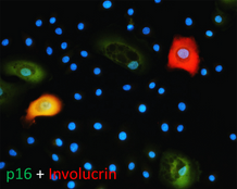
At the cellular level more is known, but from the perspective of what epigenetic ageing is not, rather than what it is. While we still do not know what cellular feature is associated with epigenetic ageing, we can now remove:

  • somatic cell differentiation

from the list of possibilities and place it with

  • cellular senescence,
  • proliferation and
  • telomere length maintenance,

which represent cellular features that are all not linked to epigenetic ageing.”


The study used several agents, including rapamycin, to investigate the hypotheses. Rapamycin isn’t a panacea, however:

“The ability of rapamycin to suppress the progression of epigenetic ageing is very encouraging for many reasons not least because it provides a valuable point-of-entry into molecular pathways that are potentially associated with it. Evidently, the target of rapamycin, the mTOR complex is of particular interest.

The convergence of the GWAS observation with the experimental system described here is a testament of the strength of the skin & blood clock in uncovering biological features that are consistent between the human level and cellular level. It lends weight to the emerging view that the mTOR pathway may be the underlying mechanism that supports epigenetic ageing.”

The limitation section ended with:

“It is important to note that it is inadvisable (actively discouraged) to directly extrapolate the studies here, especially in terms of the magnitude of age suppression, to potential effects of rapamycin on humans.”

https://www.aging-us.com/article/101976/text “Rapamycin retards epigenetic ageing of keratinocytes independently of its effects on replicative senescence, proliferation and differentiation”

Statistical inferences vs. biological realities

A 2019 UCLA study introduced a derivative of the epigenetic clock named GrimAge:

“DNAm GrimAge, a linear combination of chronological age, sex, and DNAm-based surrogate biomarkers for seven plasma proteins and smoking pack-years, outperforms all other DNAm-based biomarkers, on a variety of health-related metrics.

An age-adjusted version of DNAm GrimAge, which can be regarded as a new measure of epigenetic age acceleration (AgeAccelGrim), is associated with a host of age-related conditions, lifestyle factors, and clinical biomarkers. Using large scale validation data from three ethnic groups, we demonstrate that AgeAccelGrim stands out among pre-existing epigenetic clocks in terms of its predictive ability for time-to-death, time-to-coronary heart disease, time-to-cancer, its association with computed tomography data for fatty liver/excess fat, and early age at menopause.”

https://www.ncbi.nlm.nih.gov/pmc/articles/PMC6366976/ “DNA methylation GrimAge strongly predicts lifespan and healthspan”


A miserable attempt at reporting the study’s findings included angles of superstition, fear-of-the-future, and suspicion-by-spurious-association:

“The research has already captured the attention of the life insurance industry. After all, a solid death date could mean real savings when it comes to pricing policies.

The hope is that if and when legitimate anti-aging drugs are developed, GrimAge could be used to test their effectiveness. In a world with functional anti-aging drugs, “doctors could test [your GrimAge number] and say, ‘You know what, you’re aging too quickly. Take this,'” Horvath said.”

https://onezero.medium.com/a-new-test-predicts-when-youll-die-give-or-take-a-few-years-2d08147c8ea6 “A New Test Predicts When You’ll Die (Give or Take a Few Years)”


A detailed blog post from Josh Mitteldorf provided scientific coverage of the study:

“Methylation sites associated with smoking history predicted how long the person would live more accurately than the smoking history itself. Even stranger, the methylation marks most closely associated with smoking were found to be a powerful indication of future health even when the sample was confined to non-smokers.

The DNAm GrimAge clock was developed in two stages, a correlation of a correlation. Curiously, the indirect computation yields the better result.

Horvath’s finding that secondary methylation indicators are more accurate than the underlying primary indicator from which they were derived is provocative, and calls out for a new understanding.”

https://joshmitteldorf.scienceblog.com/2019/03/05/dnam-grimage-the-newest-methylation-clock “DNAm GrimAge—the Newest Methylation Clock”


When there are logical disconnects in findings like the above, it’s time to examine underlying premises. As noted in Group statistics don’t necessarily describe an individual, an assumption required by statistical analyses is that each measured item in the sample is interchangeable with the next.

This presumption is often false, producing individually inapplicable results. For example, Immune memory vs. immune adaptation included this description of the adaptive immune system:

“To be effective, highly specific immune response requires huge diversity of receptors and antibodies, which is achieved by somatic rearrangement of gene segments. Recombination results in millions of TCR [T cell receptor] and antibody variants able to recognize and neutralize millions of various antigens.”

Standard statistics of millions of T cell receptor and antibody variants won’t represent their individually unique properties. But individual differences are both their purpose and benefit to us.

The GrimAge study’s overreach was most apparent in stratifying educational attainment to develop correlations. As mentioned in Does a societal mandate cause DNA methylation? such statistics are poor evidence of each individual’s biological realities.

Neither derivatives of group statistics, nor correlations of correlations, seem to be the techniques needed to understand biological causes of effects. Another commentary on the GrimAge study mentioned but glossed over this point:

“It remains a mystery why exactly the epigenetic clocks work, and whether age-related changes in DNA methylation contribute to the cause of aging or are a result of it.”

Immune memory vs. immune adaptation

This 2019 Dutch/German/Romanian perspective aimed for a better understanding of immune systems:

“Based on molecular, immunological, and evolutionary arguments, we propose that innate immune memory is a primitive form of immune memory present in all living organisms, while adaptive immune memory is an advanced form of immune memory representing an evolutionary innovation in vertebrates.

Innate immune responses have the capacity to be trained, and thereby exert a new type of immunological memory upon reinfection. The central feature of trained innate immune cells is their ability to mount a qualitatively and quantitatively different transcriptional response when challenged with microbes or danger signals. Evidence supports convergence of multiple regulatory layers for mediating innate immune memory, including changes in chromatin organization, DNA methylation, and probably non-coding RNAs such as microRNAs and/or long non-coding RNAs.

Two properties of adaptive immune response are mediated by two fundamentally different types of mechanisms:

  1. Higher magnitude and speed of the response is mediated by epigenetic programming.
  2. Specificity of the response is insured by gene recombination of TCR [T cell receptor] and BCR [B cell receptor] and clonal expansion of specific cell subpopulations upon antigen recognition.

To be effective, highly specific immune response requires huge diversity of receptors and antibodies, which is achieved by somatic rearrangement of gene segments. Recombination results in millions of TCR and antibody variants able to recognize and neutralize millions of various antigens.


This paper included speculations such as “Evidence supports..probably non-coding RNAs” quoted above, and the penultimate sentence:

“One can envision that vaccines that are capable of inducing both forms of immune memory at the same time would be more effective.”

100% factual evidence is preferred. Overall information can only be as accurate as the least accurate information.

This review highlighted a goal for humans to have both a functional innate immune system and a functional adaptive immune system. The lead author coauthored A dietary supplement that trains the innate immune system and a study referenced in Eat your oats.

https://www.sciencedirect.com/science/article/pii/S1931312818306334 “Innate and Adaptive Immune Memory: an Evolutionary Continuum in the Host’s Response to Pathogens” (not freely available)

A therapy to reverse cognitive decline

This 2018 human study presented the results of 100 patients’ personalized therapies for cognitive decline:

“The first examples of reversal of cognitive decline in Alzheimer’s disease and the pre-Alzheimer’s disease conditions MCI (Mild Cognitive Impairment) and SCI (Subjective Cognitive Impairment) have recently been published..showing sustained subjective and objective improvement in cognition, using a comprehensive, precision medicine approach that involves determining the potential contributors to the cognitive decline (e.g., activation of the innate immune system by pathogens or intestinal permeability, reduction in trophic or hormonal support, specific toxin exposure, or other contributors), using a computer-based algorithm to determine subtype and then addressing each contributor using a personalized, targeted, multi-factorial approach dubbed ReCODE for reversal of cognitive decline.

An obvious criticism of the initial studies is the small number of patients reported. Therefore, we report here 100 patients, treated by several different physicians, with documented improvement in cognition, in some cases with documentation of improvement in electrophysiology or imaging, as well.”

https://www.omicsonline.org/open-access/reversal-of-cognitive-decline-100-patients-2161-0460-1000450-105387.html “Reversal of Cognitive Decline: 100 Patients”


The lead author commented on Josh Mitteldorf’s informative post A cure for Alzheimer’s? Yes, a cure for Alzheimer’s!:

  1. “We have a paper in press, due to appear 10.22.18 (open access, JADP, I’ll send a copy as soon as available), showing 100 patients with documented improvement – some with MRI volumetrics improved, others with quantitative EEG improvements, others with evoked response improvements, and all with quantitative cognitive assessment improvement. Some are very striking – 12 point improvements in MoCA [Montreal Cognitive Assessment], for example – others less so, but all also have subjective improvement. Hopefully this will address some of the criticisms that we haven’t documented improvement in enough people.
  2. We were just turned down again for a randomized, controlled clinical trial, so on the one hand, we are told repeatedly that no one will believe that this approach works until we publish a randomized, controlled study, and on the other hand, we’ve been turned down (first in 2011/12, and now in 2018), with the complaint that we are trying to address more than one variable in the trial (as if AD is a single-variable disease!). Something of a catch-22. We are now resubmitting (unfortunately, the IRBs are not populated by functional medicine physicians, so they are used to seeing old-fashioned drug studies), and we’ll see what happens.
  3. I’ve been extending the studies to other neurodegenerative diseases, and it has been impressive how much of a programmatic response there seems to be in these ‘diseases.’
  4. I agree with you that there are many features in common with aging itself.
  5. You made a good point that APP [amyloid precursor protein] is a dependence receptor, and in fact it functions as an integrating dependence receptor, responding to numerous inputs (Kurakin and Bredesen, 2015).
  6. In the book and the publications, we don’t claim it is a “cure” since we don’t have pathological evidence that the disease process is gone. What we claim is ‘reversal of cognitive decline’ since that is what we document.
  7. As I mentioned in the book, AD is turning out to be a protective response to multiple insults, and this fits well with the finding that Abeta has an antimicrobial effect (Moir and Tanzi’s work). It is a network-downsizing, protective response, which is quite effective – some people live with the ongoing degenerative process for decades.
  8. We have seen several cases now in which a clinical trial of an anti-amyloid antibody made the person much worse in a time-dependent manner (each time there was an injection, the person would get much worse for 5-10 days, then begin to improve back toward where he/she was, but over time, marked decline occurred), and this makes sense for the idea that the amyloid is actually protecting against pathogens or toxins or some other insult.
  9. It is important to note that we’ve never claimed that all people get better – this is not what we’ve seen. People very late in the process, or who don’t follow the protocol, or who don’t address the various insults, do not improve. It is also turning out to be practitioner dependent – some are getting the vast majority of people to improve, others very few, so this is more like surgery than old-fashioned prescriptive medicine – you have to do a somewhat complicated therapeutic algorithm and get it right for best results.
  10. I’m very interested in what is needed to take the next step in people who have shown improvement but who started late in the course. For example, we have people now who have increased MoCA from 0 to 9 (or 0 to 3, etc.), with marked subjective improvement but plateauing at less than normal. These people had extensive synaptic and cellular loss prior to the program. So what do we need to raise the plateau? Stem cells? Intranasal trophic support? Something else?
  11. I haven’t yet seen a mono-etiologic theory of AD or a mono-therapeutic approach that has repeatedly positive results, so although I understand that there are many theories and treatments, there doesn’t seem to be one etiology to the disease, nor does there seem to be one simple treatment that works for most. It is much more like a network failure.”

At a specific level:

  • “There doesn’t seem to be one etiology to the disease,
  • Nor does there seem to be one simple treatment that works for most.
  • We don’t have pathological evidence that the disease process is gone.”

For general concepts, however:

  • “AD is turning out to be a protective response to multiple insults.
  • It is a network-downsizing, protective response, which is quite effective.
  • The amyloid is actually protecting against pathogens or toxins or some other insult.”

For a framework of an AD cure to be valid, each source of each insult that evoked each “protective response” should be traced.

Longitudinal studies would be preferred inside this framework. These study designs would investigate evidence of each insult’s potential modifying effect on each “protective response” that could affect the cumulative disease trajectory of each individual.

In many cases, existing study designs would be adequate if they extended their periods to the end of the subjects’ natural lifetimes. One AD-relevant example would be extending the prenatally-restraint-stressed model used in:

The framework would also encourage extending studies to at least three generations to investigate evidence for transgenerational effects, as were found in:

Disproving the cholesterol paradigm

This 2018 review presented evidence that:

“For half a century, a high level of total cholesterol (TC) or low-density lipoprotein cholesterol (LDL-C) has been considered to be the major cause of atherosclerosis and cardiovascular disease (CVD), and statin treatment has been widely promoted for cardiovascular prevention. However, there is an increasing understanding that the mechanisms are more complicated and that statin treatment, in particular when used as primary prevention, is of doubtful benefit.

The authors of three large reviews recently published by statin advocates have attempted to validate the current dogma. This article delineates the serious errors in these three reviews as well as other obvious falsifications of the cholesterol hypothesis.

Our search for falsifications of the cholesterol hypothesis confirms that it is unable to satisfy any of the Bradford Hill criteria for causality and that the conclusions of the authors of the three reviews are based on:

  • Misleading statistics,
  • Exclusion of unsuccessful trials and by
  • Ignoring numerous contradictory observations.

The association between the absolute risk reduction of total mortality in 26 statin trials [squares] included in the study by Silverman et al. and in 11 ignored trials [triangles] and the year where the trial protocols were published. The vertical line indicates the year where the new trial regulations were introduced.

In 2004–2005, health authorities in Europe and the United States introduced New Clinical Trial Regulations, which specified that all trial data had to be made public. Since 2005, claims of benefit from statin trials have virtually disappeared.”


This paradigm was proven wrong eighty years ago! How much longer will its harmful consequences continue?

https://www.tandfonline.com/doi/full/10.1080/17512433.2018.1519391 “LDL-C does not cause cardiovascular disease: a comprehensive review of the current literature”

Eat your oats!

Here’s some motivation to replenish your oats supply.

From a 2013 Canadian human review:

“Review of human studies investigating the post-prandial blood-glucose lowering ability of oat and barley food products” https://www.nature.com/articles/ejcn201325

“Change in glycaemic response (expressed as incremental area under the post-prandial blood-glucose curve) was greater for intact grains than for processed foods. For processed foods, glycaemic response was more strongly related to the β-glucan dose alone than to the ratio of β-glucan to the available carbohydrate.”

The review found that people don’t have to eat a lot of carbohydrates to get the glycemic-response benefits of β-glucan. Also, eating ~3 grams of β-glucan in whole oats and barley will deliver the same glycemic-response benefits as eating ~4 grams of β-glucan in processed oats and barley.

However, the glycemic index used in the review is a very flawed measure. What’s the point of indexing healthy choices like whole grains to unhealthy choices that healthy people aren’t going to make anyway?


The reviewer somewhat redeemed herself by participating in a 2018 review:

“Processing of oat: the impact on oat’s cholesterol lowering effect” https://www.ncbi.nlm.nih.gov/pmc/articles/PMC5885279/

“For a similar dose of β-glucan:

  1. Liquid oat-based foods seem to give more consistent, but moderate reductions in cholesterol than semi-solid or solid foods where the results are more variable;
  2. The quantity of β-glucan and the molecular weight at expected consumption levels (∼3 g day) play a role in cholesterol reduction; and
  3. Unrefined β-glucan-rich oat-based foods (where some of the plant tissue remains intact) often appear more efficient at lowering cholesterol than purified β-glucan added as an ingredient.”

The review’s sections 3. Degree of processing and functionality and 4. Synergistic action of oat constituents were informative:

“Both in vitro and in vivo studies clearly demonstrated the beneficial effect of oat on cholesterolemia, which is unlikely to be due exclusively to β-glucan, but rather to a combined and synergetic action of several oat compounds acting together to reduce blood cholesterol levels.”


Another use of β-glucan is to improve immune response. Here’s a 2016 Netherlands study where the researchers used β-glucan to get a dozen people well after making them sick with lipopolysaccharide as is often done in animal studies:

β-Glucan Reverses the Epigenetic State of LPS-Induced Immunological Tolerance” https://www.ncbi.nlm.nih.gov/pmc/articles/PMC5927328/

“The innate immune ‘training stimulus’ β-glucan can reverse macrophage tolerance ex vivo.”

I’ve curated other research on β-glucan’s immune-response benefits in:

Epigenetic clock statistics and methods

This 2018 Chinese study was a series of statistical and methodological counter-arguments to a previous epigenetic clock study finding that:

“Only [CpG] sites mapping to the ELOVL2 promoter constitute cell and tissue-type independent aDMPs [age-associated differentially methylated positions].”

The study used external data sets and the newer epigenetic clock’s fibroblast data in its analyses to find:

“While we agree that specific sites mapping to ELOVL2 are special aDMPs in the sense that their effect sizes are particularly large across a number of different tissue-types, our analysis suggests that most aDMPs are valid across multiple different tissue types, suggesting that shared aDMPs are common.”

The details of each of the study’s counter-arguments were compelling. For example:

“We analyzed Illumina 850k data from an EWAS profiling blood, buccal and cervical samples from a common set of 263 women. Because blood is a complex mixture of many immune-cell subtypes, and buccal and cervical samples are highly contaminated by immune cells, we identified aDMPs in each tissue after adjustment for batch effects and cell-type heterogeneity.

Using either an FDR [false discovery rate] < 0.05 or Bonferroni adjusted P-value < 0.05 thresholds, the overlap of aDMPs between the 3 tissues was highly significant, mimicking the result obtained on blood cell subtypes. We observed a total of 2200 aDMPs in common between blood, buccal and cervix, an overlap which cannot be explained by random chance.”

The study’s Discussion section provided qualifications and limitations such as:

“It is important to point out that even if age-associated DNAm changes are widespread across the genome, downstream functional effects may be rare. While specific aDMPs may be shared between tissue-types, it is only in specific tissues or cell-types that any associated functional deregulation may be of biological and clinical significance.

https://www.aging-us.com/article/101666/text “Cell and tissue type independent age-associated DNA methylation changes are not rare but common”


The November 2018 issue of Aging also contained other articles of interest:

https://www.aging-us.com/article/101626/text “Accelerated DNA methylation age and the use of antihypertensive medication among older adults”

“DNAmAge and AA [age acceleration] may not be able to capture the preventive effects of AHMs [antihypertensive medications] that reduce cardiovascular risks and mortality.”

https://www.aging-us.com/article/101633/text “Azithromycin and Roxithromycin define a new family of senolytic drugs that target senescent human fibroblasts”

“Azithromycin preferentially targets senescent cells, removing approximately 97% of them with great efficiency. This represents a near 25-fold reduction in senescent cells.”

https://www.aging-us.com/article/101647/text “Disease or not, aging is easily treatable”

“Aging consists of progression from (pre)-pre-diseases (early aging) to diseases (late aging associated with functional decline). Aging is NOT a risk factor for these diseases, as aging consists of these diseases: aging and diseases are inseparable.”

The arrogance of a paradigm exceeding its evidence

This 2018 commentary from the American College of Emergency Physicians by 7 physicians discussed the harm that will result from imposing a mandatory paradigm of sepsis treatment. I’ll quote sections that mention evidence:

“These metrics [for pneumonia treatment] had little evidentiary basis but led to an institutional-fostered culture of overdiagnosis and overtreatment. Have we learned from this folly or does a new sepsis guideline promote similar time-based treatment strategies with little direct supporting evidence?

Like the pneumonia quality measure, this resource-heavy care flows from an overreaching interpretation of evidence. Despite that evidence consistently fails to find a benefit of a single treatment strategy, the Surviving Sepsis Campaign continues to promote recommendations that bypass the individual clinician’s judgment.

Although well intentioned, the current sepsis bundles and the potential penalties associated with noncompliance lay a heavy weight on ED [emergency department] care absent evidence that a net benefit will follow. The proposed Surviving Sepsis Campaign abbreviated bundle heightens the burden by further restricting the time allotted for the identification and treatment of patients with suspected sepsis, all without any evidence of benefit or knowledge of the logistic consequences or cost.”

The paradigm’s promoters didn’t learn the appropriate lessons in the above page regarding “the sense of embarrassment and regret once experienced with the pneumonia quality metric.”


What do you think are the root causes of the Surviving Sepsis Campaign’s agenda?

  • Did it start with lawyers? Lawsuits can force hospitals into actions for which the primary reason is to avoid “the potential penalties associated with noncompliance.”
  • Is it due to governments? Governments can force hospitals into actions “without any evidence of benefit or knowledge of the logistic consequences or cost” when the hospitals accept government reimbursement.
  • Did it start with other groups of unaccountable people who think they know better than everyone else about how others should act?

https://www.sciencedirect.com/science/article/pii/S0196064418306073 “The 2018 Surviving Sepsis Campaign’s Treatment Bundle: When Guidelines Outpace the Evidence Supporting Their Use” (not freely available)

Reversing epigenetic changes with CRISPR/Cas9

This 2018 Chinese review highlighted areas in which CRISPR/Cas9 technology has, is, and could be applied to rewrite epigenetic changes:

“CRISPR/Cas9-mediated epigenome editing holds a great promise for epigenetic studies and therapeutics.

It could be used to selectively modify epigenetic marks at a given locus to explore mechanisms of how targeted epigenetic alterations would affect transcription regulation and cause subsequent phenotype changes. For example, inducing histone methylation or acetylation at the Fosb locus in the mice brain reward region, nucleus accumbens, could affect relevant transcription network and thus control behavioral responses evoked by drug and stress.

Epigenome editing has the potential for epigenetic treatment, especially for the disorders with abnormal gene imprinting or epigenetic marks. Targeted epigenetic silencing or reactivation of the mutant allele could be a potential therapeutic approach for diseases such as Rett syndrome and Huntington’s disease.

Noncoding RNA plays important roles in gene imprinting and chromatin remodeling. CRISPR/Cas9 has been shown to be potential for manipulating noncoding RNA expression, including microRNA, long noncoding RNA, and miRNA families and clusters.

In vivo overexpression of the Yamanaka factors have proven to be able to fully or partially help somatic cells to regain pluripotency in situ. These rejuvenated cells would subsequently differentiate again to replace the lost cell types.”


The last paragraph was described in The epigenetic clock theory of aging as a promising technique:

“To date, the most effective in vitro intervention against epigenetic ageing is achieved through expression of Yamanaka factors, which convert somatic cells into pluripotent stem cells, thereby completely resetting the epigenetic clock.”

The reviewers cited three references for in vivo studies of this technique. Overall, I didn’t see that any of the review’s references were in vivo human studies.

https://www.ncbi.nlm.nih.gov/pmc/articles/PMC6079388/ “Novel Epigenetic Techniques Provided by the CRISPR/Cas9 System”

The epigenetic clock now includes skin

The originator of the 2013 epigenetic clock improved its coverage with this 2018 UCLA human study:

“We present a new DNA methylation-based biomarker (based on 391 CpGs) that was developed to accurately measure the age of human fibroblasts, keratinocytes, buccal cells, endothelial cells, skin and blood samples. We also observe strong age correlations in sorted neurons, glia, brain, liver, and bone samples.

The skin & blood clock outperforms widely used existing biomarkers when it comes to accurately measuring the age of an individual based on DNA extracted from skin, dermis, epidermis, blood, saliva, buccal swabs, and endothelial cells. Thus, the biomarker can also be used for forensic and biomedical applications involving human specimens.

The biomarker applies to the entire age span starting from newborns, e.g. DNAm of cord blood samples correlates with gestational week.

Furthermore, the skin & blood clock confirms the effect of lifestyle and demographic variables on epigenetic aging. Essentially it highlights a significant trend of accelerated epigenetic aging with sub-clinical indicators of poor health.

Conversely, reduced aging rate is correlated with known health-improving features such as physical exercise, fish consumption, high carotenoid levels. As with the other age predictors, the skin & blood clock is also able to predict time to death.

Collectively, these features show that while the skin & blood clock is clearly superior in its performance on skin cells, it crucially retained all the other features that are common to other existing age estimators.”

http://www.aging-us.com/article/101508/text “Epigenetic clock for skin and blood cells applied to Hutchinson Gilford Progeria Syndrome and ex vivo studies”


An introduction to the study highlighted several items:

“Although the skin-blood clock was derived from significantly less samples (~900) than Horvath’s clock (~8000 samples), it was found to more accurately predict chronological age, not only across fibroblasts and skin, but also across blood, buccal and saliva tissue. A potential factor driving this improved accuracy in blood could be related to the approximate 18-fold increase in genomic coverage afforded by using Illumina 450k/850k beadarrays.

It serves as a roadmap for future clock studies, pointing towards the importance of constructing tissue or cell-type specific epigenetic clocks, to more accurately measure biological aging in the given tissue/cell-type, and therefore with the potential to be more informative of disease-risk or the success of disease interventions in the tissue or cell-type of interest.”

http://www.aging-us.com/article/101533/text “Epigenetic clocks galore: a new improved clock predicts age-acceleration in Hutchinson Gilford Progeria Syndrome patients”

The role of recall neurons in traumatic memories

This 2018 Swiss rodent study found:

“Our data show that:

  • A subset of memory recall–induced neurons in the DG [dentate gyrus] becomes reactivated after memory attenuation,
  • The degree of fear reduction positively correlates with this reactivation, and
  • The continued activity of memory recall–induced neurons is critical for remote fear memory attenuation.

Although other brain areas such as the prefrontal cortex and the amygdala are likely to be implicated in remote fear memories and remain to be investigated, these results suggest that fear attenuation at least partially occurs in memory recall–induced ensembles through updating or unlearning of the original memory trace of fear.

These data thereby provide the first evidence at an engram-specific level that fear attenuation may not be driven only by extinction learning, that is, by an inhibitory memory trace different from the original fear trace.

Rather, our findings indicate that during remote fear memory attenuation both mechanisms likely coexist, albeit with the importance of the continued activity of memory recall–induced neurons experimentally documented herein. Such activity may not only represent the capacity for a valence change in DG engram cells but also be a prerequisite for memory reconsolidation, namely, an opportunity for learning inside the original memory trace.

As such, this activity likely constitutes a physiological correlate sine qua non for effective exposure therapies against traumatic memories in humans: the engagement, rather than the suppression, of the original trauma.”

The researchers also provided examples of human trauma:

“We dedicate this work to O.K.’s father, Mohamed Salah El-Dien, and J.G.’s mother, Wilma, who both sadly passed away during its completion.”


So, how can this study help humans? The study had disclosed and undisclosed limitations:

1. Humans aren’t lab rats. We can ourselves individually change our responses to experiential causes of ongoing adverse effects. Standard methodologies can only apply external treatments.

2. It’s a bridge too far to go from neural activity in transgenic mice to expressing unfounded opinions on:

“A physiological correlate sine qua non for effective exposure therapies against traumatic memories in humans.”

Human exposure therapies have many drawbacks, in addition to being applied externally to the patient on someone else’s schedule. A few others were discussed in The role of DNMT3a in fear memories:

  • “Inability to generalize its efficacy over time,
  • Potential return of adverse memory in the new/novel contexts,
  • Context-dependent nature of extinction which is widely viewed as the biological basis of exposure therapy.”

3. Rodent neural activity also doesn’t elevate recall to become an important goal of effective human therapies. Clearly, what the rodents experienced should have been translated into human reliving/re-experiencing, not recall! Terminology used in animal studies preferentially has the same meaning with humans, since the purpose of animal studies is to help humans.

4. The researchers acknowledged that:

“Other brain areas such as the prefrontal cortex and the amygdala are likely to be implicated in remote fear memories and remain to be investigated.”

A study that provided evidence for basic principles of Primal Therapy determined another brain area:

“The findings imply that in response to traumatic stress, some individuals, instead of activating the glutamate system to store memories, activate the extra-synaptic GABA system and form inaccessible traumatic memories.”

The study I curated yesterday, Organ epigenetic memory, demonstrated organ memory storage. It’s hard to completely rule out that other body areas may also store traumatic memories.

The wide range of epigenetic memory storage vehicles is one reason why effective human therapies need to address the whole person, the whole body, and each individual’s entire history.

http://science.sciencemag.org/content/360/6394/1239 “Reactivation of recall-induced neurons contributes to remote fear memory attenuation” (not freely available)

Here’s one of the researchers’ outline:


This post has somehow become a target for spammers, and I’ve disabled comments. Readers can comment on other posts and indicate that they want their comment to apply here, and I’ll re-enable comments.

Organ epigenetic memory

This 2018 Japanese review subject was the relationships of organ memory and non-communicable diseases:

“Organ memory is the engraved phenotype of altered organ responsiveness acquired by a time-dependent accumulation of organ stress responses. This phenomenon is known as “metabolic memory” or “legacy effect,” which is similar to neuronal and immune memory.

Not only is the epigenetic change of key genes involved in the formation of organ memory but the alteration of multiple factors, including low molecular weight energy metabolites, immune mediators, and tissue structures, is involved as well. These factors intercommunicate during every stress response and carry out incessant remodeling in a certain direction in a spiral fashion through positive feedback mechanisms.

The systematic review revealed that each intervention type, that is:

  • Glucose lowering,
  • Blood pressure lowering, or
  • LDL-cholesterol lowering,

possessed unique characteristics of the memory phenomenon. Most of the observational periods of these studies lasted for > 10 years. Memory phenomenon was suggested to last for a long time and is thought to have a considerable effect on the clinical course of NCDs [non-communicable diseases].

Organs cannot possess consciousness, so it might not be appropriate to consider whether a recalling process exists in organs. However, the properties of organs are incessantly altered by external stimuli loaded on organs as if it is updating.

It is clinically important to investigate whether organ memory can be updated by our behaviors. Once organ memory is established in an organ, organ memory in each organ can influence one another and affect organ memory in a different organ.

Epigenome-modification enzymes, such as histone deacetylases and DNA methyltransferases, and transcription factors seem to be essential for the epigenetic regulation of gene expression, which is involved in the generation of organ memory. Cellular metabolism can epigenetically modulate the expression of genes that are related to the progression of diseases.”

https://www.nature.com/articles/s41440-018-0081-x “Organ memory: a key principle for understanding the pathophysiology of hypertension and other non-communicable diseases” (not freely available)


1. The reviewers asserted:

“Organs cannot possess consciousness, so it might not be appropriate to consider whether a recalling process exists in organs.”

Memory studies don’t require this consciousness to investigate even brain areas and functions. Researchers observe memory by measuring stimulus/response items like neuron activation and various levels of behavior. Consciousness is an emergent property.

2. Regarding recall: An organ’s “engraved phenotype of altered organ responsiveness” may not have recall itself, but it doesn’t have a separate existence apart from its body. An organ can’t be removed from its body for very long and still be part of its body.

When an organ is in its normal state as part of a body, it has access to recall-like functions via “inter-organ communication of organ memory.” The review also mentioned:

“Organ memory in each organ can influence one another and affect organ memory in a different organ.

Evolution didn’t support unnecessary duplication for a kidney’s memory to include recall because it’s part of a body that includes a brain that has recall. Evolution didn’t duplicate functions of a kidney’s memory in a brain, either.

Prenatal programming of human HPA axis development

This 2017 UC Irvine human review subject provided details of how fetal hypothalamic-pituitary-adrenal components and systems develop, and how they are epigenetically changed by the mother’s environment:

“The developmental origins of disease or fetal programming model predicts that intrauterine exposures have life-long consequences for physical and psychological health. Prenatal programming of the fetal hypothalamic-pituitary-adrenal (HPA) axis is proposed as a primary mechanism by which early experiences are linked to later disease risk.

Development of the fetal HPA axis is determined by an intricately timed cascade of endocrine events during gestation and is regulated by an integrated maternal-placental-fetal steroidogenic unit. Mechanisms by which stress-induced elevations in hormones of maternal, fetal, or placental origin influence the structure and function of the emerging fetal HPA axis are discussed.

Human gestational physiology and fetal HPA axis development differ even from that of closely related nonhuman primates, thereby limiting the generalizability of animal models. This review will focus solely on studies of prenatal stress and fetal HPA axis development in humans.”


1. Every time I read a prenatal study I’m in awe of all that has to go right – and at the appropriate times and sequences – for a fetus to be undamaged. Add in what needs to happen at birth, during infancy, and throughout early childhood, and it seems impossible for any human to escape epigenetic damage.

2. The reviewers referenced animal studies and human research performed with postnatal subjects, despite the disclaimer:

This review will focus solely on studies of prenatal stress and fetal HPA axis development in humans.”

This led to blurring of what had been studied or not with human fetuses regarding the subject.

3. These reviewers uncritically listed many dubious human studies that had both stated and undisclosed severe limitations on their findings. Other reviewers offer informed analysis of cited studies, as Sex-specific impacts of childhood trauma summarized with cortisol:

“Findings are dependent upon variance in extenuating factors, including but not limited to, different measurements of:

  • early adversity,
  • age of onset,
  • basal cortisol levels, as well as
  • trauma forms and subtypes, and
  • presence and severity of psychopathology symptomology.”

4. The paper would have been better had it stayed on topic with its title “Developmental origins of the human hypothalamic-pituitary-adrenal axis.” Let other reviews cover animals, post-natal humans, and questionable evidence.

5. I asked the reviewers to provide a searchable file to facilitate using their work as a reference.

https://www.researchgate.net/publication/318469661_Developmental_origins_of_the_human_hypothalamic-pituitary-adrenal_axis “Developmental origins of the human hypothalamic-pituitary-adrenal axis” (registration required)

Hidden hypotheses of epigenetic studies

This 2018 UK review discussed three pre-existing conditions of epigenetic genome-wide association studies:

“Genome-wide technology has facilitated epigenome-wide association studies (EWAS), permitting ‘hypothesis-free’ examinations in relation to adversity and/or mental health problems. Results of EWAS are in fact conditional on several a priori hypotheses:

  1. EWAS coverage is sufficient for complex psychiatric problems;
  2. Peripheral tissue is meaningful for mental health problems; and
  3. The assumption that biology can be informative to the phenotype.

1. CpG sites were chosen as potentially biologically informative based on consultation with a consortium of DNA methylation experts. Selection was, in part, based on data from a number of phenotypes (some medical in nature such as cancer), and thus is not specifically targeted to brain-based, stress-related complex mental health phenotypes.

2. The assumption is often that distinct peripheral tissues are interchangeable and equally suited for biomarker detection, when in fact it is highly probable that peripheral tissues themselves correspond differently to environmental adversity and/or disease state.

3. Analyses result in general statements such as ‘neurodevelopment’ or the ‘immune system’ being involved in the aetiology of a given phenotype. Whether these broad categories play indeed a substantial role in the aetiology of the mental health problem is often hard to determine given the post hoc nature of the interpretation.”


The reviewers mentioned in item #2 the statistical flaw of assuming that measured entities are interchangeable with one another. They didn’t mention that the problem also affected item #1 methodologies of averaging CpG methylation measurements in fixed genomic bins or over defined genomic regions, as discussed in:

The reviewers offered suggestions for reducing the impacts of these three hypotheses. But will doing more of the same, only better, advance science?

Was it too much to ask of researchers whose paychecks and reputations depended on a framework’s paradigm – such as the “biomarker” mentioned a dozen and a half times – to admit the uselessness of gathering data when the framework in which the data operated wasn’t viable? They already knew or should have known this.

Changing an individual’s future behavior even before they’re born provided one example of what the GWAS/EWAS framework missed:

“When phenotypic variation results from alleles that modify phenotypic variance rather than the mean, this link between genotype and phenotype will not be detected.”

DNA methylation and childhood adversity concluded that:

“Blood-based EWAS may yield limited information relating to underlying pathological processes for disorders where brain is the primary tissue of interest.”

The truth about complex traits and GWAS added another example of how this framework and many of its paradigms haven’t produced effective explanations of “the aetiology of the mental health problem”

“The most investigated candidate gene hypotheses of schizophrenia are not well supported by genome-wide association studies, and it is likely that this will be the case for other complex traits as well.”

Researchers need to reevaluate their framework if they want to make a difference in their fields. Recasting GWAS as EWAS won’t make it more effective.

https://www.sciencedirect.com/science/article/pii/S2352250X18300940 “Hidden hypotheses in ‘hypothesis-free’ genome-wide epigenetic associations”

Restoration of a “normal” epigenetic landscape

This 2018 Texas human review subject was prostate cancer epigenetics:

“We comprehensively review the up-to-date roles of epigenetics in the development and progression of prostate cancer. We especially focus on three epigenetic mechanisms: DNA methylation, histone modifications, and noncoding RNAs. We elaborate on current models/theories that explain the necessity of these epigenetic programs in driving the malignant phenotypes of prostate cancer cells.

It is now generally accepted that epigenetics contributes to the development of nearly every stage of PCa [prostate cancer]. Considering the highly heterogeneous nature of PCa, it is quite likely that [the] effect of a particular epigenetic pattern on growth of cancer cells varies from case to case and [is] context specific.

Restoration of a “normal” epigenetic landscape holds promise as a cure for prostate cancer.”

The review’s Epigenetic Therapy section explained much of what’s going on in the above graphic. Its Table 3 was instructive for up-to-date clinical trial information on epigenetic treatments of prostate cancer.


“Restoration of a “normal” epigenetic landscape” won’t guarantee a healthy outcome once diseases start. Prevention seems desirable to avoid the situation where:

“Numerous epigenetic alterations reinforce the establishment of a context-specific transcriptional profile that favors self-renewal, survival, and invasion of PCa cells.”

http://www.ajandrology.com/article.asp?issn=1008-682X;year=2019;volume=21;issue=3;spage=279;epage=290;aulast=Liao “Epigenetic regulation of prostate cancer: the theories and the clinical implications”