Maintaining your myelin, Part 2

Continuing Part 1 with three 2024 preprint studies, starting with an investigation of neuroinflammation in high school athletes:

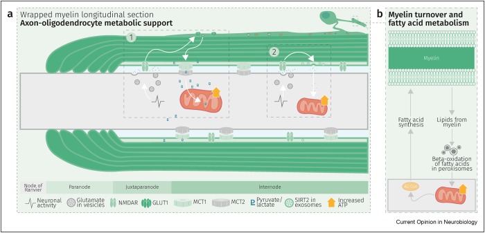
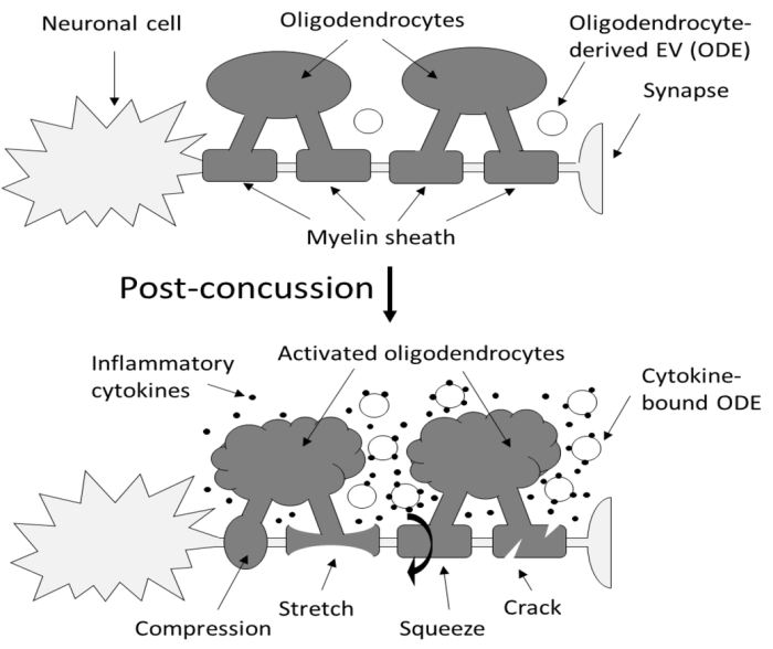
“Axons are long fibers conducting nerve impulses from nerve cells to synaptic ends. Like electric wires, axons are insulated by the myelin sheath produced by oligodendrocytes (ODC) in the brain or Schwann cells in the periphery. The myelin sheath is vulnerable to mechanical stresses after head injuries, as well as targets for autoimmune attack in multiple sclerosis and degeneration in various white matter diseases.

145850ce6289d06e5318d35f

It is challenging to definitively validate axonal neuroinflammation, because axonal neuroinflammation is only diagnosed at post-mortem autopsy, or wait for more than a decade to potentially witness progression to chronic traumatic encephalopathy, or white matter dementia. Advanced imaging analysis of computed tomography and magnetic resonance imaging are not sensitive enough to identify such microscopic abnormalities.

We developed a sandwich immunoassay detecting dual signals of myelin oligodendrocyte glycoprotein (MOG) and interleukin 1B (IL1B) in human plasma, [IL1B on MOG]. MOG is a transmembrane protein specifically expressed in ODC and Schwann cells membranes, and doesn’t freely exist in plasma. We found that serum from capillary blood is acceptable, and we tested control and athlete samples using only 5 mL samples. When we tested 63 control plasma samples, values were widely distributed over 2 logs, so we focused on longitudinal studies.

Damaged neurons are not easily detectable using conventional physical examinations, because the brain’s inherent adaptability allows it to compensate for localized damage by finding alternate routes. While this adaptability is advantageous, it also means that these concealed lesions can go unnoticed, potentially leading to future complications.

Elevation of [IL1B on MOG] was seen in some athletes who did not show concussion or traumatic brain injury (TBI). While the occurrence of concussion is relatively limited, potential prevalence of subconcussion or subconcussive condition is expected to be substantially higher.

If [IL1B on MOG] levels remain unchanged during this early post-concussion period (2-4 weeks), it may suggest that neuroinflammation has not been induced, potentially providing reassurance for the athletes to return to play. Conversely, if [IL1B on MOG] levels increase within this timeframe, it may indicate the need for intervention or closer monitoring. Thus, there is compelling potential for incorporating this test into concussion guidelines.”

https://www.researchsquare.com/article/rs-3997676/v1 “An approach for the analysis of axonal neuroinflammation by measuring dual biomarkers of oligodendrocytes and inflammatory cytokine in human plasma”


A rodent study investigated the immune system’s influence on oligodendrocyte lineage cells after TBI:

“White matter injury is thought to be a major contributor to long-term cognitive dysfunctions after TBI. This damage occurs partly due to apoptotic death of oligodendrocyte lineage cells (OLCs) after injury, triggered directly by the trauma or in response to degenerating axons.

Our data indicates that depletion of the gut microbiota after TBI impaired remyelination, reduced OLCs proliferation, and required the presence of T cells. This suggests that T cells are an important mechanistic link by which the gut microbiota modulate oligodendrocyte response and white matter recovery after TBI.

Our findings suggest that oligodendrocytes are not passive in the neuroinflammatory and degenerative environment caused by brain trauma, but instead could exert an active role in modulation of immune response.”

https://www.researchsquare.com/article/rs-4289147/v1 “Gut Microbiota Shape Oligodendrocyte Response after Traumatic Brain Injury”


A rodent study investigated whether oligodendrocyte precursor cells had myelination-independent roles in brain aging:

“OPCs, the source cells of myelin-forming cells in the central nervous system, have been linked to brain aging by their compromised differentiation and regeneration capability. Our results demonstrate that macroautophagy influx declines in aged OPCs, which results in the accumulation of senescent OPCs in aged brains. Senescent OPCs impair neuronal plasticity and exacerbate neurodegeneration, eventually leading to cognitive decline.

Inactivation of autophagy in OPCs exhibits a limited effect on myelin thickness but a loss of myelin in middle-aged mice. The loss of myelin observed is an adaptational change to suppressed neuronal plasticity. However, neither the number of OLs nor oligodendrogenesis is altered by inactivation of autophagy in adult OPCs.

The present study indicates that the intervention of senescent OPCs is an additional promising therapeutic strategy for aging and aging-related cognitive deficits. Autophagy regulates senescence by impairing protein turnover, mitochondrial homeostasis, oxidative stress, and maintaining senescence-associated secretory phenotype. Further investigation remains on whether autophagy in OPCs shares the exact mechanism to promote senescence as that in other types of cells.

Considering autophagy declines with aging, our study brings a novel mechanism in brain aging. Declined autophagy causes senescence of OPCs, which impairs neuronal plasticity and exacerbates neurodegeneration via CCL3/5-CCR5 signaling.”

https://www.researchsquare.com/article/rs-3926942/v1 “Impaired Macroautophagy in Oligodendrocyte Precursor Cells Exacerbates Aging-related Cognitive Deficits via a Senescence Associated Signaling”


PXL_20240418_104114528.MP

Maintaining your myelin, Part 1

Three papers on myelin and oligodendrocytes, starting with a 2023 review:

“Myelin is the spiral ensheathment of axons by a lipid and cholesterol-rich glial cell membrane that reduces capacitance and increases resistance of the axonal membrane. Axonal myelination speeds up nerve conduction velocity as a function of axon diameter.

While myelination proceeds rapidly after birth in the peripheral nervous system, central myelination is a spatially and temporally more regulated process. Ongoing myelination of the human brain has been documented at up to 40 years of age. This late myelination in the adult cortex is followed by exhaustion of oligodendrocyte precursor cells (OPC) with senescence and a gradual loss of myelin integrity in the aging brain.

The brain is well known for its high energy demands, specifically in gray matter areas. In white matter tracts, energy consumption is lower. Myelination poses a unique challenge for axonal energy generation where myelin sheaths cover more than 95% of the axonal surface areas.

Oligodendrocytes help support axonal integrity. Oligodendrocytes survive well in the absence of mitochondrial oxidative phosphorylation, and without signs of myelin loss, cell death, neurodegeneration or secondary inflammation.

Glycolysis products of oligodendroglial origin are readily metabolized in axonal mitochondria. Oligodendroglial metabolic support is critical for larger and faster-spiking myelinated axons that also have a higher density of mitochondria. An essential requirement for the direct transfer of energy-rich metabolites from oligodendrocytes to the myelinated axonal compartment is ‘myelinic channels’ within the myelin sheath.

Interactions of oligodendrocytes and myelin with the underlying axon are complex and exceed the transfer of energy-rich metabolites. Continuous turnover of myelin membranes by lipid degradation and fatty acid beta-oxidation in mitochondria and peroxisomes leads to recycling of acetate residues by fatty acid synthesis and membrane biogenesis.

1-s2.0-S0959438823001071-gr2_lrg

In human multiple sclerosis (MS) and its animal model myelin oligodendrocyte glycoprotein-experimental autoimmune encephalomyelitis (MOG-EAE), acute inflammatory demyelination is followed by axonal degeneration in lesion sites that is mechanistically not fully understood. It is widely thought that demyelination and the lack of an axon-protective myelin sheath in the presence of numerous inflammatory mediators are the main causes of axon loss.

But unprotected axons improve rather than worsen the overall clinical phenotype of EAE mice which exhibited the same degree of autoimmunity. Thus, ‘bad myelin is worse than no myelin’ because MS-relevant myelin injuries perturb the integrity of myelinic channels and metabolic support.

Dysfunctional or injured oligodendrocytes that do not allow for compensation by any other cell types turn the affected myelin ensheathment into a burden of the underlying axonal energy metabolism, which causes irreversible axon loss. Any loss of myelin integrity, as seen acutely in demyelinating disorders or more gradually in the aging brain, becomes a risk factor for irreversible neurodegeneration.”

https://www.sciencedirect.com/science/article/pii/S0959438823001071 “Expanding the function of oligodendrocytes to brain energy metabolism”


A 2024 review focused on myelin and oligodendrocyte plasticity:

“This review summarizes our current understanding of how myelin is generated, how its function is dynamically regulated, and how oligodendrocytes support the long-term integrity of myelinated axons.

Apart from its unique ultrastructure, there are several other exceptional features of myelin. One is certainly its molecular composition. Another is its extraordinary stability. This was compellingly illustrated when 5000-year-old myelin with almost intact ultrastructure was dissected from a Tyrolean Ice Man.

Myelin is a stable system in contrast to most membranes. However, myelin is compartmentalized into structurally and biochemically distinct domains. Noncompacted regions are much more dynamic and metabolically active than tightly compacted regions that lack direct access to the membrane trafficking machinery of oligodendrocytes.

The underlying molecular basis for stability of myelin is likely its lipid composition with high levels of saturated, long chain fatty acids, together with an enrichment of glycosphingolipids (∼20% molar percentage of total lipids) and cholesterol (∼40% of molar percentage of total lipids). In addition, myelin comprises a high proportion of plasmalogens (ether lipids) with saturated long-chain fatty acids. In fact, ∼20% of the fatty acids in myelin have hydrocarbon chains longer than 18 carbon atoms (∼1% in the gray matter) and only ∼6% of the fatty acids are polyunsaturated (∼20% in gray matter).

With maturation of oligodendrocytes, the plasma membrane undergoes major transformations of its structure. Whereas OPCs are covered by a dense layer of large and negatively charged self-repulsive oligosaccharides, compacted myelin of fully matured oligodendrocytes lacks most of these glycoprotein and complex glycolipids.

Schematic depiction of an oligodendrocyte that takes up blood-derived glucose and delivers glycolysis products (pyruvate/lactate) via monocarboxylate transporters (MCT1 and MCT2) to myelinated axons. Oligodendrocytes and myelin membranes are also coupled by gap junctions to astrocytes, and thus indirectly to the blood–brain barrier.

oligodendrocyte

Adaptive myelination refers to dynamic events in oligodendroglia driven by extrinsic factors such as experience or neuronal activity, which subsequently induces changes in circuit structure and function. Understanding how these adaptive changes in neuron-oligodendroglia interactions impact brain function remains a pressing question for the field.

Transient social isolation during adulthood results in chromatin and myelin changes, but does not induce consequent behavioral alterations. When mice undergo a social isolation paradigm during early life development, they similarly exhibit deficits in prefrontal cortex function and myelination, but these deficiencies do not recover with social reintroduction. This implicates a critical period for social deprivation effects on myelin dynamics. Experience-dependent changes in myelin dynamics may depend on not only the age, brain region, and cell type studied, but also the specific myelin structural change assessed.

Local synaptic neurotransmitter release along an axon not only affects the number of OPCs and oligodendrocytes associated with that axon and local synthesis of myelin proteins, but also drives preferential selection of active axons for myelination over the ensheathment of electrically silenced neighboring axons. Neuronal activity–induced plasticity may preferentially impact brain regions that remain incompletely myelinated compared to more fully myelinated tracts.

Whereas the myelin sheath has been regarded for a long time as an inert insulating structure, it has now become clear that myelin is metabolically active with cytoplasmic-rich pathways, myelinic channels, for movement of macromolecules into the periaxonal space. The myelin sheath and its subjacent axon need to be regarded as one functional unit, which are not only morphological but also metabolically coupled.”

https://cshperspectives.cshlp.org/content/early/2024/04/15/cshperspect.a041359 “Oligodendrocytes: Myelination, Plasticity, and Axonal Support” (not freely available) Thanks to Dr. Klaus-Armin Nave for providing a copy.


A 2024 rodent study investigated oligodendrocyte precursor cell transcriptional and epigenetic changes:

“We used single-cell RNA sequencing (scRNA-seq), single-cell ATAC sequencing (scATAC-seq), and single-cell spatial transcriptomics to characterize murine cortical OPCs throughout postnatal life. One group (active, or actOPCs) is metabolically active and enriched in white matter. The second (homeostatic, or hOPCs) is less active, enriched in gray matter, and predicted to derive from actOPCs. Relative to developing OPCs, both actOPCs and hOPCs are less active metabolically and have less open chromatin.

In adulthood, these two groups are transcriptionally but not epigenetically distinct, indicating that they may represent different states of the same OPC population. If that is the case, then one model is that the parenchymal environment maintains adult OPCs within an hOPC state, whereas those OPCs recruited into white matter or exposed to demyelinated axons may transition toward an actOPC state in preparation for making new oligodendrocytes. We do not yet know the functional ramifications of these differences, but this finding has clear implications for the development of therapeutic strategies for adult remyelination.

opcs

Another finding is that developing but not adult actOPC chromatin is preferentially open for binding motifs associated with neural stem cells, transit-amplifying precursors, and neurogenesis. Although this may simply reflect their origin as the immediate progeny of neonatal neural precursor cells, it may also explain why developing but not adult OPCs have the capacity to make neurons in culture.

If we could, at least in part, reverse the global chromatin shutdown that occurs between development and adulthood, then perhaps adult OPCs may reacquire the ability to make neurons or become better able to generate new oligodendrocytes for remyelination.”

https://www.cell.com/stem-cell-reports/fulltext/S2213-6711(24)00077-8 “Single-cell approaches define two groups of mammalian oligodendrocyte precursor cells and their evolution over developmental time”

Continued in Part 2.


PXL_20240414_103442372

What can be done today to fulfill early unmet needs?

Got agitated earlier this week watching Tucker Carlson’s freely-available interview with a maniac who thinks he’s graduated into a higher state by worshiping the Great AI (Artificial Intelligence, aka Automated Internet, inhabited solely by robots) which will dictate every aspect of what to do with his life. Nevermind that behind the Great AI curtain are the same people who have lied to billions of us, especially during every day of this decade.

Are his current set of beliefs better than previous ones he had of putting a chip into everybody’s brain? What’s wrong with getting to live your own life?

5000

What I saw expressed in the interview was an exhausting pursuit of substitutes for feeling loved. I doubt that many others saw the same, because feeling unloved is so devastating we’ll do anything to avoid it.

But re-experiencing early memories and feelings of unmet needs in a therapeutic setting is the way to keep them from subsequently running our lives. Otherwise, we’ll develop unfulfilling substitutes for what we missed, with misdirected ideas and beliefs accompanied by their unconscious act-outs.

While speaking with a mother who is doing a terrific job of meeting her six-month-old’s needs, I attempted to contrast this interview with the experiences she and her husband are giving their child. Maybe if they read this post, my poor explanation will become clearer.


Wild persimmon trees’ eclipse shadows

PXL_20240408_192336638

Changing a cancerous phenotype

A 2024 Dr. Goodenowe presentation to a professional audience. He ended the presentation by using his 86-year-old father as a case study of treatment to create an inhospitable environment for cancer.

1. Get the body ready

slide 189

2. Starve the cancer and boost the immune system

slide 190

3. Characteristics

slide 191

4. 2019 sample biochemistry

slide 192

5. 2023 biochemistry (compare HDL (33 vs. 80), see off-the-chart hsCRP, Hcy 16)

slide 193

6. Treatment details #1

slide 197

7. Treatment details #2

slide 198

https://drgoodenowe.com/tfim-2024-recording-now-available/ “Breaking Cancer: The Biochemistry of Cancer Risk Assessment, Prevention, and Treatment—Real Knowledge That You Can Use In Your Practice”


PXL_20240408_185424838

Eat broccoli sprouts to maintain your cells

Two more papers cited Precondition your defenses with broccoli sprouts, starting with a 2024 review of broccoli compounds’ influences on autophagy and cellular function:

“Promotion of autophagy has been related to lifespan expansion, tumor suppression, and maintenance of metabolic health. Alterations in this pathway have been related to human diseases or pathological states including neurodegenerative diseases, stroke, metabolic alterations, or cancer.

We describe the different types of glucosinolates (GSL), grouped depending on the structure of their side chain, with special attention to those GSL and their derived isothiocyanate (ITC) which have been suggested to be of relevance to treat or prevent human diseases, their structure, and plant source.

gsl-itc

It has been shown that SFN activates TFEB, boosting expression of genes required for autophagosome and lysosome biogenesis. SFN induced a short burst of ROS necessary for TFEB activation, and TFEB activity was required for SFN-induced NRF2 activation and protection against acute and chronic oxidative stress.

TFEB was also required for SFN-induced removal of excessive mitochondrial ROS, indicating an important role for mitophagy in SFN-induced antioxidant response. Thus, direct activation of NRF2 by SFN or other ITC can promote autophagy.

Research on autophagy has been characterized by controversies regarding autophagy mediating survival or cell death, or its role in health and disease, not only because autophagy is a complicated process with context dependent roles depending on the cell type or the step of the autophagic pathway being modulated, but also, because in occasions, autophagy is not measured correctly.

An interesting area of research would be to decipher effects of NRF2-regulated or NRF2-independent autophagy induction by ITC, and whether these effects would determine the role of the autophagic process in cellular survival or death. Also, it is needed to clarify which of the effects regulated by ITC are mediated by autophagy, and which ones are not, and the importance of autophagy induction in the therapeutic effects mediated by ITC.”

https://link.springer.com/article/10.1007/s11101-024-09944-w “Glucosinolates, isothiocyanates, and their role in the regulation of autophagy and cellular function” (not freely available)

This paper’s contact coauthor (who provided access to the full paper) is also the contact for Our model clinical trial for Changing to a youthful phenotype with broccoli sprouts.


The coauthors of Exercise substitutes? published a 2024 human cell study:

“While physical activity is an excellent inducer of mitochondrial turnover, its ability to ubiquitously activate and enhance mitochondrial turnover prevents definitive differentiation of the contribution made by each pathway. We employed three agents which are activators of important biological markers involved in antioxidant signaling, mitochondrial autophagy, and mitochondrial biogenesis.

Results suggest that early time points of treatment increase upstream pathway activity, whereas later time points represent increased phenotypic expression of related downstream markers. Findings suggest that spatiotemporal progression of these mechanisms following drug treatment is another important factor to consider when examining subcellular changes towards mitochondrial turnover in muscle.”

https://www.sciencedirect.com/science/article/pii/S2666337624000398 “Sulforaphane, Urolithin A, and ZLN005 induce time-dependent alterations in antioxidant capacity, mitophagy, and mitochondrial biogenesis in muscle cells”


PXL_20240330_175846440

Year Four of Changing to a youthful phenotype with sprouts

1. I’ve continued daily practices from Year Three to experience another year without being sick. I’ll get a set of Labcorp tests in two weeks to see if anything is sneaking up on me.

Foods are the same as Week 189 except I eat two raw eggs in the morning after Avena sativa oat sprouts. Supplements are the same except I stopped the ProdromeGlia plasmalogen precursor supplement due to it being out-of-stock.

It’s annoying because after a few days, my sense of smell and taste improvements reverted without ProdromeGlia. I’ve continued ProdromeNeuro, but it seems that its combination with ProdromeGlia was essential for stopping my left ulnar nerve elbow pain, which returned after a week without ProdromeGlia.

2. You may have noticed that earlier this month, a U.S. government agency was forced by a lawsuit to delete their 2021 propaganda pieces against a medication that’s safer than acetaminophen. I had a prescription that local pharmacies suddenly wouldn’t fill in August 2021.

Plenty of workarounds have been available, though. I hadn’t mentioned it before, but a prophylactic weekly intake may have played a part in me not being sick even one day this decade.

Another part was that my living and working in the Washington DC area for 30+ years through 2017 taught me, as an initial response, to not believe a single word of what a government employee said. I’ve since extended that to many other types of compromised people, such as medical professionals.

3. Our ancestors evolved to deal with everyday bacteria, viruses, and other pathogens. Train your immune system every day! disclosed that I was in Milan, Italy on the same February 22-23, 2020 weekend that ten towns were closed south of Milan. I still haven’t experienced any symptoms.

  • One factor in immune response was that fifteen years previous, I’d taken daily steps with yeast cell wall β-glucan to guard against the phenotypical immune system collapse of old age.
  • Another factor was that I’d ridden the filthy Washington DC Metro twice a day to-and-from work for years, and had already been exposed to who knows what.

Treat your gut microbiota well. Give them what they want – including cruciferous sprouts – and expect reciprocity.


what

Ergothioneine dosing, Part 2

Continuing Part 1 with a 2024 rodent healthspan and lifespan study:

“We investigated the effects of daily oral supplementation of ergothioneine (ERGO) dissolved in drinking water on lifespan, frailty, and cognitive impairment in male mice from 7 weeks of age to the end of their lives. Ingestion of 4 ~ 5 mg/kg/day of ERGO remarkably extended the lifespan of male mice.

11357_2024_1111_Fig1_HTML

The ERGO group showed significantly lower age-related declines in weight, fat mass, and average and maximum movement velocities at 88 weeks of age. This was compatible with dramatic suppression by ERGO of age-related increments in plasma biomarkers. ERGO also rescued age-related impairments in learning and memory ability.

Ingestion of ERGO may promote longevity and healthy aging in male mice, possibly through multiple biological mechanisms.”

https://link.springer.com/article/10.1007/s11357-024-01111-5 “Ergothioneine promotes longevity and healthy aging in male mice”

Subjects’ plasma ergothioneine levels of an estimated 4 ~ 5 mg/kg daily dose were:

11357_2024_1111_Fig3_HTML

A human equivalent daily dose is an estimated 22 mg to 28 mg (4 or 5 mg x .081 x 70 kg).

The third paper in Part 1 cited a 2017 clinical trial that provided 5 mg and 25 mg ergothioneine doses for 7 days, resulting in these plasma ergothioneine levels:

figure 3

The first paper of Part 1 referenced a 2020 human study where the dose was 5 mg/day for 12 weeks, but I don’t have access to it. It’s unclear whether humans could continually raise ergothioneine levels by daily consumption throughout our lives as did this rodent study.


A 2024 paper reviewed the importance of ergothioneine to humans:

“We propose that the diet-derived compound ergothioneine (ET) is an important nutrient in the human body, especially for maintenance of normal brain function, and that low body ET levels predispose humans to significantly increased risks of neurodegenerative and possibly other age-related diseases.

Work by multiple groups has established that low ET levels in humans are associated not only with cognitive impairment/AD but also with other age-related conditions, including frailty, Parkinson’s disease, vascular dementia, chronic renal disease, cardiovascular disease, and macular degeneration. Low ET levels also correlate with increased risk of developing preeclampsia in pregnant women [53].

Plasma ET levels from healthy (age-matched) vs unhealthy individuals in Singapore – Mild cognitive impairment (MCI); Alzheimer’s disease (AD); vascular dementia (VaD); Parkinson’s disease (PD); age-related macular degeneration (AMD):

1-s2.0-S0891584924001357-gr2_lrg

  • Does low ET cause or contribute to age-related neurodegeneration, or
  • Does disease cause low ET, or
  • Low ET and increased disease risk are both caused by something else, as yet unidentified?

Prevention of neurodegeneration is especially important, since by the time dementia is usually diagnosed damage to the brain is extensive and likely irreversible.”

https://www.sciencedirect.com/science/article/pii/S0891584924001357 “Are age-related neurodegenerative diseases caused by a lack of the diet-derived compound ergothioneine?”

Whether or not the healthy individuals ate mushrooms daily in the above graphic was lost while conglomerating multiple studies.

Note that scales of the above two human graphics are a thousand times smaller than the above rodent graphic. I thought that maybe the rodent study made a plasma ergothioneine calculation error, but didn’t see one in the provided Supplementary data.


Reference 53 of the second paper is a 2023 human study:

“We analysed early pregnancy samples from a cohort of 432 first time mothers. Of these 432 women, 97 went on to develop pre-term or term pre-eclampsia (PE).

If a threshold was set at the 90th percentile of the reference range in the control population (≥462 ng/ml), only one of these 97 women (1%) developed PE, versus 96/397 (24.2%) whose ergothioneine level was below this threshold. One possible interpretation of these findings, consistent with previous experiments in a reduced uterine perfusion model in rats, is that ergothioneine may indeed prove protective against PE in humans.”

https://portlandpress.com/bioscirep/article/43/7/BSR20230160/233119/Relationship-between-the-concentration-of “Relationship between the concentration of ergothioneine in plasma and the likelihood of developing pre-eclampsia”

Eyeballing the Healthy individuals in the above graphic, none of those 544 people were below this study’s 462 ng threshold.


A 2023 companion article analyzed the third paper’s unusual findings:

“These results suggest that there might be a dichotomized association between ergothioneine concentrations and preeclampsia; and only a high ergothioneine level over 90th percentile of the control population could be protective against preeclampsia.

Univariable results showed that ergothioneine had a significant non-linear association with preeclampsia and it would start to offer protective effect from 300 ng/ml onward. Analysis also confirmed that body mass index was significantly associated with an increased risk of preeclampsia.

A large observational study could strengthen the causal association between ergothioneine and preeclampsia. If confirmed, a randomized controlled trial (RCT) assessing whether ergothioneine supplementation can reduce risk of preeclampsia will be imminently feasible. Ideally, such RCT should compare placebo with a range of different doses of ergothioneine to identify the best or minimal effective dose, given its good safety records, including in pregnancy, with a no-observed-adverse-effect level (NOAEL) of 800 mg/kg body weight per day.”

https://portlandpress.com/bioscirep/article/43/8/BSR20231076/233395/Dose-related-relationship-between-ergothioneine “Dose-related relationship between ergothioneine concentrations and risk of preeclampsia”

My daily mushroom ergothioneine dose is around 7 mg, and I weigh about 70 kg. I don’t think a daily 800 mg/kg ergothioneine dose would be desirable for anybody, regardless of what experts say.

How many times have public health employees been wrong this decade? Would you bet your or your child’s health on their advice?


PXL_20240316_182330822

Sulforaphane’s effects on autism and liver disease

Here are two more papers that cited Precondition your defenses with broccoli sprouts, starting with a 2024 human / rodent study investigating gut microbiota / sulforaphane’s effects on autism:

“Sulforaphane (SFN) has been found to alleviate complications linked with several diseases by regulating gut microbiota (GM), while the effect of GM on SFN for autism spectrum disorders (ASD) has not been studied. We evaluated therapeutic effects of SFN on maternal immune activation (MIA)-induced ASD-like rat model and pediatric autism patients aged 4–7 years.

OSU-SO for social interactive OSU behavioral subscores, OSU-CO for non-verbal communicative OSU behavioral [significant] subscores, and OSU-ST for repetitive or ritualistic OSU behavioral subscores:

fnut-10-1294057-g0005

Although gut microbiota composition was significantly altered in SFN-treated ASD-like rats, alteration of GM was not evident in ASD patients after 12 weeks of SFN treatment. Limitations in this study:

  1. Studies were conducted in male rats and boys only;
  2. The sample size of our clinical study is relatively small [6 SFN-treated boys] and needs to be further expanded in the future; and
  3. This study only uncovered a potential link between gut flora and the therapeutic effects of SFN on ASD.

SFN treatment alleviates social deficits in MIA-induced ASD-like rats and ASD patients, and improvements might be associated with gut microbiota.”

https://www.frontiersin.org/articles/10.3389/fnut.2023.1294057/full “Therapeutic efficacy of sulforaphane in autism spectrum disorders and its association with gut microbiota: animal model and human longitudinal studies”

The 2022 Efficacy of Sulforaphane in Treatment of Children with Autism Spectrum Disorder: A Randomized Double-Blind Placebo-Controlled Multi-center Trial (not freely available) was referenced for sulforaphane (actually, glucoraphanin with myrosinase enzyme) doses:

“Dosing was weight-based:

  • Two tablets/day for 10–29 lb;
  • Three tablets/day for 30–49 lb;
  • Four tablets/day for 50–69 lb.

An estimated delivery of approximately 24, 36, and 48 μmol of sulforaphane daily was expected in the respective SF dosage groups.”

Weights of the above μmol estimated dose amounts per https://pubchem.ncbi.nlm.nih.gov/compound/sulforaphane are 4.3, 6.4, and 8.5 mg, respectively. An average weight of a 4-year-old boy is 36 lbs / 16.3 kg, and a 7-year-old boy is 51.1 lbs / 23.2 kg.

This study’s maternal immune activation was done by injecting lipopolysaccharide into pregnant rats. Would injecting pregnant women with immune-activating substances have similar harmful effects on the fetus? We don’t have evidence because unbiased and unconflicted studies looking for such effects weren’t sponsored and/or published before immune-activating substances’ deployments.


A 2024 rodent study investigated sulforaphane’s effects on diabetic liver damage:

“We investigated whether sulforaphane, an Nrf2 activator and antioxidant, prevents diabetes-induced hepatic ferroptosis, and the mechanisms involved. Results showed that diabetes-induced inactivation of Nrf2 and decreased expression of its downstream antiferroptotic molecules critical for:

  • Antioxidative defense (catalase, superoxide dismutases, thioredoxin reductase);
  • Iron metabolism (ferritin heavy chain (FTH1), ferroportin 1);
  • Glutathione (GSH) synthesis (cystine-glutamate antiporter system, cystathionase, glutamate-cysteine ligase catalitic subunit, glutamate-cysteine ligase modifier subunit, glutathione synthetase); and
  • GSH recycling – glutathione reductase (GR)

were reversed/increased by sulforaphane treatment.

Diabetes-induced increases in serum glucose and triglyceride levels were also significantly reduced by sulforaphane. Taken together, our results demonstrate a potent effect of SFN in inhibiting ferroptotic death of hepatocytes under diabetic conditions in vivo, thereby alleviating liver injury.

This is the first study to demonstrate the protective role of SFN against ferroptosis in the liver of diabetic mice. This nominates sulforaphane as a promising phytopharmaceutical for the prevention/alleviation of ferroptosis in diabetes-related pathologies.”

https://iubmb.onlinelibrary.wiley.com/doi/10.1002/biof.2042 “Sulforaphane prevents diabetes-induced hepatic ferroptosis by activating Nrf2 signaling axis”

Improving peroxisomal function

A 2024 review provided details about “mysteries” in peroxisome research:

“Peroxisomes are key metabolic organelles with essential functions in cellular lipid metabolism (e.g., β-oxidation of fatty acids and synthesis of ether phospholipids, which contribute to myelin sheath formation), and metabolism of reactive oxygen species (ROS), particularly hydrogen peroxide. Loss of peroxisomal function causes severe metabolic disorders in humans.

Additional non-metabolic roles of peroxisomes have been revealed in cellular stress responses, regulation of cellular redox balance and healthy ageing, pathogen and antiviral defence, and as cellular signalling platforms. New findings also point to a role in regulation of immune responses.

In our previous reviews, we addressed the role of peroxisomes in the brain, in neurological disorders, in development of cancer, and in antiviral defence. To avoid repetition, we refer to those articles where appropriate, and to more specialised recent reviews on peroxisome biology.

418_2023_2259_Fig5

Proper functioning of peroxisomes in metabolism requires the concerted interaction with other subcellular organelles, including the endoplasmic reticulum (ER), mitochondria, lipid droplets, lysosomes, and the cytosol. A striking example of peroxisome-ER metabolic cooperation is de novo biosynthesis of ether phospholipids.

Metabolic activities of peroxisomes, such as ɑ- and β-oxidation of fatty acids, plasmalogen synthesis, and ROS/reactive nitrogen species metabolism, have been linked to numerous immune-related pathways. Roles for peroxisomes in immune and defence mechanisms have opened a new field of peroxisome research, and highlight once more how important peroxisomes are for human health and disease.

It is still not fully understood how peroxisomal functions and abundance are regulated, what kinases/phosphatases are involved, or how peroxisomes are linked to cellular signalling pathways and how they act as signalling platforms.”

https://link.springer.com/article/10.1007/s00418-023-02259-5 “The peroxisome: an update on mysteries 3.0”


Last Friday was Day 90 of a 90-day trial of plasmalogens coincident with improving peroxisomal function via resistance exercise and time-restricted eating. A sticking point has been leg resistance exercises. Ankle issues are interfering with progress, although beach walks aren’t similarly affected. I’m almost back to an upper body exercise routine of five years ago, and I’ve added a half-dozen abs exercises.

I’ll continue taking the two Prodrome plasmalogen precursor supplements (ProdromeGlia and ProdromeNeuro) and with efforts to improve peroxisomal function. Since achieving effective resistance exercise levels is taking longer than expected, and my crystal ball is out-of-commission, I don’t have a realistic end time estimate for stopping the supplements.

Get a little stress into your life

Two reviews on beneficial effects of mild stress, starting with a 2024 paper coauthored by the lead researcher of Sulforaphane in the Goldilocks zone:

“This paper addresses how long lifespan can be extended via multiple interventions, such as dietary supplements, pharmaceutical agents, caloric restriction, intermittent fasting, exercise, and other activities. This evaluation was framed within the context of hormesis, a biphasic dose response with specific quantitative features describing the limits of biological/phenotypic plasticity for integrative biological endpoints.

Human maximum longevity has remained relatively constant in the 110–120 year time period. Yet, research with C. elegans indicates that hormetic processes increase both average (median/mean) and maximum lifespans. These observations were consistently shown by different research teams using highly diverse stressors but with generally similar experimental methods. Thus, lifespan can be increased in an overall average manner but also within the context of the maximum lifespan potential via hormetic processes, which has not been shown to occur in human population studies.

In multiple experimental and epidemiological contexts, antioxidants have prevented lifespan extension of numerous hormetic agents and blocked human health benefits (e.g., exercise), supporting the hypothesis that oxidative stress is necessary for healthspan improvements and lifespan extension.

Maximum lifespan may be prolonged by extending the lifespan of healthy subjects. Median lifespan would be enhanced by protecting those who are susceptible to genetic/environmental diseases.

Most experimental studies indicate that maximum hormetic lifespan benefits are in the 15 – 25% range when responses are optimized. Human-based benefits could be expected to be less than this maximum range. The issue of hormetic synergies is important to consider, but the available data to date indicates that these benefits are also constrained by limits of biological plasticity.”

https://www.sciencedirect.com/science/article/abs/pii/S1568163723003409 “Hormesis determines lifespan” (not freely available) Thanks to Dr. Evgenios Agathokleous for providing a copy.


A 2023 review of nematode studies was cited three times:

“While stress response pathways are important in allowing organisms to survive acute and chronic stresses, these pathways also contribute to longevity under unstressed conditions. Multiple stress response pathways are required for normal lifespan in wild-type worms, and all of the stress response pathways discussed in this review contribute to the longevity of long-lived mutants.

Four stress response pathways were consistently required for longevity:

  1. The FOXO transcription factor DAF-16-mediated stress response;
  2. The Nrf2 homolog SKN-1-mediated oxidative stress response;
  3. The cytoplasmic unfolded protein response (cyto-UPR); and
  4. The endoplasmic reticulum unfolded protein response (ER-UPR)

are required for normal lifespan, and may contribute to the extended lifespan of long-lived mutants. Developing strategies to activate these pathways, at the right time(s) and in the right tissue(s), may help to promote healthy aging and ameliorate age-onset disease.”

https://www.sciencedirect.com/science/article/pii/S1568163723001009 “Biological resilience and aging: Activation of stress response pathways contributes to lifespan extension”


PXL_20240114_184135203

Eat broccoli sprouts for your liver, Part 2

A 2023 review cited Part 1 and caught up other relevant research on sulforaphane effects through early 2023:

“A growing number of studies have reported that sulforaphane (SFN) could significantly ameliorate hepatic steatosis and prevent development of fatty liver, improve insulin sensitivity, attenuate oxidative damage and liver injury, induce apoptosis, and inhibit proliferation of hepatoma cells through multiple signaling pathways.

SFN inhibits lipogenesis and oxidative stress while enhancing lipid droplet degradation through modulating expression of genes involved in lipid synthesis, metabolism, and oxidation. SFN modulates autophagy, lipolysis, mitochondrial function, and ER stress to alleviate fatty liver through AMPK-, AHR-, PGC1α-, and FGF21-mediated pathways.

fphar-14-1256029-g001

There is still a gap between basic research and clinical application of SFN. More efficient delivery systems and precise dose schedules of SFN are expected to be developed in future studies, which would improve its solubility, stability, and bioavailability, and reduce inter-individual variations in humans.”

https://www.frontiersin.org/articles/10.3389/fphar.2023.1256029/full “Therapeutic potential of sulforaphane in liver diseases: a review”


These reviewers did alright gathering papers. That’s only part of what needed to be done, with the other part being reading, understanding, and interpreting these papers.

First example: Sulforaphane in the Goldilocks zone was cited [reference 12], but applicability to this review with its main point “The stimulatory zone for in vitro studies proved to be consistently in the 1-10 μM range” as in Figure 10 “Effects of R-sulforaphane on phase II enzyme activation in precision-cut liver slices of young adult male Albino Wistar rats” wasn’t understood:

figure 10

These reviewers complained:

“Few dose-response studies on SFN have been reported, and the range of its effective doses is unclear. Doses used in most animal studies have exceeded the highest dose of SFN used in humans.”

So it might have taken a little bit more effort, but these reviewers could have highlighted studies where sulforaphane liver treatments were in the 1-10 μM potentially therapeutic range.

Another example: these reviewers said “The half-life of SFN is very short due to its rapid metabolism in the human body.” They missed a point that the second paper in How much sulforaphane is suitable for healthy people? [reference 46] made in section 6.4. “NQO1 Pharmacokinetics following SFN Ingestion:”

“Maximal induction of NQO1 occurred at around 24 hours, declining thereafter (Figure 8). This peak represents an approximate 2.8-fold induction over baseline.

These findings are useful when considering the effect of SFN as an intervention material in acute compared with chronic conditions. A significant increase in NQO1 occurred between 6 and 12 hours, a timeframe that may not be sufficiently responsive for management of an acute state, leaving one to conclude that NQO1 induction is best suited to chronic conditions where a rapid response may not be necessary.”

OMCL2019-2716870.008

Sulforaphane’s effects of inducing NQO1 for its cytoprotective, antioxidant, and other functions lasts for days, regardless of when sulforaphane leaves the bloodstream.

Taurine’s effects on healthspan and lifespan, Part 2

Four 2023 papers that cited Part 1, starting with a review of hypothetical parameters for taurine clinical trials that aren’t going to happen because:

  • Drug companies can’t make money from a research area that’s cheap, not patentable, and readily accessible.
  • Government sponsors are likewise not incentivized to act in the public’s interest per their recent behavior.

“We propose the rationale that an adequately powered randomized-controlled-trial (RCT) is needed to confirm whether taurine can meaningfully improve metabolic and microbiome health, and biological age.

taurine hypothetical trial

Using long-term survival as a primary outcome is desirable but difficult; any demonstrable difference in this outcome will require a substantial sample size with prolonged follow-up (e.g., 5 years or longer) if the effect size is relatively small (or modest at best). Biological age based on DNA methylation biomarkers according to the Levine PhenoAge or newer biological age models is increasingly being recognized as an important dynamic health parameter, and hence it can also be used as a surrogate outcome in assessing benefits of taurine supplementation.

The recent taurine trial on nonhuman primates used an equivalent dose that was between 3 and 6 g per day for an 80-kg person, and this could represent a reasonable dose range for any human RCTs. We believe that a 6-month or longer interventional period matching what was successfully done on nonhuman primates will be an acceptable time frame in assessing potential efficacy of taurine on human metabolic health in a RCT.”

https://www.sciopen.com/article/10.26599/1671-5411.2023.11.004 “Flattening the biological age curve by improving metabolic health: to taurine or not to taurine, that’s the question”

A six-month duration and a 6 grams per day dose were in the above table’s desirable features column, but epigenetic clock measurements weren’t included as an outcome. I’d guess that its omission reflected disagreements among coauthors, because the desirability of using epigenetic clocks as surrogate measures of human healthspan and lifespan was mentioned several times.


Another review:

“As described in the first half of this review, recent advances in omics analysis technology have led to research to detect the causative gene of dilated cardiomyopathy. It has been found that rare mutations in the taurine transporter gene contribute to the development of dilated cardiomyopathy in humans. It is unlikely that a taurine-deficient diet is a factor in dilated cardiomyopathy, but taurine intake may have positive cardiovascular effects.

The second half summarizes the relationship between taurine and healthspan and lifespan. It is difficult to summarize the effect of age in whole body taurine content, which may vary in species, strain, sex, and age of animal models. Future human studies will clarify the relationship between dietary taurine intake and healthy life expectancy.”

https://www.sciencedirect.com/science/article/pii/S1347861323000749 “Taurine deficiency associated with dilated cardiomyopathy and aging”


A human study investigated brain chemicals that fluctuate with our circadian rhythm:

“We conducted a MRS study at 7 T, where occipital NAD content, lactate, and other metabolites were assessed in two different morning and afternoon diurnal states in healthy participants. Salivary cortisol levels were determined to confirm that the experiment was done in two circadian different physiological conditions.

Although no significant differences in NAD+, NADH, and NAD+/NADH were detected between the morning and afternoon sessions, there was a significant variance difference in NAD+/NADH, with a higher variance of NAD+/NADH redox ratio in the morning.

None of the over 30 measured brain metabolites were significantly affected by the circadian rhythm (CR) except for taurine, which decreased in the afternoon. Further CR studies should consider the prospective measurement of taurine levels in different regions of the human brain, and explore how taurine supplements could impact brain CR metabolism in health and diseases.”

https://www.frontiersin.org/articles/10.3389/fphys.2023.1285776/full “Effect of circadian rhythm on NAD and other metabolites in human brain”

I omitted findings regarding this study’s pathetic Balloon Analogue Risk Task (BART) test. Older studies that drew spurious findings from this video game include:


A rodent study modeled human childhood cataracts:

“Our analysis identified targets that are required for early normal differentiation steps and altered in cataractous lenses, particularly metabolic pathways involving glutathione and amino acids. Glutathione and taurine were spatially altered, and both taurine and the ratio of reduced glutathione to oxidized glutathione, two indicators of redox status, were differentially compromised in lens biology.

1-s2.0-S2213231723002707-ga1_lrg

Dietary amino acid supplementation has been shown to prevent cataract development, and dietary intake of taurine was protective in a glutathione depletion-derived opacity model. This opens up the possibility that dietary supplementation of taurine could be used as a strategy to prevent human congenital cataracts.

Our findings shed light on molecular mechanisms associated with congenital cataracts, and point out that unbalanced redox status due to reduced levels of taurine and glutathione, metabolites already linked to age-related cataracts, could be a major underlying mechanism behind lens opacities that appear early in life.”

https://www.sciencedirect.com/science/article/pii/S2213231723002707 “Unbalanced redox status network as an early pathological event in congenital cataracts”


PXL_20240103_191340418

Take acetyl-L-carnitine if you are healthy

Eight 2023 acetyl-L-carnitine / L-carnitine papers, starting with three healthy human studies:

“Thirty healthy volunteers aged between 19 and 52 years were divided randomly into two equal groups, one of which received 1000 mg of L-carnitine (LC) per day over a 12-week period. Total cholesterol and HDL-C increased significantly after supplementation. LC could be useful in impeding development of heart diseases in subjects with low HDL-C.”

https://journaljammr.com/index.php/JAMMR/article/view/5166 “L-Carnitine Increases High Density Lipoprotein-Cholesterol in Healthy Individuals: A Randomized Trial”

Rationale for dose selection wasn’t provided, and the possibility of limited results due to poor study design wasn’t mentioned.


“This study examined effects of 12 weeks of LC supplementation on bone mineral density (BMD) and selected blood markers involved in bone metabolism of postmenopausal women participating in a resistance training (RT) program. Participants’ diets were supplemented with either 1 g of LC-L-tartrate and 3 g of leucine per day (LC group) or 4 g of leucine per day as a placebo (PLA group), in a double-blind fashion.

Because the study protocol consisted of both exercise and supplementation, some favorable changes in the BMD could be expected. However, it was not possible to detect them in the short study period. No significant modification in BMDs of the spine, hip, and total skeleton and no differences between groups in one-repetition maximum could be due to the relatively short duration of the RT intervention.”

https://nutritionandmetabolism.biomedcentral.com/articles/10.1186/s12986-023-00752-1 “Effect of a 3-month L-carnitine supplementation and resistance training program on circulating markers and bone mineral density in postmenopausal women: a randomized controlled trial”

Same comments as the first study regarding no rationale for dose selection, and no mention that limited results were possibly due to an inadequate dose.


In a letter to the editor, a researcher took issue with a study’s methodology:

“Based on finding that intravenous provision with carnitine alone does not increase muscle carnitine accretion, and on the above-reevaluated data, it appears that the basis for carnitine with caffeine being able to increase muscle carnitine levels, and thereby manipulation of muscle metabolism and exercise performance, is uncertain.

Carnitine bioavailability in any group would have been 9.5%. This assessment would be in line with previously recorded values of 5%–18% carnitine bioavailability. It is firmly believed that low carnitine bioavailability is attributable to the inability of kidneys to reabsorb carnitine when the threshold concentration for tubular reabsorption (about 40–60 μmol/L) has passed this value.

The authors’ proposed long-term use of carnitine supplementation as an aid to improve fat oxidation in type II diabetes also seems to lack provision.”

https://physoc.onlinelibrary.wiley.com/doi/10.14814/phy2.15736 “LTE: Does caffeine truly raise muscle carnitine in humans?”


Two genetic studies:

“Our findings suggest that humans have lost a gene involved in carnitine biosynthesis. Hydroxytrimethyllysine aldolase (the second enzyme of carnitine biosynthesis) activity of serine hydroxymethyl transferase partially compensates for its function.”

https://www.researchsquare.com/article/rs-3295520/v1 “One substrate-many enzymes virtual screening uncovers missing genes of carnitine biosynthesis in human and mouse”


“Reported prevalence of primary carnitine deficiency (PCD) in the Faroe Islands of 1:300 is the highest in the world. The Faroese PCD patient cohort has been closely monitored and we now report results from a 10-year follow-up study of 139 PCD patients.

PCD is an autosomal recessive disorder that affects the function of organic cation transporter 2 (OCTN2) high-affinity carnitine transporters, that localizes to the cell membrane and transport carnitine actively inside the cell. Without proper functioning OCTN2 carnitine transporters, renal reabsorption of carnitine is impaired, and as a consequence, patients suffering from PCD have low plasma levels of carnitine. This can disturb cellular energy production and cause fatigue, but also in extreme cases lead to cellular dysfunction and severe symptoms of coma and sudden death.

PCD patients seem to adhere well to L-carnitine treatment, even though they have to ingest L-carnitine tablets at least three times a day. Overall mean L-carnitine dosage was 66.3 mg/kg/day.”

https://onlinelibrary.wiley.com/doi/10.1002/jmd2.12383 “Patients with primary carnitine deficiency treated with L-carnitine are alive and doing well—A 10-year follow-up in the Faroe Islands”

The average daily dose is (66.3 mg x 70 kg) = 4,641 mg. A third of this dose would be about 1.5 g.

The first study of Acetyl-L-carnitine dosing also suggested dosing L-carnitine three times a day because of 10-20% bioavailability.


A study with unhealthy humans:

“This retrospective study analyzed medical records of adult patients between March 2007 and April 2019, with presenting complaints of fatigue and lethargy. Acetyl-L-carnitine has physiological functions similar to L-carnitine but has higher bioavailability and antioxidant properties. This study confirmed that a triple combination therapy with γ-linolenic acid, V. vinifera extract, and acetyl-L-carnitine can improve arterial stiffness in patients.

Our study had some limitations:

  1. The study population may not be representative of the entire Korean adult population.
  2. The study did not have a medication-free control group. Instead, the comparison group comprised patients with medication compliance <80%.
  3. Drop-out rate of the triple-combination therapy (46.2%, 147/318) was relatively high, indicating the possibility of bias due to loss to follow-up.
  4. The study did not consider lifestyle factors such as smoking, diet, and physical activity level, which may affect arterial stiffness.
  5. The study did not examine interactions among drugs comprising the combination therapy, although all drugs are known to positively impact blood vessels.”

https://onlinelibrary.wiley.com/doi/10.1111/jch.14708 “Efficacy of γ-linolenic acid, Vitis vinifera extract, and acetyl-L-carnitine combination therapy for improving arterial stiffness in Korean adults: Real-world evidence”

This study’s acetyl-L-carnitine dose was 500 mg three times a day.


Wrapping up with two rodent studies:

“Acetyl L-carnitine (ALCAR) has proved useful in treatment of different types of chronic pain with excellent tolerability. The present work aimed at evaluating the anti-hyperalgesic efficacy of ALCAR in a model of persistent visceral pain associated with colitis.

The acetyl group in the ALCAR molecule can enhance cholinergic signalling by promoting synthesis of neurotransmitter acetylcholine, which plays an important role in both the enteric and central nervous systems. Acetylcholine signalling has significant antinociceptive effects in development of visceral pain, so it has been proposed as a therapeutic target.

ijms-24-14841-g001

ALCAR significantly reduced establishment of visceral hyperalgesia in DNBS-treated animals, though the interventive protocol showed a greater efficacy than the preventive one.

  • The interventive protocol partially reduced colon damage in rats, counteracting enteric glia and spinal astrocyte activation resulting from colitis.
  • The preventive protocol effectively protected enteric neurons from inflammatory insult.

These findings suggest the putative usefulness of ALCAR as a food supplement for patients suffering from inflammatory bowel diseases.”

https://www.mdpi.com/1422-0067/24/19/14841 “Anti-Hyperalgesic Efficacy of Acetyl L-Carnitine (ALCAR) Against Visceral Pain Induced by Colitis: Involvement of Glia in the Enteric and Central Nervous System

This study cited multiple animal studies that found acetyl-L-carnitine was effective for different types of pain. I’ve taken it every day for nineteen years, and haven’t noticed that effect.


“Repetitive mild traumatic brain injuries (rmTBI) may contribute to development of neurodegenerative diseases through secondary injury pathways. Acetyl-L-carnitine (ALC) shows neuroprotection through anti-inflammatory effects, and via regulation of neuronal synaptic plasticity by counteracting post-trauma excitotoxicity. This study aimed to investigate mechanisms implicated in etiology of neurodegeneration in rmTBI mice treated with ALC.

ALC is an endogenously produced carnitine metabolite present in tissue and plasma, and readily crosses the blood brain barrier, unlike its unacetylated form. ALC is also a commonly available nutritional supplement, with a known safety profile, and had been well-studied for its role in aiding β-oxidation of long chain fatty acids in the mitochondria.

While some studies have shown promise for improving clinical and psychometric outcomes in individuals with probable Alzheimer’s disease (AD) and mild cognitive impairment, other studies that included participants with moderate AD progression were less conclusive. It may be that this lack of improvement is related to a therapeutic window of opportunity. Once neurodegenerative mechanisms have commenced, a reversal of these processes is not attainable.

There is currently a lack of evidence for safe therapeutics that can be administered long-term to reduce the risk of individuals developing cognitive and neuropsychological deficits after rmTBIs. Prophylactic ALC treatment in a paradigm of neurotrauma may be a way to maximize its therapeutic potential.

While brain structures display differential vulnerability to insult as evidenced by location specific postimpact disruption of key genes, this study shows correlative mRNA neurodegeneration and functional impairment that was ameliorated by ALC treatment in several key genes. ALC may mitigate damage inflicted in various secondary neurodegenerative cascades – confirmed by improvements in behavioral and cognitive function – and contribute to functional protection following rmTBI.”

https://www.frontiersin.org/articles/10.3389/fphar.2023.1254382/full “Repetitive mild traumatic brain injury-induced neurodegeneration and inflammation is attenuated by acetyl-L-carnitine in a preclinical model”

I read many traumatic brain injury papers earlier this year, but only curated two in Brain endothelial cells. I came away thinking that there’s no permanent recovery from TBIs, as just symptoms are effectively treated.

Most TBIs happen to old people who have diminished brain reserves. I didn’t see studies that factored in evidence of what happened earlier in injured people’s lives that created TBI susceptibility but wasn’t remembered.

Unlike other years, I haven’t watched any football this season. It’s unsettling that transient entertainment value continues to take precedence over permanent effects on players’ lives.


PXL_20231223_175628957

Oat sprout stressors

Two 2023 Avena sativa oat sprout studies, starting with one that found different effects during germination from varying temperature and relative humidity:

“This study evaluated effects of temperature (20, 25, and 30°C) and relative humidity (RH, 55, 60, and 65%) as abiotic stressors during oat germination. We identified eighty polyphenols, nine avenanthramides, twelve lignans, and five phytosterols.

  • 100% germination was achieved at 25°C/60% RH from day 3, yielding the longest radicle size.
  • The highest content of most phenolic acids, avenanthramides, and lignans occurred at 30°C/65% RH, where 100% germination was attained by day 5, but with a shorter radicle size.
  • The best flavonoid and phytosterol profile was obtained at 20°C/55% RH, achieving only a 67% germination rate by day 5.

sprouted oat temp rh

By considering germination conditions, end-users can harness the versatility of oat sprouting to meet their specific needs and maximize potential benefits of this promising cereal crop. For instance, manufacturers of functional foods and beverages could consider using sprouts from conditions that yield high polyphenol content for products targeting antioxidant benefits, whereas nutraceutical manufacturers could focus on sprouting conditions that result in elevated levels of avenanthramides, well-known for their health-promoting properties.”

https://www.sciencedirect.com/science/article/abs/pii/S0308814623027917Impact of temperature and humidity conditions as abiotic stressors on the phytochemical fingerprint of oat (Avena sativa L.) sprouts” (not freely available) Thanks to Dr. Iza F. Pérez-Ramírez for providing a copy.


Another study compared and contrasted eight sprouted grains to their ungerminated grains and to each other. I’ll highlight oat sprout results:

“The method used was germination for up to 72 h at temperatures ranging from 19–23°C. Oat germination rate was 80%.

Linoleic acid (omega-6) was the predominant fatty acid in oat grain powder, followed by similar amounts of oleic and palmitic acids and smaller amounts of stearic and linolenic (omega-3) acids. Since omega-6 content remained unchanged and omega-3 quantity increased slightly in sprouted oats, the omega-6/omega-3 ratio decreased.”

https://www.mdpi.com/2304-8158/12/17/3306 “Effect of Germination on Fatty Acid Composition in Cereal Grains”


My kitchen cupboard’s oat sprouting conditions are closer to this second study’s temperature, where relative humidity wasn’t specified. I doubt that kitchen winter-time relative humidity ever rises to the 55% lower threshold of the first study for more than a few minutes.

At this time of year in Sprouting hulled oats, I got a 97% germination rate over three days with an estimated 21°C (70°F) and a relative humidity closer to 30% than 55%. Couldn’t tell you why the first study’s germination rate with 20°C/55% RH was only 67% at day 5, or why the second study’s germination rate was only 80% at day 3 with 19–23°C.


PXL_20231223_175744134

What you expect may not be what you find

I’m halfway through a 90-day trial of plasmalogens coincident with improving peroxisomal function via resistance exercise and time-restricted eating. I haven’t curated related 2023 papers I’ve read concerning plasmalogens, peroxisomes, sphingolipids, ceramides, and mitochondrial interactions with these, mainly because I haven’t seen human-pertinent aspects similar to Dr. Goodenowe’s efforts.

The 2023 papers I’ve read have more to do with researcher incentives rather than actual human benefits. I’d guess that researchers care about these related subjects to the extent that they want to be the first to publish arcane details about them, like peroxisomes in the parotid salivary gland.

One area I expected to see a difference at the regimen’s beginning was in my peripheral nervous system Schwann cells. Instead, I had taste and smell improvements in my primary olfactory nervous system olfactory ensheathing cells, which are highly similar to Schwann cells. I was also happy to experience an immediate halt to my ulnar nerve elbow pain after what I interpret as ProdromeNeuro effects and perhaps coincident ProdromeGlia effects on items upstream of Schwann cells.

Here are three papers on Schwann cells that I haven’t yet seen as applicable to my current regimen, starting with a 2022 review:

“We summarise contributions of neurotransmitter receptors in regulation of morphogenetic events of glial cells, with particular attention paid to the role of acetylcholine receptors in Schwann cell physiology. This redundant and complex integrated regulation system could be explained as a mechanism of preserving glial cell physiology. In case of a single receptor signalling dysfunction, other neurotransmitters can overcome the deficit, preserving functions of glia and health of the nervous system.

Increased knowledge in medicinal chemistry and in bioinformatics accompanied by drug delivery studies might open a fascinating therapeutic perspective for cholinergic mimetics for treatment of several nervous system pathologies, and in reducing neuroinflammation both in the central and peripheral nervous systems.”

https://www.mdpi.com/2227-9059/11/1/41 “Emerging Roles of Cholinergic Receptors in Schwann Cell Development and Plasticity”


A 2023 study investigated the vagus nerve’s Schwann cells’ impact with gut function:

“The vagus nerve is the longest extrinsic cranial nerve in the body. It regulates gut physiology through the intrinsic nervous system (myenteric and submucosal plexus) and enteric glial cells interactions, which participate in controlling intestinal absorption, secretion, immune homeostasis, and motility.

Normal intestinal motility is critical for nutrition assimilation and several biological functions. The loss of normal gut function aggravates inflammation, oxidative stress, and other cellular stressors.”

https://bmcbiotechnol.biomedcentral.com/articles/10.1186/s12896-023-00781-x “A critical role for erythropoietin on vagus nerve Schwann cells in intestinal motility”


I haven’t curated a Buck Institute for Research on Aging sponsored study for a while, since their 2015 A study of how “age” itself wasn’t a causal factor for wound-healing differences detracted from science and their 2020 Linear thinking about biological age clocks wasted resources.

This 2023 rodent study couldn’t investigate anything outside of Buck’s limited paradigm’s echo chamber. This sponsor would rather break their arms patting themselves on their backs pretending they’re advancing science than fund relevant human research successes that do advance science:

“Following peripheral nerve injury, successful axonal growth and functional recovery require Schwann cell (SC) reprogramming into a reparative phenotype. This work provides the first characterization of senescent SCs and their influence on axonal regeneration in aging and chronic denervation.”

https://www.embopress.org/doi/full/10.15252/emmm.202317907 “Senescent Schwann cells induced by aging and chronic denervation impair axonal regeneration following peripheral nerve injury”


PXL_20231207_185309349