Random events

This floated into my backyard overnight:


Later, from Turn of a Friendly Card:

Time flowing like a river
Time beckoning me
Who knows when we shall meet again
If ever
But time keeps flowing like a river
To the sea

Goodbye my love
Maybe for forever
Goodbye my love
The tide waits for me
Who knows when we shall meet again
If ever

But time keeps flowing like a river
On and on to the sea
To the sea
Til it’s gone forever
Gone forever
Gone forevermore

Goodbye my friend
Maybe for forever
Goodbye my friend
The stars wait for me
Who knows where we shall meet again
If ever

But time keeps flowing like a river
On and on to the sea
To the sea
Til it’s gone forever
Gone forever
Gone forevermore

Epigenetic causes of sexual orientation and handedness?

This 2018 Austrian human study subject was various associations of prenatal testosterone levels to fetal development:

“The available evidence suggests, albeit not conclusively, that prenatal testosterone levels may be one cause for the association of sexual orientation with handedness. Associations among women were consistent with predictions of the Geschwind–Galaburda theory (GGT), whereas those among men were consistent with predictions of the callosal hypothesis. However, research on the associations between sexual orientation and handedness appears to be compromised by various methodological and interpretational problems which need to be overcome to arrive at a clearer picture.

The GGT posits that high prenatal testosterone levels cause a delay in the fetal development of the left cerebral hemisphere which results in a right-hemisphere dominance and hence in a tendency for left-handedness. According to the GGT, high prenatal testosterone levels entail not only a masculinization of the female fetus, but also a feminization of the male fetus (contrary to neurohormonal theory). Overall, the male fetus is subjected to higher levels of intrauterine testosterone than the female fetus. The GGT is thus consistent with the higher prevalence of left-handedness among men than among women.

The callosal hypothesis applies to men only and assumes, in line with neurohormonal theory, that low prenatal testosterone levels are associated with later homosexuality. According to the CH, high prenatal testosterone enhances processes of cerebral lateralization through mechanisms of axonal pruning, thereby resulting in stronger left-hemisphere dominance and a smaller corpus callosum. Consistent with this, women have a larger corpus callosum than men.”


The study’s Limitations section included the following:

  1. “Limitations of the current study pertain to the self-report nature of our data. Behavioral data may provide differing results from those obtained here.
  2. Assessment of sexual orientation relied on a single-item measure. Utilization of rating scales (e.g., the Kinsey Sexual Orientation Scale) or of multi-item scales, and assessing different components of sexual orientation, would have allowed for a more fine-grained analysis and for a cross-validation of sexual orientation ratings with sexual attraction.
  3. Albeit both our samples were large, the proportions of bisexual and homosexual individuals were, expectedly, only small, as were effects of lateral preferences. Thus, in analysis we could not differentiate bisexual from homosexual individuals. Bisexual and homosexual individuals may differ with regard to the distribution of lateral preferences.
  4. Some effect tests in this study have been underpowered. Independent replications with even larger samples are still needed.”

The largest unstated limitation was no fetal measurements. When a fetus’ epigenetic responses and adaptations aren’t considered, not only can the two competing hypotheses not be adequately compared, but causes for the studied phenotypic programming and other later-life effects will also be missed.

https://link.springer.com/article/10.1007/s10508-018-1346-9 “Associations of Bisexuality and Homosexuality with Handedness and Footedness: A Latent Variable Analysis Approach”

Burying human transgenerational epigenetic evidence

The poor substitutes for evidence in this 2018 US study guaranteed that human transgenerational epigenetically inherited effects wouldn’t be found in the generations that followed after prenatal diethylstilbestrol (DES) exposure:

“A synthetic, nonsteroidal estrogen, DES was administered to pregnant women under the mistaken belief it would reduce pregnancy complications and losses. From the late 1930s through the early 1970s, DES was given to nearly two million pregnant women in the US alone.

Use of DES in pregnancy was discontinued after a seminal report showed a strong association with vaginal clear cell adenocarcinoma in prenatally exposed women. A recent analysis of the US National Cancer Institute (NCI) DES Combined Cohort Follow-up Study showed elevated relative risks of twelve adverse health outcomes.

We do not have sufficient data concerning the indication for DES in the grandmother to determine whether adverse pregnancy outcomes in the third generation might resemble those of their grandmothers. Fourth generation effects of prenatal exposures in humans have not been reported.”

https://www.sciencedirect.com/science/article/pii/S0890623818304684 “Reproductive and Hormone-Related Outcomes in Women whose Mothers were Exposed in utero to Diethylstilbestrol (DES): A Report from the US National Cancer Institute DES Third Generation Study” (not freely available)


This study had many elements in common with its poor-quality reference [25] “Transgenerational effects of prenatal exposure to the 1944–45 Dutch famine” which is freely available at https://obgyn.onlinelibrary.wiley.com/doi/full/10.1111/1471-0528.12136.

That study’s Methods section showed:

  1. Its non-statistical data was almost all unverified self-reports by a self-selected sample of the F2 grandchildren, average age 37.
  2. No detailed physical measurements or samples were taken of the F2 grandchildren, or of their F1 parents, or of their F0 grandparents, all of which are required as baselines for any transgenerational epigenetic inheritance findings.
  3. No detailed physical measurements or samples were taken of the F3 great-grandchildren, which is the generation that may provide transgenerational evidence if the previous generations also have detailed physical baselines.

That study’s researchers drew enough participants (360) such that their statistics package allowed them to impute and assume into existence a LOT of data. But the scientific method constrained them to make factual statements of what the evidence actually showed. They admitted:

“In conclusion, we did not find a transgenerational effect of prenatal famine exposure on the health of grandchildren in this study.”


The current study similarly used the faulty methods 1-3 above to produce results such as:

“We do not have sufficient data concerning the indication for DES in the [F0] grandmother to determine whether adverse pregnancy outcomes in the [F2] third generation might resemble those of their grandmothers.

Fourth [F3] generation effects of prenatal exposures in humans have not been reported.

Zero studies of probably more than 10,000,000 F3 great-grandchildren of DES-exposed women just here in the US?

Who is against funding these studies? Who is afraid of what such studies may find?

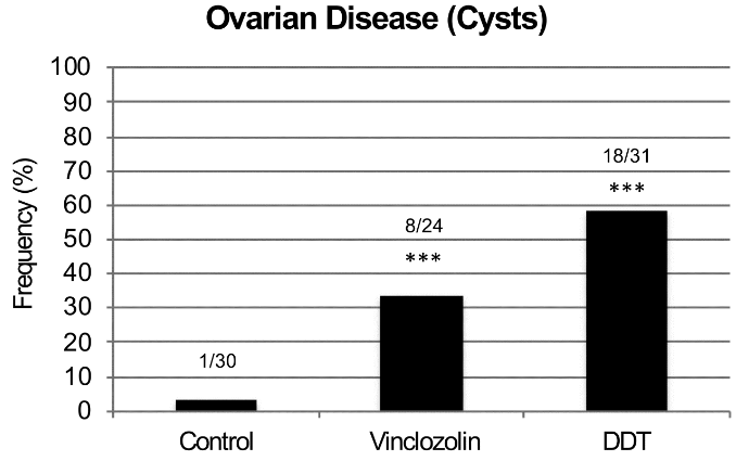
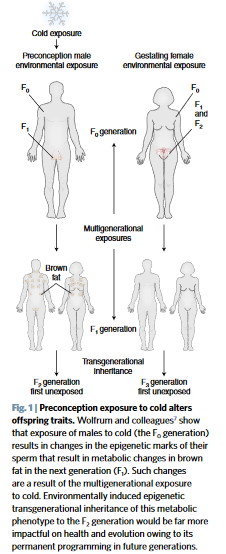
One plausible hypothesis of these human studies would be of inherited effects that skipped generations! The rodent studies Epigenetic transgenerational inheritance mechanisms that lead to prostate disease and Epigenetic transgenerational inheritance of ovarian disease found inherited diseases that didn’t manifest until the F3 great-grand offspring:

The F3 generation can have disease while the F1 and F2 generations do not.

Ancestral exposure to toxicants is a risk factor that must be considered in the molecular etiology of ovarian disease.

For the current study:

  • What could be expected from a study design that didn’t include F3 women and men, which is the only generation that didn’t have direct DES exposure?
  • What a nonsensical study design to permit NON-evidence like educational level!

Human studies of possible intergenerational and transgenerational epigenetic inheritance are urgently needed. There will be abundant evidence to discover if researchers will take their fields seriously.

Fear of feeling?

Here’s a 2018 article from two researchers involved in the Dunedin (New Zealand) Longitudinal Study. They coauthored many studies, including People had the same personalities at age 26 that they had at age 3.

The paper’s grand hypothesis was:

“A single dimension is able to measure a person’s liability to mental disorder, comorbidity among disorders, persistence of disorders over time, and severity of symptoms.”

The coauthors partially based this on:

“Repeated diagnostic interviews carried out over 25 years, when the research participants were 11, 13, 15, 18, 21, 26, 32, and 38 years old, and include information about seven diagnostic groups: anxiety, depression, attention deficit hyperactivity disorder, conduct disorder, substance dependence, bipolar disorder, and schizophrenia.”


https://ajp.psychiatryonline.org/doi/full/10.1176/appi.ajp.2018.17121383 “All for One and One for All: Mental Disorders in One Dimension” (not freely available)


More about the coauthors:

Two psychologists followed 1000 New Zealanders for decades. Here’s what they found about how childhood shapes later life

“Dunedin and other studies show that most people have at least one episode of mental illness during their lifetime.”


What compels people to manufacture “universal” truths? Aren’t such beliefs poor substitutes for feeling? For understanding historical, factual, personal truths?

What if the price we pay for avoiding and pressuring down our feelings is: A wasted life?

What if the grand hypothesis worth proving is: For one’s life to have meaning, each individual has to regain their feelings?

Fitting data

Let’s start out the new year with a repost of a cautionary reminder:

“Both “predict and “explain” imply that investigators have uncovered a reliable structure to phenomena, the latter involving hypotheses describing unseen mechanisms, leading to a new ability to control events and produce formerly unpredicted/unpredictable outcomes. This is clearly not a fair description of post hoc correlation-fishing.

The current publication system almost forces authors to make causal statements using filler verbs (e.g. to drive, alter, promote) as a form of storytelling (Gomez-Marin, 2017); without such a statement they are often accused of just collecting meaningless facts.”

https://mythsofvisionscience.wordpress.com/2018/12/30/neuroscience-newspeak-or-how-to-publish-meaningless-facts/ “Neuroscience Newspeak, Or How to Publish Meaningless Facts”

Eat your oats!

Here’s some motivation to replenish your oats supply.

From a 2013 Canadian human review:

“Review of human studies investigating the post-prandial blood-glucose lowering ability of oat and barley food products” https://www.nature.com/articles/ejcn201325

“Change in glycaemic response (expressed as incremental area under the post-prandial blood-glucose curve) was greater for intact grains than for processed foods. For processed foods, glycaemic response was more strongly related to the β-glucan dose alone than to the ratio of β-glucan to the available carbohydrate.”

The review found that people don’t have to eat a lot of carbohydrates to get the glycemic-response benefits of β-glucan. Also, eating ~3 grams of β-glucan in whole oats and barley will deliver the same glycemic-response benefits as eating ~4 grams of β-glucan in processed oats and barley.

However, the glycemic index used in the review is a very flawed measure. What’s the point of indexing healthy choices like whole grains to unhealthy choices that healthy people aren’t going to make anyway?


The reviewer somewhat redeemed herself by participating in a 2018 review:

“Processing of oat: the impact on oat’s cholesterol lowering effect” https://www.ncbi.nlm.nih.gov/pmc/articles/PMC5885279/

“For a similar dose of β-glucan:

  1. Liquid oat-based foods seem to give more consistent, but moderate reductions in cholesterol than semi-solid or solid foods where the results are more variable;
  2. The quantity of β-glucan and the molecular weight at expected consumption levels (∼3 g day) play a role in cholesterol reduction; and
  3. Unrefined β-glucan-rich oat-based foods (where some of the plant tissue remains intact) often appear more efficient at lowering cholesterol than purified β-glucan added as an ingredient.”

The review’s sections 3. Degree of processing and functionality and 4. Synergistic action of oat constituents were informative:

“Both in vitro and in vivo studies clearly demonstrated the beneficial effect of oat on cholesterolemia, which is unlikely to be due exclusively to β-glucan, but rather to a combined and synergetic action of several oat compounds acting together to reduce blood cholesterol levels.”


Another use of β-glucan is to improve immune response. Here’s a 2016 Netherlands study where the researchers used β-glucan to get a dozen people well after making them sick with lipopolysaccharide as is often done in animal studies:

β-Glucan Reverses the Epigenetic State of LPS-Induced Immunological Tolerance” https://www.ncbi.nlm.nih.gov/pmc/articles/PMC5927328/

“The innate immune ‘training stimulus’ β-glucan can reverse macrophage tolerance ex vivo.”

I’ve curated other research on β-glucan’s immune-response benefits in:

Adverse epigenetic effects of prenatal and perinatal anesthesia

This 2018 Chinese animal review subject was prenatal and perinatal anesthesia’s adverse epigenetic effects on a fetus/neonate:

“Accumulating evidence from rodent and primate studies has demonstrated that in utero or neonatal exposure to commonly used inhaled and intravenous general anesthetics is associated with neural degeneration and subsequent neurocognitive impairments, manifested in learning and memory disabilities.

So far, conflicting data exist about the effect of anesthetic agents on neurodevelopment in humans and no definite conclusion has been given yet.”

The inhibitors in the above graphic counter anesthesia’s effects on the fetus/neonate, summarized as:

“Epigenetic targeting of DNA methyltransferases and/or histone deacetylases may have some therapeutic value.”


Do physicians consider possible epigenetic alterations of a newborn’s chromatin structure and gene expression when they administer anesthesia to mothers during childbirth?

https://www.ncbi.nlm.nih.gov/pmc/articles/PMC6079265/ “Epigenetic Alterations in Anesthesia-Induced Neurotoxicity in the Developing Brain”

A top-down view of biological goal-directed mechanisms

This 2016 US/Italy article was written from a perspective of regenerative bioengineering:

“Higher levels beyond molecular can have their own unique dynamics that offer better (e.g. more parsimonious and potent) explanatory power than models made at lower levels. Biological systems may be best amenable to models that include information structures (organ shape, size, topological arrangements and complex anatomical metrics) not defined at molecular or cellular level but nevertheless serving as the most causally potent ‘knobs’ regulating large-scale outcomes.

Top-down models can be as quantitative as familiar bottom-up systems biology examples, but they are formulated in terms of building blocks that cannot be defined at the level of gene expression and treat those elements as bona fide causal agents (which can be manipulated by interventions and optimization techniques). The near-impossibility of determining which low-level components must be tweaked in order to achieve a specific system-level outcome is a problem that plagues most complex systems.

The current paradigm in biology of exclusively tracking physical measurable and ignoring internal representation and information structures in patterning contexts quite resemble the ultimately unsuccessful behaviourist programme in psychology and neuroscience. For example, even if stem cell biologists knew how to make any desired cell type from an undifferentiated progenitor, the task of assembling them into a limb would be quite intractable.

Current state of the art in the field of developmental bioelectricity is that it is known, at the cellular level, how resting potentials are transduced into downstream gene cascades, as well as which transcriptional and epigenetic targets are sensitive to change in developmental bioelectrical signals. What is largely missing however is a quantitative understanding of how global dynamics of bioelectric circuits make decisions that orchestrate large numbers of individual cells, spread out over considerable anatomical distances, towards specific pattern outcomes.”


Regenerative research is gathering evidence for goal-directed memory and learning that doesn’t meet current definitions. For example:

salamander

“A tail grafted to the flank of a salamander slowly remodels to a limb, a structure more appropriate for its new location, illustrating shape homeostasis towards a normal amphibian body plan. Even tail tip cells (in red) slowly become fingers, showing that remodelling is not driven by only local information.”

These reviewers compared their findings to several existing research and real-world-operations domains. Other models may also benefit from concepts of:

“Quantitative, predictive, mechanistic understanding of goal-directed morphogenesis.”

https://royalsocietypublishing.org/doi/full/10.1098/rsif.2016.0555 “Top-down models in biology: explanation and control of complex living systems above the molecular level”


I came across this article as a result of its citation in The Body Electric blog post.

“Levin drops a hint that there are photo-sensitive drugs that can control ion gates that can be used to translate a projected geometric image into a pattern of membrane potentials. He argues that the patterns encode ‘blueprints’ rather than a ‘construction manual’ based on the fact that the program is adaptive in the face of physical barriers and disruptions.”

Epigenetic clock statistics and methods

This 2018 Chinese study was a series of statistical and methodological counter-arguments to a previous epigenetic clock study finding that:

“Only [CpG] sites mapping to the ELOVL2 promoter constitute cell and tissue-type independent aDMPs [age-associated differentially methylated positions].”

The study used external data sets and the newer epigenetic clock’s fibroblast data in its analyses to find:

“While we agree that specific sites mapping to ELOVL2 are special aDMPs in the sense that their effect sizes are particularly large across a number of different tissue-types, our analysis suggests that most aDMPs are valid across multiple different tissue types, suggesting that shared aDMPs are common.”

The details of each of the study’s counter-arguments were compelling. For example:

“We analyzed Illumina 850k data from an EWAS profiling blood, buccal and cervical samples from a common set of 263 women. Because blood is a complex mixture of many immune-cell subtypes, and buccal and cervical samples are highly contaminated by immune cells, we identified aDMPs in each tissue after adjustment for batch effects and cell-type heterogeneity.

Using either an FDR [false discovery rate] < 0.05 or Bonferroni adjusted P-value < 0.05 thresholds, the overlap of aDMPs between the 3 tissues was highly significant, mimicking the result obtained on blood cell subtypes. We observed a total of 2200 aDMPs in common between blood, buccal and cervix, an overlap which cannot be explained by random chance.”

The study’s Discussion section provided qualifications and limitations such as:

“It is important to point out that even if age-associated DNAm changes are widespread across the genome, downstream functional effects may be rare. While specific aDMPs may be shared between tissue-types, it is only in specific tissues or cell-types that any associated functional deregulation may be of biological and clinical significance.

https://www.aging-us.com/article/101666/text “Cell and tissue type independent age-associated DNA methylation changes are not rare but common”


The November 2018 issue of Aging also contained other articles of interest:

https://www.aging-us.com/article/101626/text “Accelerated DNA methylation age and the use of antihypertensive medication among older adults”

“DNAmAge and AA [age acceleration] may not be able to capture the preventive effects of AHMs [antihypertensive medications] that reduce cardiovascular risks and mortality.”

https://www.aging-us.com/article/101633/text “Azithromycin and Roxithromycin define a new family of senolytic drugs that target senescent human fibroblasts”

“Azithromycin preferentially targets senescent cells, removing approximately 97% of them with great efficiency. This represents a near 25-fold reduction in senescent cells.”

https://www.aging-us.com/article/101647/text “Disease or not, aging is easily treatable”

“Aging consists of progression from (pre)-pre-diseases (early aging) to diseases (late aging associated with functional decline). Aging is NOT a risk factor for these diseases, as aging consists of these diseases: aging and diseases are inseparable.”

Chronological age by itself is an outdated clinical measurement

This 2018 editorial in the New England Journal of Medicine concerned a clinical trial of an osteoporosis treatment:

“When measurement of bone density was first introduced 25 years ago, absolute bone mineral density (g per square centimeter) was considered as too onerous for clinicians to understand. Ultimately, these events led to a treatment gap in patients who had strong clinical risk factors for an osteoporotic fracture (particularly age) but had T scores in the osteopenic range.

The average age of the participants in the current trial was approximately 3.5 years older than that in the Fracture Intervention Trial. Owing to the interaction between age and bone mineral density, the results of the current trial should not be extrapolated to younger postmenopausal women (50 to 64 years of age) with osteopenia.

This trial reminds us that risk assessment and treatment decisions go well beyond bone mineral density and should focus particularly on age and a history of previous fractures.”

https://www.nejm.org/doi/pdf/10.1056/NEJMe1812434 “A Not-So-New Treatment for Old Bones”


This editorial provided some history of how a still-generally-accepted set of diagnostic measurements were selected for their relative convenience instead of chosen for their efficacy. Add chronological age to such ineffective measurements.

Let’s recognize better aging and diagnostic measurements, then incorporate them. How else will we advance past the above uninformative averaging and unhelpful recommendation based on chronological age?

The time has passed for physicians and clinicians to consider only chronological age when evaluating a patient’s clinical age. More effective human age measurements covering the entire person as well as their body’s components include:

F2.large

A slanted view of the epigenetic clock

The founder of the epigenetic clock technique was interviewed for MIT Technology Review:

“We need to find ways to keep people healthier longer,” he says. He hopes that refinements to his clock will soon make it precise enough to reflect changes in lifestyle and behavior.”


The journalist attempted to dumb the subject down “for the rest of us” with distortions such as the headline. The varying correlation of epigenetic age to chronological age was somewhat better reported in the story:

“The epigenetic clock is more accurate the younger a person is. It’s especially inaccurate for the very old.”

The journalist inappropriately used luck as a synonym for randomness/stochasticity:

“He estimates that about 40% of the ticking rate is determined by genetic inheritance, and the rest by lifestyle and luck.”

A third example of less-than-straightforward journalism started with:

“Such personalization raises questions about fairness. If your epigenetic clock is ticking faster through no fault of your own..”

Were MIT Technology Review readers unable to comprehend a straightforward story on the epigenetic clock? What was the purpose of slants and distortions in an introductory article?

https://www.technologyreview.com/s/612256/want-to-know-when-youre-going-to-die/ “Want to know when you’re going to die?”

Reductionism vs. reductionism

This 2004 essay by an evolutionary biologist reviewed his field’s direction in the current century:

“Science is impelled by two main factors, technological advance and a guiding vision (overview). A properly balanced relationship between the two is key to the successful development of a science.

Without the proper technological advances the road ahead is blocked. Without a guiding vision there is no road ahead; the science becomes an engineering discipline, concerned with temporal practical problems.

Empirical reductionism is in essence methodological; it is simply a mode of analysis, the dissection of a biological entity or system into its constituent parts in order better to understand it. Empirical reductionism makes no assumptions about the fundamental nature, an ultimate understanding, of living things.

Fundamentalist reductionism (the reductionism of 19th century classical physics), on the other hand, is in essence metaphysical. It is ipso facto a statement about the nature of the world: living systems (like all else) can be completely understood in terms of the properties of their constituent parts.

This is a view that flies in the face of what classically trained biologists tended to take for granted, the notion of emergent properties. Whereas emergence seems to be required to explain numerous biological phenomena, fundamentalist reductionism flatly denies its existence: in all cases the whole is no more than the sum of its parts.”

Regarding cellular evolution:

“Modern concepts of cellular evolution are effectively petrified versions of 19th century speculations. Try to imagine a biology released from the intellectual shackles of mechanism, reductionism, and determinism.

Evolution, as a complex dynamic process, will encounter critical points in its course, junctures that result in phase transitions (drastic changes in the character of the system as a whole). Human language is a development that has set Homo sapiens worlds apart from its otherwise very close primate relatives, adding new dimensions to the phase space within which human evolution occurs. Another good critical-point candidate is the advent of (eucaryotic) multicellularity.

Nowhere in thinking about a symbiotic origin of the eucaryotic cell has consideration been given to the fact that the process as envisioned would involve radical change in the designs of the cells involved. You can’t just tear cell designs apart and willy-nilly construct a new type of design from the parts.

The organization of the mitochondrial endosymbiont is radically changed during its evolution, but that change is a degeneration to a far simpler “cell-like” design. The mitochondrial design could never evolve back to the level of complexity that its free-living [bacterial] ancestor had.

A common thread that links language and multicellularity is communication (interaction at a distance). In each case a complex, sophisticated network of interactions forms the medium within which the new level of organization (entities) comes into existence.

Our experience with variation and selection in the modern context does not begin to prepare us for understanding what happened when cellular evolution was in its very early, rough-and-tumble phase(s) of spewing forth novelty. Cellular evolution began in a highly multiplex fashion, from many initial independent ancestral starting points, not just a single one.”

https://mmbr.asm.org/content/68/2/173 “A New Biology for a New Century”


I came across this review by it being referenced in this researcher’s blog post:

Chinese Longevity Herb
I often don’t agree with him, but I subscribe to his blog because it’s interesting.

The arrogance of a paradigm exceeding its evidence

This 2018 commentary from the American College of Emergency Physicians by 7 physicians discussed the harm that will result from imposing a mandatory paradigm of sepsis treatment. I’ll quote sections that mention evidence:

“These metrics [for pneumonia treatment] had little evidentiary basis but led to an institutional-fostered culture of overdiagnosis and overtreatment. Have we learned from this folly or does a new sepsis guideline promote similar time-based treatment strategies with little direct supporting evidence?

Like the pneumonia quality measure, this resource-heavy care flows from an overreaching interpretation of evidence. Despite that evidence consistently fails to find a benefit of a single treatment strategy, the Surviving Sepsis Campaign continues to promote recommendations that bypass the individual clinician’s judgment.

Although well intentioned, the current sepsis bundles and the potential penalties associated with noncompliance lay a heavy weight on ED [emergency department] care absent evidence that a net benefit will follow. The proposed Surviving Sepsis Campaign abbreviated bundle heightens the burden by further restricting the time allotted for the identification and treatment of patients with suspected sepsis, all without any evidence of benefit or knowledge of the logistic consequences or cost.”

The paradigm’s promoters didn’t learn the appropriate lessons in the above page regarding “the sense of embarrassment and regret once experienced with the pneumonia quality metric.”


What do you think are the root causes of the Surviving Sepsis Campaign’s agenda?

  • Did it start with lawyers? Lawsuits can force hospitals into actions for which the primary reason is to avoid “the potential penalties associated with noncompliance.”
  • Is it due to governments? Governments can force hospitals into actions “without any evidence of benefit or knowledge of the logistic consequences or cost” when the hospitals accept government reimbursement.
  • Did it start with other groups of unaccountable people who think they know better than everyone else about how others should act?

https://www.sciencedirect.com/science/article/pii/S0196064418306073 “The 2018 Surviving Sepsis Campaign’s Treatment Bundle: When Guidelines Outpace the Evidence Supporting Their Use” (not freely available)

Epigenetic transgenerational inheritance of ovarian disease

This 2018 Washington rodent study investigated ovarian disease in F3 great-granddaughters caused by their F0 great-grandmothers’ exposures to DDT or vinclozolin while pregnant:

“Two of the most prevalent ovarian diseases affecting women’s fertility and health are Primary Ovarian Insufficiency (POI) and Polycystic Ovarian Syndrome (PCOS). POI is characterized by a marked reduction in the primordial follicle pool of oocytes and the induction of menopause prior to age 40. POI currently affects approximately 1% of female population. While genetic causes can be ascribed to a minority of patients, around 90% of POI cases are considered idiopathic, with no apparent genetic link nor known cause.

PCOS is a multi-faceted disease that affects 6-18% of women. It is characterized by infrequent ovulation or anovulation, high androgen levels in the blood, and the presence of multiple persistent ovarian cysts.

For both PCOS and POI other underlying causes such as epigenetic transgenerational inheritance of disease susceptibility have seldom been considered. Epigenetic transgenerational inheritance is defined as “the germline transmission of epigenetic information and phenotypic change across generations in the absence of any continued direct environmental exposure or genetic manipulation.” Epigenetic factors include:

    • DNA methylation,
    • Histone modifications,
    • Expression of noncoding RNA,
    • RNA methylation, and
    • Alterations in chromatin structure.

The majority of transgenerational studies have examined sperm transmission of epigenetic changes due to limitations in oocyte numbers for efficient analysis.

There was no increase in ovarian disease in direct fetal exposed F1 [grandmothers] or germline exposed F2 [mothers] generation vinclozolin or DDT lineage rats compared to controls.

F3 generation ovarian disease

The transgenerational molecular mechanism is distinct and involves the germline (sperm or egg) having an altered epigenome that following fertilization may modify the embryonic stem cells epigenome and transcriptome. This subsequently impacts the epigenetics and transcriptome of all somatic cell types derived from these stem cells.

Therefore, all somatic cells in the transgenerational [F3] animal have altered epigenomes and transcriptomes and those sensitive to this alteration will be susceptible to develop disease. The F3 generation can have disease while the F1 and F2 generations do not, due to this difference in the molecular mechanisms involved.

The epimutations and gene expression differences observed are present in granulosa cells in the late pubertal female rats at 22-24 days of age, which is long before any visible signs of ovarian disease are detectable. This indicates that the underlying factors that can contribute to adult-onset diseases like PCOS and POI appear to be present early in life.

Ancestral exposure to toxicants is a risk factor that must be considered in the molecular etiology of ovarian disease.”


1. The study highlighted a great opportunity for researchers of any disease that frequently has an “idiopathic” diagnosis. It said a lot about research priorities that “around 90% of POI cases are considered idiopathic, with no apparent genetic link nor known cause.”

It isn’t sufficiently explanatory for physicians to continue using categorization terminology from thousands of years ago. Science has progressed enough with measured evidence to discard the “idiopathic” category and express probabilistic understanding of causes.

2. One of this study’s coauthors made a point worth repeating in The imperative of human transgenerational studies: What’s keeping researchers from making a significant difference in their fields with human epigenetic transgenerational inheritance studies?

3. Parts of the study’s Discussion section weren’t supported by its evidence. The study didn’t demonstrate:

  • That “all somatic cells in the transgenerational animal have altered epigenomes and transcriptomes”; and
  • The precise “molecular mechanisms involved” that exactly explain why “the F3 generation can have disease while the F1 and F2 generations do not.”

https://www.tandfonline.com/doi/abs/10.1080/15592294.2018.1521223 “Environmental Toxicant Induced Epigenetic Transgenerational Inheritance of Ovarian Pathology and Granulosa Cell Epigenome and Transcriptome Alterations: Ancestral Origins of Polycystic Ovarian Syndrome and Primary Ovarian Insuf[f]iency” (not freely available)

The imperative of human transgenerational studies

The coauthor of:

pointed out the opportunity for the researchers of A seasonal epigenetic effect of conception on BMI to have their work make a difference in their field:

“The ability of environmental epigenetics to promote an adaptive phenotype to cold has impacts on evolution. However, the impacts would be far greater if the phenomenon was transgenerational.

Future studies are now needed to determine whether the cold-induced thrifty metabolic phenotype is transmitted to subsequent generations. If exposure not only impacts the health of offspring, but also of all subsequent generations, the impact is significant.”


Every human alive today has observable lasting epigenetic effects caused by environmental factors:

  • During the earliest parts of our lives;
  • From our parents’ exposures and experiences before we’re conceived – many of which are inadequately researched; and
  • Potentially from some of our earlier ancestors’ exposures and experiences.

Aren’t animal studies’ evidence for epigenetic transgenerational inheritance sufficient to compel serious human follow-on research efforts by research sponsors and study designers?

The same comments about epigenetic effects caused by temperature potentially inherited by multiple human generations can also be made about other environmental factors, such as:

  • Nutrition,
  • Toxins – the commentator’s usual area of study, and
  • Stress.

I hope that these researchers value their professions enough to make a difference with this or other areas of their expertise. And that sponsors won’t thwart researchers’ desires for difference-making science by putting them into endless funding queues.

https://www.nature.com/articles/s41591-018-0187-3 “Preconception cold-induced epigenetic inheritance” (not freely available)