Stuck in the wrong paradigm

This 2019 article questioned the paradigm of determining substance carcinogenicity:

“In the absence of robust epidemiological data, the final arbiter of whether a chemical is considered to be a carcinogen or not has been based on the outcome of long-term rodent bioassays. This approach is incompatible with the current knowledge of the etiology of cancer. The current view of the etiology of cancer suggests that it is not useful to consider carcinogenicity as a single hazardous property with its own hazard category.

There is no bright line between carcinogens and non-carcinogens but rather there is a continuum with some chemicals having high potential, some having no potential, and others having potential at a point along the continuum. This continuum exists alongside other adverse effects. One problem is being stuck in the old practice of wishing to reproduce the binary “carcinogen/non-carcinogen” results of the long-term bioassay rather than move to a new paradigm in assessing the chemical’s position on the spectrum of carcinogenic potential.

The two-year bioassay has such high variability (because of the variability of the carcinogenic process it is trying to measure and the interplay between dose limiting toxicity and cell proliferation inducing toxicity) that the outcome of the assay for compounds with low to intermediate carcinogenic potential is little more than a lottery. After half a century, it has only been used to evaluate less than 5% of chemicals that are in use. It is not reproducible because of the probabalistic nature of the process it is evaluating combined with dose limiting toxicity, dose selection, and study design.”


Unscientific research paradigms will eventually collapse because they can’t withstand the scrutiny of the scientific method. Too bad the coauthors didn’t kill off this one while they were still in positions at the US Environmental Protection Agency, World Health Organization, etc.

https://www.sciencedirect.com/science/article/pii/S0273230019300248 “Chemical carcinogenicity revisited 2: Current knowledge of carcinogenesis shows that categorization as a carcinogen or non-carcinogen is not scientifically credible” (not freely available)

Burying human transgenerational epigenetic evidence

The poor substitutes for evidence in this 2018 US study guaranteed that human transgenerational epigenetically inherited effects wouldn’t be found in the generations that followed after prenatal diethylstilbestrol (DES) exposure:

“A synthetic, nonsteroidal estrogen, DES was administered to pregnant women under the mistaken belief it would reduce pregnancy complications and losses. From the late 1930s through the early 1970s, DES was given to nearly two million pregnant women in the US alone.

Use of DES in pregnancy was discontinued after a seminal report showed a strong association with vaginal clear cell adenocarcinoma in prenatally exposed women. A recent analysis of the US National Cancer Institute (NCI) DES Combined Cohort Follow-up Study showed elevated relative risks of twelve adverse health outcomes.

We do not have sufficient data concerning the indication for DES in the grandmother to determine whether adverse pregnancy outcomes in the third generation might resemble those of their grandmothers. Fourth generation effects of prenatal exposures in humans have not been reported.”

https://www.sciencedirect.com/science/article/pii/S0890623818304684 “Reproductive and Hormone-Related Outcomes in Women whose Mothers were Exposed in utero to Diethylstilbestrol (DES): A Report from the US National Cancer Institute DES Third Generation Study” (not freely available)


This study had many elements in common with its poor-quality reference [25] “Transgenerational effects of prenatal exposure to the 1944–45 Dutch famine” which is freely available at https://obgyn.onlinelibrary.wiley.com/doi/full/10.1111/1471-0528.12136.

That study’s Methods section showed:

  1. Its non-statistical data was almost all unverified self-reports by a self-selected sample of the F2 grandchildren, average age 37.
  2. No detailed physical measurements or samples were taken of the F2 grandchildren, or of their F1 parents, or of their F0 grandparents, all of which are required as baselines for any transgenerational epigenetic inheritance findings.
  3. No detailed physical measurements or samples were taken of the F3 great-grandchildren, which is the generation that may provide transgenerational evidence if the previous generations also have detailed physical baselines.

That study’s researchers drew enough participants (360) such that their statistics package allowed them to impute and assume into existence a LOT of data. But the scientific method constrained them to make factual statements of what the evidence actually showed. They admitted:

“In conclusion, we did not find a transgenerational effect of prenatal famine exposure on the health of grandchildren in this study.”


The current study similarly used the faulty methods 1-3 above to produce results such as:

“We do not have sufficient data concerning the indication for DES in the [F0] grandmother to determine whether adverse pregnancy outcomes in the [F2] third generation might resemble those of their grandmothers.

Fourth [F3] generation effects of prenatal exposures in humans have not been reported.“

Zero studies of probably more than 10,000,000 F3 great-grandchildren of DES-exposed women just here in the US?

Who is against funding these studies? Who is afraid of what such studies may find?

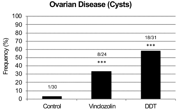
One plausible hypothesis of these human studies would be of inherited effects that skipped generations! The rodent studies Epigenetic transgenerational inheritance mechanisms that lead to prostate disease and Epigenetic transgenerational inheritance of ovarian disease found inherited diseases that didn’t manifest until the F3 great-grand offspring:

The F3 generation can have disease while the F1 and F2 generations do not.

Ancestral exposure to toxicants is a risk factor that must be considered in the molecular etiology of ovarian disease.

For the current study:

  • What could be expected from a study design that didn’t include F3 women and men, which is the only generation that didn’t have direct DES exposure?
  • What a nonsensical study design to permit NON-evidence like educational level!

Human studies of possible intergenerational and transgenerational epigenetic inheritance are urgently needed. There will be abundant evidence to discover if researchers will take their fields seriously.

The arrogance of a paradigm exceeding its evidence

This 2018 commentary from the American College of Emergency Physicians by 7 physicians discussed the harm that will result from imposing a mandatory paradigm of sepsis treatment. I’ll quote sections that mention evidence:

“These metrics [for pneumonia treatment] had little evidentiary basis but led to an institutional-fostered culture of overdiagnosis and overtreatment. Have we learned from this folly or does a new sepsis guideline promote similar time-based treatment strategies with little direct supporting evidence?

Like the pneumonia quality measure, this resource-heavy care flows from an overreaching interpretation of evidence. Despite that evidence consistently fails to find a benefit of a single treatment strategy, the Surviving Sepsis Campaign continues to promote recommendations that bypass the individual clinician’s judgment.

Although well intentioned, the current sepsis bundles and the potential penalties associated with noncompliance lay a heavy weight on ED [emergency department] care absent evidence that a net benefit will follow. The proposed Surviving Sepsis Campaign abbreviated bundle heightens the burden by further restricting the time allotted for the identification and treatment of patients with suspected sepsis, all without any evidence of benefit or knowledge of the logistic consequences or cost.”

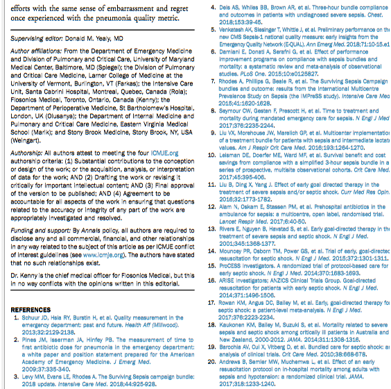
The paradigm’s promoters didn’t learn the appropriate lessons in the above page regarding “the sense of embarrassment and regret once experienced with the pneumonia quality metric.”


What do you think are the root causes of the Surviving Sepsis Campaign’s agenda?

  • Did it start with lawyers? Lawsuits can force hospitals into actions for which the primary reason is to avoid “the potential penalties associated with noncompliance.”
  • Is it due to governments? Governments can force hospitals into actions “without any evidence of benefit or knowledge of the logistic consequences or cost” when the hospitals accept government reimbursement.
  • Did it start with other groups of unaccountable people who think they know better than everyone else about how others should act?

https://www.sciencedirect.com/science/article/pii/S0196064418306073 “The 2018 Surviving Sepsis Campaign’s Treatment Bundle: When Guidelines Outpace the Evidence Supporting Their Use” (not freely available)

Epigenetic transgenerational inheritance of ovarian disease

This 2018 Washington rodent study investigated ovarian disease in F3 great-granddaughters caused by their F0 great-grandmothers’ exposures to DDT or vinclozolin while pregnant:

“Two of the most prevalent ovarian diseases affecting women’s fertility and health are Primary Ovarian Insufficiency (POI) and Polycystic Ovarian Syndrome (PCOS). POI is characterized by a marked reduction in the primordial follicle pool of oocytes and the induction of menopause prior to age 40. POI currently affects approximately 1% of female population. While genetic causes can be ascribed to a minority of patients, around 90% of POI cases are considered idiopathic, with no apparent genetic link nor known cause.

PCOS is a multi-faceted disease that affects 6-18% of women. It is characterized by infrequent ovulation or anovulation, high androgen levels in the blood, and the presence of multiple persistent ovarian cysts.

For both PCOS and POI other underlying causes such as epigenetic transgenerational inheritance of disease susceptibility have seldom been considered. Epigenetic transgenerational inheritance is defined as “the germline transmission of epigenetic information and phenotypic change across generations in the absence of any continued direct environmental exposure or genetic manipulation.” Epigenetic factors include:

    • DNA methylation,
    • Histone modifications,
    • Expression of noncoding RNA,
    • RNA methylation, and
    • Alterations in chromatin structure.

The majority of transgenerational studies have examined sperm transmission of epigenetic changes due to limitations in oocyte numbers for efficient analysis.

There was no increase in ovarian disease in direct fetal exposed F1 [grandmothers] or germline exposed F2 [mothers] generation vinclozolin or DDT lineage rats compared to controls.

F3 generation ovarian disease

The transgenerational molecular mechanism is distinct and involves the germline (sperm or egg) having an altered epigenome that following fertilization may modify the embryonic stem cells epigenome and transcriptome. This subsequently impacts the epigenetics and transcriptome of all somatic cell types derived from these stem cells.

Therefore, all somatic cells in the transgenerational [F3] animal have altered epigenomes and transcriptomes and those sensitive to this alteration will be susceptible to develop disease. The F3 generation can have disease while the F1 and F2 generations do not, due to this difference in the molecular mechanisms involved.

The epimutations and gene expression differences observed are present in granulosa cells in the late pubertal female rats at 22-24 days of age, which is long before any visible signs of ovarian disease are detectable. This indicates that the underlying factors that can contribute to adult-onset diseases like PCOS and POI appear to be present early in life.

Ancestral exposure to toxicants is a risk factor that must be considered in the molecular etiology of ovarian disease.”


1. The study highlighted a great opportunity for researchers of any disease that frequently has an “idiopathic” diagnosis. It said a lot about research priorities that “around 90% of POI cases are considered idiopathic, with no apparent genetic link nor known cause.”

It isn’t sufficiently explanatory for physicians to continue using categorization terminology from thousands of years ago. Science has progressed enough with measured evidence to discard the “idiopathic” category and express probabilistic understanding of causes.

2. One of this study’s coauthors made a point worth repeating in The imperative of human transgenerational studies: What’s keeping researchers from making a significant difference in their fields with human epigenetic transgenerational inheritance studies?

3. Parts of the study’s Discussion section weren’t supported by its evidence. The study didn’t demonstrate:

  • That “all somatic cells in the transgenerational animal have altered epigenomes and transcriptomes”; and
  • The precise “molecular mechanisms involved” that exactly explain why “the F3 generation can have disease while the F1 and F2 generations do not.”

https://www.tandfonline.com/doi/abs/10.1080/15592294.2018.1521223 “Environmental Toxicant Induced Epigenetic Transgenerational Inheritance of Ovarian Pathology and Granulosa Cell Epigenome and Transcriptome Alterations: Ancestral Origins of Polycystic Ovarian Syndrome and Primary Ovarian Insuf[f]iency” (not freely available)

The imperative of human transgenerational studies

The coauthor of:

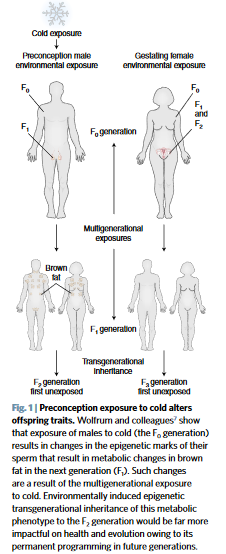
pointed out the opportunity for the researchers of A seasonal epigenetic effect of conception on BMI to have their work make a difference in their field:

“The ability of environmental epigenetics to promote an adaptive phenotype to cold has impacts on evolution. However, the impacts would be far greater if the phenomenon was transgenerational.

Future studies are now needed to determine whether the cold-induced thrifty metabolic phenotype is transmitted to subsequent generations. If exposure not only impacts the health of offspring, but also of all subsequent generations, the impact is significant.”


Every human alive today has observable lasting epigenetic effects caused by environmental factors:

  • During the earliest parts of our lives;
  • From our parents’ exposures and experiences before we’re conceived – many of which are inadequately researched; and
  • Potentially from some of our earlier ancestors’ exposures and experiences.

Aren’t animal studies’ evidence for epigenetic transgenerational inheritance sufficient to compel serious human follow-on research efforts by research sponsors and study designers?

The same comments about epigenetic effects caused by temperature potentially inherited by multiple human generations can also be made about other environmental factors, such as:

  • Nutrition,
  • Toxins – the commentator’s usual area of study, and
  • Stress.

I hope that these researchers value their professions enough to make a difference with this or other areas of their expertise. And that sponsors won’t thwart researchers’ desires for difference-making science by putting them into endless funding queues.

https://www.nature.com/articles/s41591-018-0187-3 “Preconception cold-induced epigenetic inheritance” (not freely available)

Reversing epigenetic changes with CRISPR/Cas9

This 2018 Chinese review highlighted areas in which CRISPR/Cas9 technology has, is, and could be applied to rewrite epigenetic changes:

“CRISPR/Cas9-mediated epigenome editing holds a great promise for epigenetic studies and therapeutics.

It could be used to selectively modify epigenetic marks at a given locus to explore mechanisms of how targeted epigenetic alterations would affect transcription regulation and cause subsequent phenotype changes. For example, inducing histone methylation or acetylation at the Fosb locus in the mice brain reward region, nucleus accumbens, could affect relevant transcription network and thus control behavioral responses evoked by drug and stress.

Epigenome editing has the potential for epigenetic treatment, especially for the disorders with abnormal gene imprinting or epigenetic marks. Targeted epigenetic silencing or reactivation of the mutant allele could be a potential therapeutic approach for diseases such as Rett syndrome and Huntington’s disease.

Noncoding RNA plays important roles in gene imprinting and chromatin remodeling. CRISPR/Cas9 has been shown to be potential for manipulating noncoding RNA expression, including microRNA, long noncoding RNA, and miRNA families and clusters.

In vivo overexpression of the Yamanaka factors have proven to be able to fully or partially help somatic cells to regain pluripotency in situ. These rejuvenated cells would subsequently differentiate again to replace the lost cell types.”


The last paragraph was described in The epigenetic clock theory of aging as a promising technique:

“To date, the most effective in vitro intervention against epigenetic ageing is achieved through expression of Yamanaka factors, which convert somatic cells into pluripotent stem cells, thereby completely resetting the epigenetic clock.”

The reviewers cited three references for in vivo studies of this technique. Overall, I didn’t see that any of the review’s references were in vivo human studies.

https://www.ncbi.nlm.nih.gov/pmc/articles/PMC6079388/ “Novel Epigenetic Techniques Provided by the CRISPR/Cas9 System”

Epigenetic factors affecting female rat sexual behavior

This 2018 Baltimore/Montreal rodent study found:

“If sexually naïve females have their formative sexually rewarding experiences paired with the same male, they will recognize that male and display mate-guarding behavior towards him in the presence of a female competitor. Female rats that display mate-guarding behavior also show enhanced activation of oxytocin and vasopressin neurons in the supraoptic and paraventricular hypothalamic nucleus.

We examined the effect of a lysine-specific demethylase-1 inhibitor to block the action of demethylase enzymes and maintain the methylation state of corresponding genes. Female rats treated with the demethylase inhibitor failed to show any measure of mate guarding, whereas females treated with vehicle displayed mate guarding behavior. Demethylase inhibitor treatment also blocked the ability of familiar male cues to activate oxytocin and vasopressin neurons, whereas vehicle-treated females showed this enhanced activation.”

General principles and their study-specific illustrations were:

“Histone modifications are a key element in gene regulation through chromatin remodeling. Histone methylation / demethylation does not have straightforward transcriptional outcomes as do other histone modifications, like acetylation, which is almost invariably associated with transcriptional activation.

What is of vital importance in regards to histone methylation / demethylation is the pattern of methylation that is established. Patterns of methylation incorporate both methylated and demethylated residues, and are what ultimately play a role in transcriptional outcomes.

In the present study, inhibiting LSD1 demethylase enzymes disrupted the ability of cells to properly establish histone methylation / demethylation patterns, thus creating a deficit in the cells’ ability to transcribe the gene products necessary for the enhanced induction of OT, AVP, and the subsequent mate-guarding behaviors we observed. This study is the first to demonstrate a definitive role of epigenetic histone modifications in a conditioned sexual response.”

https://www.sciencedirect.com/science/article/pii/S0031938418303421 “Inhibition of lysine-specific demethylase enzyme disrupts sexually conditioned mate guarding in the female rat” (not freely available)

The role of recall neurons in traumatic memories

This 2018 Swiss rodent study found:

“Our data show that:

  • A subset of memory recall–induced neurons in the DG [dentate gyrus] becomes reactivated after memory attenuation,
  • The degree of fear reduction positively correlates with this reactivation, and
  • The continued activity of memory recall–induced neurons is critical for remote fear memory attenuation.

Although other brain areas such as the prefrontal cortex and the amygdala are likely to be implicated in remote fear memories and remain to be investigated, these results suggest that fear attenuation at least partially occurs in memory recall–induced ensembles through updating or unlearning of the original memory trace of fear.

These data thereby provide the first evidence at an engram-specific level that fear attenuation may not be driven only by extinction learning, that is, by an inhibitory memory trace different from the original fear trace.

Rather, our findings indicate that during remote fear memory attenuation both mechanisms likely coexist, albeit with the importance of the continued activity of memory recall–induced neurons experimentally documented herein. Such activity may not only represent the capacity for a valence change in DG engram cells but also be a prerequisite for memory reconsolidation, namely, an opportunity for learning inside the original memory trace.

As such, this activity likely constitutes a physiological correlate sine qua non for effective exposure therapies against traumatic memories in humans: the engagement, rather than the suppression, of the original trauma.”

The researchers also provided examples of human trauma:

“We dedicate this work to O.K.’s father, Mohamed Salah El-Dien, and J.G.’s mother, Wilma, who both sadly passed away during its completion.”


So, how can this study help humans? The study had disclosed and undisclosed limitations:

1. Humans aren’t lab rats. We can ourselves individually change our responses to experiential causes of ongoing adverse effects. Standard methodologies can only apply external treatments.

2. It’s a bridge too far to go from neural activity in transgenic mice to expressing unfounded opinions on:

“A physiological correlate sine qua non for effective exposure therapies against traumatic memories in humans.”

Human exposure therapies have many drawbacks, in addition to being applied externally to the patient on someone else’s schedule. A few others were discussed in The role of DNMT3a in fear memories:

  • “Inability to generalize its efficacy over time,
  • Potential return of adverse memory in the new/novel contexts,
  • Context-dependent nature of extinction which is widely viewed as the biological basis of exposure therapy.”

3. Rodent neural activity also doesn’t elevate recall to become an important goal of effective human therapies. Clearly, what the rodents experienced should have been translated into human reliving/re-experiencing, not recall! Terminology used in animal studies preferentially has the same meaning with humans, since the purpose of animal studies is to help humans.

4. The researchers acknowledged that:

“Other brain areas such as the prefrontal cortex and the amygdala are likely to be implicated in remote fear memories and remain to be investigated.”

A study that provided evidence for basic principles of Primal Therapy determined another brain area:

“The findings imply that in response to traumatic stress, some individuals, instead of activating the glutamate system to store memories, activate the extra-synaptic GABA system and form inaccessible traumatic memories.”

The study I curated yesterday, Organ epigenetic memory, demonstrated organ memory storage. It’s hard to completely rule out that other body areas may also store traumatic memories.

The wide range of epigenetic memory storage vehicles is one reason why effective human therapies need to address the whole person, the whole body, and each individual’s entire history.

http://science.sciencemag.org/content/360/6394/1239 “Reactivation of recall-induced neurons contributes to remote fear memory attenuation” (not freely available)

Here’s one of the researchers’ outline:


This post has somehow become a target for spammers, and I’ve disabled comments. Readers can comment on other posts and indicate that they want their comment to apply here, and I’ll re-enable comments.

Flawed epigenetic measurements of behavioral experiences

This 2018 New York rodent study not only wasted resources but also speciously attempted to extrapolate animal study findings to humans:

“While it is clear that behavioral experience modulates epigenetic profiles, it is less evident how the nature of that experience influences outcomes and whether epigenetic/genetic “biomarkers” could be extracted to classify different types of behavioral experience.

Male and female mice were subjected to either:

  • a Fixed Interval (FI) schedule of food reward, or
  • a single episode of forced swim followed by restraint stress, or
  • no explicit behavioral experience

after which global expression levels of two activating (H3K9ac and H3K4me3) and two repressive (H3K9me2 and H3k27me3) post-translational histone modifications (PTHMs), were measured in hippocampus (HIPP) and frontal cortex (FC).

A random subset of 5 of the 12 animals from each sex/behavioral experience group were used for these analyses. FC and HIPP were dissected from each of those 5 brains and homogenized for subsequent analyses. Thus, sample size for PTHM expression levels was n = 5 for each region/sex/behavioral treatment group and all PTHM expression level analyses utilized the homogenized tissue.

The specific nature of the behavioral experience differentiated profiles of PTHMs in a sex- and brain region-dependent manner, with all 4 PTHMs changing in parallel in response to different behavioral experiences. Global PTHMs may provide a higher-order pattern recognition function.”


The researchers knew or should have known that measuring “global expression levels” in “homogenized tissue” of “n = 5” subjects was flawed, and they did it anyway. They acknowledged some of the numerous study design defects with qualifiers such as:

“Even though these were global levels of histone modifications (and thus not indicative of changes at specific genes or sites on genes)..

As FS-RS behavioral experience was completed before FI behavioral experience, a longer overall post-behavior experience time (approximately 1 week) elapsed for this group, resulting in some differences in overall timing between these experiences and global PTHM assessment. However, extending the duration of the FS-RS experience (i.e., repeated exposures) would also have led to habituation..”

Did they purposely make these mistakes because of the “biomarkers” paradigm?

What would they have found if they had followed their judgments and training to design a better study? Experience-dependent histone modifications that differed by gender and brain region was certainly a promising research opportunity.

As for extrapolating the cited animal study findings to humans? Ummm..NO!

https://www.ncbi.nlm.nih.gov/pmc/articles/PMC6060276/ “Different Behavioral Experiences Produce Distinctive Parallel Changes in, and Correlate With, Frontal Cortex and Hippocampal Global Post-translational Histone Levels”

Going off the rails with the biomarker paradigm

This 2018 US government rodent study used extreme dosages to achieve its directed goals of demonizing nicotine and extolling the biomarker paradigm:

“This study examined whether adolescent nicotine exposure alters adult hippocampus-dependent learning, involving persistent changes in hippocampal DNA methylation and if choline, a dietary methyl donor, would reverse and mitigate these alterations.

Mice were chronically treated with nicotine (12.6mg/kg/day) starting at post-natal day 23 (pre-adolescent), p38 (late adolescent), or p54 (adult) for 12 days followed by a 30-day period during which they consumed either standard chow or chow supplemented with choline (9g/kg).

Our gene expression analyses support this model and point to two particular genes involved in chromatin remodeling, Smarca2 and Bahcc1. Both Smarca2 and Bahcc1 showed a similar inverse correlation pattern between promoter methylation and gene expression.

Our findings support a role for epigenetic modification of hippocampal chromatin remodeling genes in long-term learning deficits induced by adolescent nicotine and their amelioration by dietary choline supplementation.”


Let’s use the average weight of a US adult male – published by the US Centers for Disease Control as 88.8 kg – to compare the study’s dosages with human equivalents:

  1. Nicotine at ((“12.6mg/kg/day” x .081) x 88.8 kg) = 90.6 mg.
  2. Choline at ((“9g/kg” x .081) x 88.8 kg) = 65 g. The US National Institutes of Health published the Tolerable Upper Intake Levels for Choline as 3.5 g!!

Neither of these dosages are even remotely connected to human realities:

  1. The human-equivalent dosage of nicotine used in this study would probably kill an adult human before the end of 12 days.
  2. What effects would an adult human suffer from exceeding the choline “Tolerable Upper Intake Level” BY 18 TIMES for 30 days?

Isn’t the main purpose of animal studies to help humans? What’s the justification for performing animal studies simply to promote an agenda?


A funding source of this study was National Institute on Drug Abuse (NIDA) Identification of Biomarkers for Nicotine Addiction award (T-DA-1002 MG). Has the biomarker paradigm been institutionalized to the point where research proposals that don’t have biomarkers as goals aren’t funded?

https://www.sciencedirect.com/science/article/pii/S107474271830193X “Choline ameliorates adult learning deficits and reverses epigenetic modification of chromatin remodeling factors related to adolescent nicotine exposure” (not freely available)

Measuring epigenetic changes at a single-cell level

This 2018 Canadian cell study described the development of a single-cell protocol to:

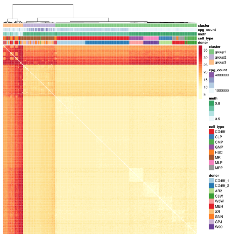
“Profile primitive hematopoietic cells of mouse and human origin to identify epigenetically distinct subpopulations. Deep sampling of the CpG content of individual HSCs allowed for the near complete reconstitution of regulatory states from epigenetically defined subpopulations of HSCs and revealed a high level of redundancy of CpG methylation states within these phenotypically defined hematopoietic cell types.

Hematopoietic stem cells (HSCs) are functionally defined cells that display evidence of extensive self-renewal of their ability to generate mature blood cells for the lifetime of the organism and following transplantation into myelosuppressed permissive hosts. Most of the epigenetic measurements underpinning these observations represent consensus values experimentally derived from thousands of cells partially enriched in HSCs or their progeny, thus failing to discern distinct epigenetic states within HSCs.

Current analytical strategies for single-cell DNA methylation measurements average DNA methylation in fixed genomic bins or over defined genomic regions.

However, inference across cells (as well as sequence context) assumes homogeneity across cells, which is at cross-purposes with the generation of single-cell molecular measurements through the potential to mask rare subpopulations.

We identified donor as a significant source of consistent epigenetic heterogeneity, which was reduced but not eliminated by correcting for personal genetic variants. This observation is consistent with previous reports that showed genetic diversity as related to but not accountable for all DNA methylation differences and suggests that in utero environmental differences may be encoded within the HSC compartment.”


The study advanced science not only by measuring CpG methylation within each HSC, but also by producing another data point “that in utero environmental differences may be encoded within the HSC compartment.”

The paragraph with “assumes homogeneity across cells” bold text provided another example of the statistical analysis flaw that gives individually inapplicable results per Group statistics don’t necessarily describe an individual. The above graphic of human hematopoietic phenotypes demonstrated that the researchers have potentially solved this problem by measuring individual cells.

The researchers discussed another aspect of the study that’s similar to the epigenetic clock methodology:

“Phenotype-specific methylation signatures are characterized by extensive redundancy such that distinct epigenetic states can be accurately described by only a small fraction of single-CpG methylation states. In support of such a notion, the unique components of a DNA methylation “age” signature are contained in ∼353 CpGs sites, presumably representing a random sample of a total age signature that involves many more sites not detected using the reduced representation strategies from which these signatures have been derived.”

Also, in The epigenetic clock theory of aging the originator of the epigenetic clock characterized HSCs as an effective intervention against epigenetic aging:

“In vivo, haematopoietic stem cell therapy resets the epigenetic age of blood of the recipient to that of the donor.”

https://www.cell.com/stem-cell-reports/article/S2213-6711(18)30308-4/fulltext “High-Resolution Single-Cell DNA Methylation Measurements Reveal Epigenetically Distinct Hematopoietic Stem Cell Subpopulations”

Epigenetic effects of breast cancer treatments

This 2018 UC San Diego review subject was the interplay between breast cancer treatments and their effects on aging:

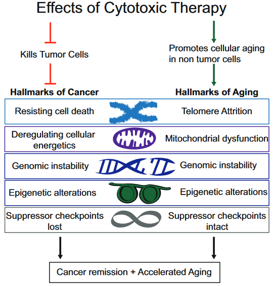
“Although current breast cancer treatments are largely successful in producing cancer remission and extending lifespan, there is concern that these treatments may have long lasting detrimental effects on cancer survivors, in part, through their impact on non-tumor cells. It is unclear whether breast cancer and/or its treatments are associated with an accelerated aging phenotype.

In this review, we have highlighted five of nine previously described cellular hallmarks of aging that have been described in the context of cytotoxic breast cancer treatments:

  1. Telomere attrition;
  2. Mitochondrial dysfunction;
  3. Genomic instability;
  4. Epigenetic alterations; and
  5. Cellular senescence.”


The review was full of caveats weakening the above graphic’s associations:

  1. “Telomere attrition – Blood TL [telomere length] was not associated with chemotherapy in three out of four studies;
  2. Mitochondrial dysfunction – How cancer therapies affect cellular energetics as they relate to rate of aging is unclear;
  3. Genomic instability – Potentially contributing to accelerated aging;
  4. Epigenetic alterations – Although some of the key regulators of these processes have begun to be identified, including DNA and histone methylases and demethylases, histone acetylases and de-acetylases and chromatin remodelers, how they regulate the changes in aging through alteration of global transcriptional programs, remains to be elucidated; and
  5. Cellular senescence – Dysregulated pathways can be targeted by cytotoxic chemotherapies, resulting in preferential cell death of tumor cells, but how these treatments also affect normal cells with intact pathways is unclear.”

To their credit, these reviewers at least presented some of the contrary evidence, and didn’t continue on with a directed narrative as other reviewers are prone to do.

https://www.sciencedirect.com/science/article/pii/S1879406818301176 “Breast cancer treatment and its effects on aging” (not freely available)


The originator of the epigenetic clock methodology was a coauthor of the review. Only one of his works was cited in the Epigenetic alterations subsection:

https://link.springer.com/article/10.1007%2Fs10549-017-4218-4 “DNA methylation age is elevated in breast tissue of healthy women”

This freely-available 2017 study quoted below highlighted that epigenetic clock measurements as originally designed were tissue-specific:

“To our knowledge, this is the first study to demonstrate that breast tissue epigenetic age exceeds that of blood tissue in healthy female donors. In addition to validating our earlier finding of age elevation in breast tissue, we further demonstrate that the magnitude of the difference between epigenetic age of breast and blood is highest in the youngest women in our study (age 20–30 years) and gradually diminishes with advancing age. As women approach the age of the menopausal transition, we found that the epigenetic of age of blood approaches that of the breast.”

Additional caution was justified in both interpreting age measurements and extending them into “cellular hallmarks” when the tissue contained varying cell types:

“Our studies were performed on whole breast tissue. Diverse types of cells make up whole breast tissue, with the majority of cells being adipocytes. Other types of cells include epithelial cells, cuboidal cells, myoepithelial cells, fibroblasts, inflammatory cells, vascular endothelial cells, preadipocytes, and adipose tissue macrophages.

This raises the possibility that the magnitude of the effects we observe, of breast tissue DNAm age being greater than other tissues, might be an underestimation, since it is possible that not all of the cells of the heterogenous sample have experienced this effect. Since it is difficult to extract DNA from adipose tissue, we suspect that the majority of DNA extracted from our whole breast tissues was from epithelial and myoepithelial cells.”

Allergies and epigenetic histone modifications

This 2018 German review provided short summaries of 44 studies on the contribution of histone modifications to allergies. An overall summary of their search results was:

“There are at least two levels at which the role of histone modifications is manifested.

  • One is the regulation of cells that contribute to the allergic inflammation (T cells and macrophages) and those that participate in airway remodeling.
  • The other is the direct association between histone modifications and allergic phenotypes.

Inhibitors of histone-modifying enzymes may potentially be used as anti-allergic drugs. Furthermore, epigenetic patterns may provide novel tools in the diagnosis of allergic disorders.”


This type of search is what’s expected of researchers who will perform either:

  • A meta-analysis of studies selected from the search results; or
  • Their own study.

These reviewers didn’t indicate that they were proceeding along either path.

The review was fine for the purpose of presenting current studies of the subject. But this was just the preparatory stage of research.

https://aacijournal.biomedcentral.com/articles/10.1186/s13223-018-0259-4 “Histone modifications and their role in epigenetics of atopy and allergic diseases”

Epigenetic variations in metabolism

This 2018 German review was comprehensive for its subject, epigenetic control of variation and stochasticity in metabolic disease. I’ll focus on one aspect, phenotypic variation:

“Phenotypic [Mendelian] variation can result both from gain- and loss-of-function mutations. Because of the extreme interconnectivity of cell regulatory networks, even at the cellular level, predicting the impact of a sequence variant is difficult as the resultant variation acts:

  • In the context of all other variants and
  • Their potential additive, synergistic and antagonistic interactions.

This phenomenon is known as epistasis.

∼98.5% of our genome is non-protein-coding: it is pervasively transcribed, and its transcripts can support regulatory function. Among the best functionally characterized non-coding RNAs (ncRNAs) arising from these sequences are microRNAs (miRNAs).

Environmental [non-Mendelian] variation or ‘stimuli’ occurring during critical windows of susceptibility can elicit lifelong alterations in an individual’s phenotype. Intergenerational metabolic reprogramming [in fruit flies] results from global alterations in chromatin state integrity, particularly from reduced H3K27me3 and H3K9me3 [histone] domains.

The broad variation of fingerprints in humans is thought to depend to a large degree on stochastic variation in mechanical forces. These clear examples of inducible multi-stable or stochastic variation highlight how little we know about the landscape of potential phenotypic variation itself.

Consensus estimates of heritability for obesity and T2D are ∼70% and ∼35% respectively. The remaining, unexplained component is known to involve gene–environment interactions as well as non-Mendelian players.”


Although the above graphic displays transgenerational inheritance for humans, the reviewers didn’t cite any human studies that adequately demonstrated causes for and effects of transgenerational epigenetic inheritance.

I’ve read the cited Swedish and Dutch studies. Their designs, methods, and “correlate with” / “was associated with” results didn’t provide incontrovertible evidence from the F0 great-grandparents, F1 grandparents, F2 parents, and F3 children. It’s necessary to thoroughly study each generation to confirm definitive transgenerational epigenetic inheritance causes and effects.

As noted in How to hijack science: Ignore its intent and focus on the 0.0001%, there aren’t any such published studies to cite. Researchers urgently need to do this human research, and stop using these poor substitutes [1] to pretend there are already adequately evidenced transgenerational epigenetic inheritance human results.

I downgraded the review for treating research of this and other subjects as faits accomplis. It’s opposite ends of the evidential spectrum to state “how little we know about the landscape of potential phenotypic variation,” and in the same review, speciously extrapolate animal experiments into putative human results.

https://www.sciencedirect.com/science/article/pii/S2212877818301984 “Epigenetic control of variation and stochasticity in metabolic disease”


[1] As an example of the poor substitutes for evidence, a researcher referred me to the 2013 “Transgenerational effects of prenatal exposure to the 1944–45 Dutch famine” which is freely available at https://obgyn.onlinelibrary.wiley.com/doi/full/10.1111/1471-0528.12136 as a study finding human transgenerational epigenetic inheritance.

The Methods section showed:

  • The study’s non-statistical data was almost all unverified self-reports by a self-selected sample of the F2 generation, average age 37.
  • No detailed physical measurements or samples were taken of them, nor of the F1 generation, nor of the F0 generation, all of which are required as baselines for any transgenerational epigenetic inheritance findings.
  • No detailed physical measurements or samples were taken of the F3 generation, which is the generation that may provide transgenerational evidence if the previous generations also have detailed physical baselines.

The study’s researchers drew enough participants (360) such that their statistics package allowed them to impute and assume into existence a LOT of data. But the scientific method constrained them to make factual statements of what the evidence actually showed. They admitted:

“In conclusion, we did not find a transgenerational effect of prenatal famine exposure on the health of grandchildren in this study.”

Yet this study is somehow cited for evidence of human transgenerational epigenetically inherited causes and effects!

Group statistics don’t necessarily describe an individual

I’m curating this 2018 UC Berkeley/Drexel/Netherlands analysis of human studies via its press coverage. The authors:

“Collaborated to analyze data on hundreds of adults – some mentally or physically sound, others suffering from various conditions such as depression, anxiety, or post-traumatic stress disorder. Participants had completed surveys about their mental health and had their heart rates monitored via electrocardiogram.

Researchers used the data to conduct six different experiments. They sought to find out whether the conclusions of each study would successfully apply to participants individually.

One study that focused on how frequently depression sufferers reported feeling worried. Results tallied from the pool of participants showed that depressed people worry a significant amount.

But when the analysis was applied individually, the results were all over the map. Some participants worried hardly at all, while others were notably beyond the group average.

Another experiment that centered around the link between fear and avoidance showed a strong correlation when measured as a group. Yet a significant number of participants who experienced fear had no issues with avoiding various activities.

Across all six experiments, the authors could not show that what was concluded for the group applied to most individuals.”


http://www.pnas.org/content/early/2018/06/15/1711978115.full “Lack of group-to-individual generalizability is a threat to human subjects research”


Other studies such as the below have addressed problems with statistical analysis techniques. These issues aren’t limited to human studies:


The current study highlighted the fact that people aren’t interchangeable. Assuming ergodicity is a statistical analysis flaw that produces individually inapplicable results for many measurements of fruit flies, cells, humans, you name the organism.

When this presumption makes a study’s statistics useless for an individual, researchers can’t cure the analysis by invoking an “individual differences” meme. Neither is the flaw fixed by spinning a tale about “This is how we can truly personalize medicine.”

The current study needed to provide evidence for its proposed solution.


Regarding worrying, Dr. Arthur Janov said it best as I quoted in How well can catastrophes be predicted?:

“Worrying is not a problem, it is the symptom of something that is occurring physiologically within the brain. What causes worrying is the problem.

Constant worry is anticipating catastrophe. But what we don’t realize is that the catastrophe already has happened; we simply have no access to it.

We are actually worried about the past, not the future.”