Beneficial epigenetic effects of mild stress with social support during puberty

This 2016 Pennsylvania rodent study found:

“Stress in the context of social support experienced over the pubertal window can promote epigenetic reprogramming in the brain to increase resilience to age-related cognitive decline in females.

These findings are actually consistent with previous studies showing that some amount of adversity, or adversity under more favorable circumstances such as social support or a protective gene polymorphism, provides a measure of ‘grit’ in coping with later life challenges.

Our findings provide a unique perspective on this relationship, as they highlight the important link between experience during the pubertal window and cognitive health during aging.”

These researchers made efforts to further investigate causes of unexpected results, such as:

“Peripubertal stress alone did not significantly alter Barnes maze performance in aging compared to aged Controls. Mice that had experienced stress with concurrent social support (CVS + SI) actually performed better than Control aged mice, specifically in learning the reversal task faster.

Peripubertal stress had no effect on corticosterone levels in response to an acute restraint stress or in sensorimotor gating and baseline startle reactivity.”

Their investigations led to epigenetic findings:

“Consistent with our behavioral findings, stress in the context of social interaction resulted in long-term reprogramming of gene expression in the PFC [prefrontal cortex]. While there were no differentially expressed genes between Control and CVS females, there were 88 genes that were significantly different between Control and CVS + SI groups. Of genes that were downregulated, a large portion (23 genes; 35%) were microRNAs.

We found that the PFC transcriptome of CVS + SI aged females was significantly enriched for predicted targets of the 23 microRNAs that were downregulated in the PFC in these mice. This suggests that microRNAs represent a mode of regulation capable of enacting far-reaching programmatic effects, and are a critical epigenetic gene expression regulatory mechanism.”

Applicability to humans was suggested by associations such as:

“A single microRNA can target more than a hundred different mRNA targets, and more than 45,000 conserved microRNA binding sites have been annotated in the 3′ UTR of 60% of human genes.”


A few limitations were noted:

“Given that mice at this age (1 year) are commonly compared to ‘late middle aged’ humans, later aging time points may yield differences in this group. Alternatively, it is possible that there was an effect of peripubertal stress that was not long-lasting due to the mild nature of our chronic stress model.

To include early neglect as a part of the stressor experience, CVS females were weaned one week earlier (PN21) than Control and CVS + SI mice. Addition of stress of this earlier weaning likely poses a significant contribution to programming of the PFC.”

One of the study coauthors was also a coauthor of:

https://www.ncbi.nlm.nih.gov/pmc/articles/PMC4870871/ “Peripubertal stress with social support promotes resilience in the face of aging”
A review that inadvertently showed how memory paradigms prevented relevant research

This 2016 Swiss review of enduring memories demonstrated what happens when scientists’ reputations and paychecks interfered with them recognizing new research and evidence in their area but outside their paradigm: “A framework containing the basic assumptions, ways of thinking, and methodology that are commonly accepted by members of a scientific community.”

A. Most of the cited references were from decades ago that established these paradigms of enduring memories. Fine, but the research these paradigms excluded was also significant.

B. All of the newer references were continuations of established paradigms. For example, a 2014 study led by one of the reviewers found:

“Successful reconsolidation-updating paradigms for recent memories fail to attenuate remote (i.e., month-old) ones.

Recalling remote memories fails to induce histone acetylation-mediated plasticity.”

The researchers elected to pursue a workaround of the memory reconsolidation paradigm when the need for a new paradigm of enduring memories directly confronted them!

C. None of the reviewers’ calls for further investigations challenged existing paradigms. For example, when the reviewers suggested research into epigenetic regulation of enduring memories, they somehow found it best to return to 1984, a time when dedicated epigenetics research had barely begun:

“Whether memories might indeed be ‘coded in particular stretches of chromosomal DNA’ as originally proposed by Crick [in 1984] and if so what the enzymatic machinery behind such changes might be remain unclear. In this regard, cell population-specific studies are highly warranted.”


Two examples of relevant research the review failed to consider:

1. A study that provided evidence for basic principles of Primal Therapy went outside existing paradigms to research state-dependent memories:

“If a traumatic event occurs when these extra-synaptic GABA receptors are activated, the memory of this event cannot be accessed unless these receptors are activated once again.

It’s an entirely different system even at the genetic and molecular level than the one that encodes normal memories.”

What impressed me about that study was the obvious nature of its straightforward experimental methods. Why hadn’t other researchers used the same methods decades ago? Doing so could have resulted in dozens of informative follow-on study variations by now, which is my point in Item A. above.

2. A relevant but ignored 2015 French study What can cause memories that are accessible only when returning to the original brain state? which supported state-dependent memories:

“Posttraining/postreactivation treatments induce an internal state, which becomes encoded with the memory, and should be present at the time of testing to ensure a successful retrieval.”


The review also showed the extent to which historical memory paradigms depend on the subjects’ emotional memories. When it comes to human studies, though, designs almost always avoid studying emotional memories.

It’s clearly past time to Advance science by including emotion in research.

http://www.hindawi.com/journals/np/2016/3425908/ “Structural, Synaptic, and Epigenetic Dynamics of Enduring Memories”

Epigenetic effects of diet, and reversing DNA methylation

This 2015 French review focused on:

“The role of maternal health and nutrition in the initiation and progression of metabolic and other disorders.

The effects of various in utero exposures and maternal nutritional status may have different effects on the epigenome. However, critical windows of exposure that seem to exist during development need to be better defined.

The epigenome can be considered as an interface between the genome and the environment that is central to the generation of phenotypes and their stability throughout the life course.”

The reviewer used the term “transgenerational” to refer to effects that were more appropriately termed parental or intergenerational. Per the definition in A review of epigenetic transgenerational inheritance of reproductive disease, for the term to apply there needed to be evidence in at least the next 2 male and/or 3 female generations of:

“Altered epigenetic information between generations in the absence of continued environmental exposure.”

The review had separate sections for animal and human studies.

http://www.ncbi.nlm.nih.gov/pmc/articles/PMC4663595/ “Impact of Maternal Diet on the Epigenome during In Utero Life and the Developmental Programming of Diseases in Childhood and Adulthood”


I arrived at the above review as a result of it citing the 2014 Harvard Reversing DNA Methylation: Mechanisms, Genomics, and Biological Functions. I’ll quote a few items from that review’s informative “Role of DNA demethylation in neural development” section:

“Distinct parts of mammalian brains, including frontal cortex, hippocampus, and cerebellum, all exhibit age-dependent acquisition of 5hmC [an oxidized derivative of 5mC [methylation of the fifth position of cytosine]].

In fact, the genome of mature neurons in adult central nervous system contains the highest level of 5hmC of any mammalian cell-type (~40% as abundant as 5mC in Purkinje neurons in cerebellum). These observations indicate that 5mC oxidation and potentially DNA demethylation may be functionally important for neuronal differentiation and maturation processes.

A comprehensive base-resolution analyses of 5mC and 5hmC in mammalian frontal cortex in both fetal and adult stages indicate that non-CpG methylation (mCH) and CpG hydroxymethylation (hCG) drastically build up in cortical neurons after birth, coinciding with the peak of synaptogenesis and synaptic pruning in the cortex. This study demonstrated that mCH could become a dominant form of cytosine modifications in adult brains, accounting for 53% in adult human cortical neuronal genome.

In mature neurons, intragenic mCH is preferentially enriched at inactive non-neuronal lineage-specific genes, indicating a role in negative regulation of the associated transcripts. By contrast, genic hCG is positively correlated with gene expression levels.”

What’s the underlying question for every brain study to answer?

Is the underlying question for every brain study to answer:

  • How do our brains internally represent the external world?

Is it:

  • How did we learn what we know?
  • How do we forget or disregard what we’ve learned?
  • What keeps us from acquiring and learning newer or better information?

How about:

  • What affects how we pay attention to our environments?
  • How do our various biochemical states affect our perceptions, learning, experiences, and behavior?
  • How do these factors in turn affect our biology?

Or maybe:

  • Why do we do what we do?
  • How is our behavior affected by our experiences?
  • How did we become attracted and motivated toward what we like?
  • How do we develop expectations?
  • Why do we avoid certain situations?

Not to lose sight of:

  • How do the contexts affect all of the above?
  • What happens over time to affect all of the above?

This 2015 UCLA paper reviewed the above questions from the perspective of Pavlovian conditioning:

“The common definition of Pavlovian conditioning, that via repeated pairings of a neutral stimulus with a stimulus that elicits a reflex the neutral stimulus acquires the ability to elicit that the reflex, is neither accurate nor reflective of the richness of Pavlovian conditioning. Rather, Pavlovian conditioning is the way we learn about dependent relationships between stimuli.

Pavlovian conditioning is one of the few areas in biology in which there is direct experimental evidence of biological fitness.”


The most important question unanswered by the review was:

  • How can its information be used to help humans?

How can Pavlovian conditioning answer: What can a human do about the thoughts, feelings, behavior, epigenetic effects – the person – the phenotype – that they’ve been shaped into?

One example of the unanswered question: the review pointed out in a section about fear extinction that this process doesn’t involve unlearning. Fear extinction instead inhibits the symptoms of fear response. The fear memory is still intact, awaiting some other context to be reactivated and expressed.

How can this information be used to help humans?

  • Is inhibiting the symptoms and leaving the fear memory in place costless with humans?
  • Or does this practice have both potential and realized adverse effects?
  • Where’s the human research on methods that may directly address a painful emotional memory?

One relevant hypothesis of Dr. Arthur Janov’s Primal Therapy is that a person continues to be their conditioned self until they address the sources of their pain. A corollary is that efforts to relieve symptoms seldom address causes.

How could it be otherwise? A problem isn’t cured by ameliorating its effects.

http://cshperspectives.cshlp.org/content/8/1/a021717.full “The Origins and Organization of Vertebrate Pavlovian Conditioning”

Use it or lose it: the interplay of new brain cells, age, and activity

This 2015 German review was of aging and activity in the context of adult neurogenesis:

“Adult neurogenesis might be of profound functional significance because it occurs at a strategic bottleneck location in the hippocampus.


Age-dependent changes essentially reflect a unidirectional development in that everything builds on what has occurred before. In this sense, aging can also be seen as continued or lifelong development. This idea has limitations but is instructive with regard to adult neurogenesis, because adult neurogenesis is neuronal development under the conditions of the adult brain.

The age-related alterations of adult neurogenesis themselves have quantitative and qualitative components. So far, most research has focused on the quantitative aspects. But there can be little doubt that qualitative changes do not simply follow quantitative changes (e.g., in cell or synapse numbers), but emerge on a systems level and above when an organism ages. With respect to adult neurogenesis, only one multilevel experiment including morphology and behavior has been conducted, and, even in that study, only three time points were investigated.

In old age, adult neurogenesis occurs at only a small fraction of the level in early adulthood. The decline does not seem to be ‘regulated’ but rather the by-product of many age-related changes of other sorts.


From a behavioral level down to a synaptic level, activity increases adult neurogenesis. This regulation does not seem to occur in an all-or-nothing fashion but rather influences different stages of neuronal development differently. Both cell proliferation and survival are influenced by or even depend on activity.

The effects of exercise and environmental enrichment are additive, which indicates that increasing the potential for neurogenesis is sufficient to increase the actual use of the recruitable cells in the case of cognitive stimulation. Physical activity would not by itself provide specific hippocampus-relevant stimuli that induce net neurogenesis but be associated with a greater chance to encounter specific relevant stimuli.


Adult hippocampal neurogenesis might contribute to a structural or neural reserve that if appropriately trained early in life might provide a compensatory buffer of brain plasticity in the face of increasing neurodegeneration or nonpathological age-related functional losses. There is still only limited information on the activity-dependent parameters that help to prevent the age-dependent decrease in adult neurogenesis and maintain cellular plasticity.

The big question is what the functional contribution of so few new neurons over so long periods can be. Any comprehensive concept has to bring together the acute functional contributions of newly generated, highly plastic neurons and the more-or-less lasting changes they introduce to the network.”

I’ve quoted quite a lot, but there are more details that await your reading. A few items from the study referenced in the first paragraph above:

“The hippocampus represents a bottleneck in processing..adult hippocampal neurogenesis occurs at exactly the narrowest spot.

We have derived the theory that the function of adult hippocampal neurogenesis is to enable the brain to accommodate continued bouts of novelty..a mechanism for preparing the hippocampus for processing greater levels of complexity.”


The role of the hippocampus in emotion was ignored as it so often is. The way to address many of the gaps mentioned by the author may be to Advance science by including emotion in research.

For example, from the author’s The mystery of humans’ evolved capability for adults to grow new brain cells:

“Adult neurogenesis is already effective early in life, actually very well before true adulthood, and is at very high levels when sexual maturity has been reached. Behavioral advantages associated with adult neurogenesis must be relevant during the reproductive period.”

When human studies are designed to research how “behavioral advantages associated with adult neurogenesis must be relevant” what purpose does it serve to exclude emotional content?

http://cshperspectives.cshlp.org/content/7/11/a018929.full “Activity Dependency and Aging in the Regulation of Adult Neurogenesis”

A problematic study of oxytocin receptor gene methylation, childhood abuse, and psychiatric symptoms

This 2016 Georgia human study found:

“A role for OXTR [oxytocin receptor gene] in understanding the influence of early environments on adult psychiatric symptoms.

Data on 18 OXTR CpG sites, 44 single nucleotide polymorphisms, childhood abuse, and adult depression and anxiety symptoms were assessed in 393 African American adults. The Childhood Trauma Questionnaire (CTQ), a retrospective self-report inventory, was used to assess physical, sexual, and emotional abuse during childhood.

While OXTR CpG methylation did not serve as a mediator to psychiatric symptoms, we did find that it served as a moderator for abuse and psychiatric symptoms.”

From the Limitations section:

  1. “Additional insight will likely be gained by including a more detailed assessment of abuse timing and type on the development of biological changes and adverse outcomes.
  2. The degree to which methylation remains fixed following sensitive developmental time periods, or continues to change in response to the environment, is still a topic of debate and is not fully known.
  3. Comparability between previous findings and our study is limited given different areas covered.
  4. Our study was limited to utilizing peripheral tissue [blood]. OXTR methylation should ideally be assessed in the tissues that are known to express OXTR and directly involved in psychiatric symptoms. The degree to which methylation of peripheral tissues can be used to study methylation changes in response to the environment or in association with behavioral outcomes is currently a topic of debate.
  5. Our study did not evaluate gene expression and thus cannot explore the role of study CpG sites on regulation and expression.”

Addressing the study’s limitations:

  1. Early-life epigenetic regulation of the oxytocin receptor gene demonstrated – with no hint of abuse – how sensitive an infant’s experience-dependent oxytocin receptor gene DNA methylation was to maternal care. Treating prenatal stress-related disorders with an oxytocin receptor agonist provided evidence for prenatal oxytocin receptor gene epigenetic changes.
  2. No human’s answers to the CTQ, Adverse Childhood Experiences, or other questionnaires will ever be accurate self-reports of their prenatal, infancy, and early childhood experiences. These early development periods were likely when the majority of the subjects’ oxytocin receptor gene DNA methylation took place. The CTQ self-reports were – at best – evidence of experiences at later times and places, distinct from earlier experience-dependent epigenetic changes.
  3. As one example of incomparability, the 2009 Genomic and epigenetic evidence for oxytocin receptor deficiency in autism was cited in the Introduction section and again in the Limitations section item 4. Since that study was sufficiently relevant to be used as a reference twice, the researchers needed to provide a map between its findings and the current study.
  4. Early-life epigenetic regulation of the oxytocin receptor gene answered the question of whether or not an individual’s blood could be used to make inferences about their brain oxytocin receptor gene DNA methylation. The evidence said: NO, it couldn’t.
  5. It’s assumed that oxytocin receptor gene DNA methylation directly impacted gene expression such that increased levels of methylation were associated with decreased gene transcription. The study assumed but didn’t provide evidence that higher levels of methylation indicated decreased ability to use available oxytocin due to decreased receptor expression. The study also had no control group.

To summarize the study’s limitations:

  1. The study zeroed in on childhood abuse, and disregarded evidence for more relevant factors determining an individual’s experience-dependent oxytocin receptor gene DNA methylation. That smelled like an agenda.
  2. The study used CTQ answers as determinants, although what happened during the subjects’ earliest life was likely when the majority of epigenetic changes to the oxytocin receptor gene took place. If links existed between the subjects’ early-life DNA methylation and later-life conditions, they weren’t evidenced by CTQ answers about later life that couldn’t self-report relevant experiences from conception through age three that may have caused DNA methylation.
  3. There was no attempt to make findings comparable with cited studies. That practice and the lack of a control group reminded me of Problematic research with telomere length.
  4. The researchers tortured numbers until they confessed “that CpG methylation may interact with abuse to predict psychiatric symptoms.” But there was no direct evidence that each subject’s blood oxytocin gene receptor DNA methylation interacted as such! Did the “may interact” phrase make the unevidenced inferences more plausible, or permit contrary evidence to be disregarded?
  5. See Testing the null hypothesis of oxytocin’s effects in humans for examples of what happens when researchers compound assumptions and unevidenced inferences.

The study’s institution, Emory University, and one of the study’s authors also conducted Conclusions without evidence regarding emotional memories. That 2015 study similarly disregarded relevant evidence from other research, and made statements that weren’t supported by that study’s evidence.

The current study used “a topic of debate” and other disclaimers to provide cover for unconvincing methods and analyses in pursuit of..what? What overriding goals were achieved? Who did the study really help?

http://onlinelibrary.wiley.com/enhanced/doi/10.1111/cdev.12493/ “Oxytocin Receptor Genetic and Epigenetic Variations: Association With Child Abuse and Adult Psychiatric Symptoms”


This post has somehow become a target for spammers, and I’ve disabled comments. Readers can comment on other posts and indicate that they want their comment to apply here, and I’ll re-enable comments.

Early-life epigenetic regulation of the oxytocin receptor gene

This 2015 US/Canadian rodent study investigated the effects of natural variation in maternal care:

“The effects of early life rearing experience via natural variation in maternal licking and grooming during the first week of life on behavior, physiology, gene expression, and epigenetic regulation of Oxtr [oxytocin receptor gene] across blood and brain tissues (mononucleocytes, hippocampus, striatum, and hypothalamus).

Rats reared by high licking-grooming (HL) and low licking-grooming (LL) rat dams exhibited differences across study outcomes:

  • LL offspring were more active in behavioral arenas,
  • Exhibited lower body mass in adulthood, and
  • Showed reduced corticosterone responsivity to a stressor.

Oxtr DNA methylation was significantly lower at multiple CpGs in the blood of LL versus HL males, but no differences were found in the brain. Across groups, Oxtr transcript levels in the hypothalamus were associated with reduced corticosterone secretion in response to stress, congruent with the role of oxytocin signaling in this region.

Methylation of specific CpGs at a high or low level was consistent across tissues, especially within the brain. However, individual variation in DNA methylation relative to these global patterns was not consistent across tissues.

These results suggest that:

  • Blood Oxtr DNA methylation may reflect early experience of maternal care, and
  • Oxtr methylation across tissues is highly concordant for specific CpGs, but
  • Inferences across tissues are not supported for individual variation in Oxtr methylation.

nonsignificance

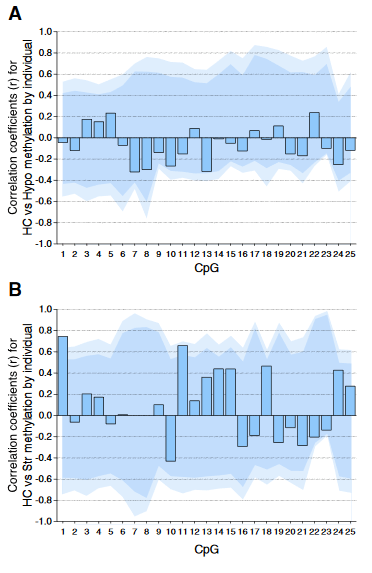
Individual DNA methylation values were not correlated across brain tissues, despite tissue concordance at the group level.

For each CpG, we computed the Pearson correlation coefficient r between methylation values for matched samples in pairs of brain regions (bars). Dark and light shaded regions represent 95% and 99% thresholds, respectively, of distributions of possible correlation coefficients determined from 10,000 permutations of the measured values among the individuals. These distributions represent the null hypothesis that an individual DNA methylation value in one brain region does not help to predict the value in another region in the same animal.

(A) Correlations based on pyrosequencing data for matched samples passing validation in both hippocampus (HC) and hypothalamus (Hypo). Correlations for individuals at each CpG were either weak (.2 < r < .3) or absent (r < .2), and none were significant, even prior to correction for multiple comparisons.

(B) Correlations for matched samples passing validation in both hippocampus and striatum (Str). Two correlations (CpG 1 and 11) were individually significant prior to but not following correction, and this result could be expected by chance.

Correlations between hippocampus and blood (described in the text) yielded similar results, and no particular CpG yielded consistently high correlation across multiple tissues.”


The study focused on whether or not an individual’s experience-dependent oxytocin receptor gene DNA methylation in one of the four studied tissues could be used to infer a significant effect in the three other tissues. The main finding was NO, it couldn’t!

The researchers’ other findings may have been strengthened had they also examined causes for the observed effects. The “natural variation in maternal licking and grooming” developed from somewhere, didn’t it?

The subjects’ mothers were presumably available for the same tests as the subjects, but nothing was done with them. Investigating at least one earlier generation may have enabled etiologic associations of “the effects of early life rearing experience” and “individual variation in DNA methylation.”

https://www.sciencedirect.com/science/article/abs/pii/S0018506X1500118X “Natural variation in maternal care and cross-tissue patterns of oxytocin receptor gene methylation in rats” (not freely available)

The effects of imposing helplessness

This 2016 New York rodent study found:

“By using unbiased and whole-brain imaging techniques, we uncover a number of cortical and subcortical brain structures that have lower activity in the animals showing helplessness than in those showing resilience following the LH [learned helplessness] procedure. We also identified the LC [locus coeruleus] as the sole subcortical area that had enhanced activity in helpless animals compared with resilient ones.

Some of the brain areas identified in this study – such as areas in the mPFC [medial prefrontal cortex], hippocampus, and amygdala – have been previously implicated in clinical depression or depression-like behavior in animal models. We also identified novel brain regions previously not associated with helplessness. For example, the OT [olfactory tubercle], an area involved in odor processing as well as high cognitive functions including reward processing, and the Edinger–Westphal nucleus containing centrally projecting neurons implicated in stress adaptation.

The brains of helpless animals are locked in a highly stereotypic pathological state.”

Concerning the study’s young adult male subjects:

“To achieve a subsequent detection of neuronal activity related to distinct behavioral responses, we used the c-fosGFP transgenic mice expressing c-FosGFP under the control of a c-fos promoter. The expression of the c-fosGFP transgene has been previously validated to faithfully represent endogenous c-fos expression.

Similar to wild-type mice, approximately 22% (32 of 144) of the c-fosGFP mice showed helplessness.”

The final sentence of the Introduction section:

“Our study..supports the view that defining neuronal circuits underlying stress-induced depression-like behavior in animal models can help identify new targets for the treatment of depression.”


Helplessness is both a learned behavior and a cumulative set of experiences during every human’s early life. Therapeutic approaches to detrimental effects of helplessness can be different with humans than with rodents in that we can address causes.

The researchers categorized activity in brain circuits as causal in the Discussion section:

“Future studies aimed at manipulating these identified neural changes are required for determining whether they are causally related to the expression of helplessness or resilience.”

Studying whether or not activity in brain circuits induces helplessness in rodents may not inform us about causes of helplessness in humans. Our experiences are often the ultimate causes of helplessness effects. Many of our experiential “neural changes” are only effects, as demonstrated by this and other studies’ induced phenotypes such as “Learned Helplessness” and “Prenatally Restraint Stressed.”

Weren’t the researchers satisfied that the study confirmed what was known and made new findings? Why attempt to extend animal models that only treat effects to humans, as implied in the Introduction above and in the final sentence of the Discussion section:

“Future studies aimed at elucidating the specific roles of these regions in the pathophysiology of depression as well as serve as neural circuit-based targets for the development of novel therapeutics.”

http://journal.frontiersin.org/article/10.3389/fncir.2016.00003/full “Whole-Brain Mapping of Neuronal Activity in the Learned Helplessness Model of Depression” (Thanks to A Paper a Day Keeps the Scientist Okay)

Does shame keep you up at night?

This 2016 Netherlands human study found:

“Restless REM [rapid eye movement] sleep reflects a process that interferes with the overnight resolution of distress. Its accumulation may promote the development of chronic hyperarousal.

We use the term “restless REM sleep” here to refer to REM sleep with a high number of phasic events, including arousals and eye movements.

The present study focused on shame, because it may interfere the most with healthy psychological functioning and was shown to be predictive of developing depression and PTSD symptoms, including hyperarousal. By obstructing effective coping mechanisms, shame often hinders therapeutic progress, to the point that it may even lead to a negative therapeutic outcome.

A dedicated assessment of the subjective duration of distress after a shameful experience was complemented by assessments on nocturnal mentation, insomnia severity, hyperarousal, and major life events, as well as an Internet-implemented structured interview on health.”

From the Limitations section:

  1. “Restless REM sleep was not directly quantified but approximated by means of a validated questionnaire rating of thought-like nocturnal mentation.
  2. Non-REM sleep has also been implicated in the resolution of emotional distress.
  3. A third limitation regards the observational nature of the present study..a more definite conclusion will require studies using experimental manipulation of emotions and sleep.
  4. Whereas there was good reason to focus first on distress induced by shame in our innovative approach to the role of sleep in self-conscious emotions rather than the basic emotions usually studied, our findings should not be interpreted as supporting a unique role for shame or self-conscious emotions. Future studies could address whether the duration of distress elicited by other self-conscious and basic emotions has a similar two-factor structure.”

I applaud the inclusion of emotion in research. I’m not convinced that studying shame will lead to etiologic advances in science, though.

How does shame arise in our lives? Is it a biologic human need on the same level as nourishment, protection, and socialization?

Shame is a symptom along with “nocturnal mentation, insomnia severity, hyperarousal.” If a person’s thoughts, feelings, behavior, and sleep are adversely affected by shame, a resolution should be achieved by addressing the underlying causes, not by tamping down the symptoms.

http://www.pnas.org/content/113/9/2538.full “Slow dissolving of emotional distress contributes to hyperarousal”

Epigenetic memories of stress as therapeutic targets

This 2015 Swedish rodent study found:

Histone modifications induced by glucose are associated with activation of TXNIP gene [a proinflammatory gene involved in diabetic kidney disease] transcription.

Glucose-stimulated TXNIP gene expression can be

  • reversed by inhibition of histone acetyltransferase (HAT), or
  • enhanced by inhibition of histone deacetylase (HDAC).”

A 2016 Japanese commentary expounded on the study:

“Epigenetic changes accumulate as cell memory, and this epigenetic memory plays a crucial role in the long-term consequences of adult-onset diseases and aging.

The first stimulus, which might be high glucose levels or hypoxia, changes the condition of histone modification or chromosomal conformations. The changes are then memorized as epigenetic memory in the cells, which could help to maintain epigenetic status in response to the first stimulus.

Consequently, when a second stimulus occurs, cells with epigenetic memory respond to the stimulus promptly by the upregulation of downstream genes through binding transcriptional factors. The cells without epigenetic memory take longer to upregulate the expression of downstream target genes.

High glucose levels that are sustained for long periods appear to change histone modification, resulting in the prompt response of TXNIP gene upregulation. Considering that TXNIP is an important proinflammatory gene, this prompt response increases the likelihood of diabetic complications. TXNIP is reported to be augmented by high glucose levels and to promote oxidative stress.”

The study and commentary provided specific examples of the wide-ranging forms of physiological memory induced by stress.

http://www.sciencedirect.com/science/article/pii/S0085253815000927 “Epigenetic regulation of the thioredoxin-interacting protein (TXNIP) gene by hyperglycemia in kidney”

Lifelong effects of stress

A 2016 commentary A trilogy of glucocorticoid receptor actions that included two 2015 French rodent studies started out:

Glucocorticoids (GCs) belong to a class of endogenous, stress-stimulated steroid hormones. They have wide ranging physiologic effects capable of impacting metabolism, immunity, development, stress, cognition, and arousal.

GCs exert their cellular effects by binding to the GC receptor (GR), one of a 48-member (in humans) nuclear receptor superfamily of ligand-activated transcription factors.”

The French studies were exceedingly technical. The first GR SUMOylation and formation of an SUMO-SMRT/NCoR1-HDAC3 repressing complex is mandatory for GC-induced IR nGRE-mediated transrepression:

“GCs acting through binding to the GR are peripheral effectors of circadian and stress-related homeostatic functions fundamental for survival.

Unveils, at the molecular level, the mechanisms that underlie the GC-induced GR direct transrepression function mediated by the evolutionary conserved inverted repeated negative response element. This knowledge paves the way to the elucidation of the functions of the GR at the submolecular levels and to the future educated design and screening of drugs, which could be devoid of undesirable debilitating effects on prolonged GC therapy.”

The companion study Glucocorticoid-induced tethered transrepression requires SUMOylation of GR and formation of a SUMO-SMRT/NCoR1-HDAC3 repressing complex stated:

“GCs have been widely used to combat inflammatory and allergic disorders. However, multiple severe undesirable side effects associated with long-term GC treatments, as well as induction of glucocorticoid resistance associated with such treatments, limit their therapeutic usefulness.”

Even when researchers study causes, they often justify their efforts in terms of outcomes that address effects. Is an etiologic advancement in science somehow unsatisfactory in and of itself?


Once in a while I get a series of personal revelations while reading scientific publications. Paradoxically, understanding aspects of myself has seldom been sufficient to address historical problems.

Thoughts are only where some of the effects of problems show up, and clarifying my understanding can – at most – tamp down these effects. The causes are elsewhere, and addressing them at the source is what ultimately needs to happen.

A few glucocorticoid-related items to ponder:

  • How has stress impacted my life? When and where did it start?
  • Why do I feel wonderful after taking prednisone or other anti-inflammatories? What may be the originating causes of such effects?
  • Why have prolonged periods of my life been characterized by muted responses to stress? How did I get that way?
  • Have I really understood why I’ve reflexively put myself into stressful situations? What will break me out of that habit?
  • Why do the feelings I experience while under stressful situations feel familiar? Does my unconsciousness of their origins have something to do with “homeostatic functions fundamental for survival?”
  • Why haven’t I noticed that symptoms of stress keep showing up in my life? There are “physiologic effects capable of impacting metabolism, immunity,” etc. but I don’t do something about it?
  • How else may stress impact my biology? Brain functioning? Ideas and beliefs? Behavior?

Treating prenatal stress-related disorders with an oxytocin receptor agonist

This 2015 French/Italian rodent study found:

“Chronic systemic treatment with carbetocin [unavailable in the US] in PRS [prenatally restraint stressed] rats corrected:

  • the defect in glutamate release,
  • anxiety– and depressive-like behavior,

and abnormalities:

  • in social behavior,
  • in the HPA response to stress, and
  • in the expression of stress-related genes in the hippocampus and amygdala.

These findings disclose a novel function of oxytocin receptors in the hippocampus, and encourage the use of oxytocin receptor agonists in the treatment of stress-related psychiatric disorders in adult life.”

carbetocin

The adult male subjects were:

“PRS rats..the offspring of dams exposed to repeated episodes of restraint stress during pregnancy.

These rats display anxiety- and depressive-like behaviors and show an excessive glucocorticoid response to acute stress, which is indicative of a dysregulation of the hypothalamus-pituitary-adrenal (HPA) axis caused by an impaired hippocampal glucocorticoid negative feedback.

PRS rats show a selective reduction in glutamate release in the ventral hippocampus.”

The researchers cited several other studies they have performed with the PRS phenotype. In the current study:

“Carbetocin treatment had no effect on these behavioral and neuroendocrine parameters in prenatally unstressed (control) rats, with the exception of a reduced expression of the oxytocin receptor gene in the amygdala.

Carbetocin displayed a robust therapeutic activity in PRS rats, but had no effect in unstressed rats, therefore discriminating between physiological and pathological conditions.”


The PRS phenotype showed the ease with which a child can be epigenetically changed – even before they’re born – to be less capable over their entire life. Just stress the pregnant mother-to-be.

https://www.sciencedirect.com/science/article/abs/pii/S0306453015002395 “Activation of presynaptic oxytocin receptors enhances glutamate release in the ventral hippocampus of prenatally restraint stressed rats” (not freely available) Thanks to coauthor Dr. Eleonora Gatta for providing the full study.

Are hormone ratios useful in explaining health? Behavior? Neurobiology? Anything?

This 2015 Zurich human review addressed:

“A remarkable lack of discussion on the meaning and interpretation of frequently used hormone ratios.

The interpretation of hormone ratios is complicated and in many cases not sufficiently supported from a theoretical point of view.

Based on the assumption that the balance between two interdependent hormones determines their eventual effects on brain and other tissues, this index has been commonly interpreted as an indicator of the balance between two endocrine systems.

The ratio is typically calculated by simply dividing the raw value of one hormone by the raw value of a second hormone. However, endocrine parameters may fluctuate considerably within individuals across short periods of time on the basis of circadian rhythms or contextual factors. Nevertheless, the ratio method has so far only rarely been applied in the context of repeated endocrine assessments.”

The researchers made a non-exhaustive list of three dozen studies that used hormone ratios among cortisol, dehydroepiandrosterone sulfate (DHEA-S), estradiol, progesterone, testosterone, triiodothyronine (T3), thyroxine (T4), etc., to explain various outcome measures such as:

  • “Health status
  • Aggressive behavior
  • Psychopathy
  • Marital violence
  • fMRI response to angry and happy faces
  • Early life adversity
  • Depression
  • Chronic stress
  • Alexithymia”

Their 2015 study on “endocrine correlates of pro-environmental behavior” was used as an illustrative example. It had 229 male subjects between ages 19 and 77. Salivary cortisol (C) and testosterone (T) was sampled with these results:

“T/C and C/T ratios produce different means, standard deviations and distributional properties which significantly deviate from normality.

Height is not significantly associated with either T/C or C/T. In fact, looking at the original variables, C correlates positively with height while T shows no association.

When we include age as a covariate (assuming that it is associated with both height and hormone status) the partial correlation between T/C and height then is significant while the association between C/T and height is non-significant, even though both ratios are based on the exact same data.

Looking at the negative association between age and T/C the observed age-related ratio decline is mainly due to the fact that the T value in the numerator decreases with age while the C value in the denominator remains relatively constant. In this case, the analysis of the individual variables therefore offers more information and a more accurate picture of the underlying relationships.


A few previous studies have standardized the two underlying hormone distributions before calculating the ratio in order to account for the fact that two hormones often exhibit very different means and standard deviations. Standardization leads to values that express each subject’s hormone concentration relative to the sample mean.

A ratio calculated on the basis of such standardized hormones takes on a different meaning. In particular, the ratio no longer merely represents the proportion of the two hormones within the individual but also incorporates how high the two hormone concentrations are with respect to the sample distributions.”


Practices to improve the use and interpretation of hormone measurements included:

“Regression techniques employed on the original variables constitute a better suited alternative devoid of the problems associated with the ratio method. Moderation analysis, in particular, is a useful approach, which often provides more detailed insight into the relationships of interest.

Ratios should either be analyzed with non-parametric techniques, or be log-transformed before parametric statistical methods are applied.”

Set points and variations in an individual’s hormone balances are usually effects of underlying causes. Researchers will hopefully pay more attention to effectively dealing with ultimate causes as the preferred methods of managing an individual’s health, behavioral, and neurobiological effects.

https://www.sciencedirect.com/science/article/abs/pii/S0306453015009531 “How to use and interpret hormone ratios” (not freely available)

Stress consequences on gut bacteria, behavior, immune system, and neurologic function

This 2015 Canadian rodent study found:

“Chronic social defeat induced behavioral changes that were associated with reduced richness and diversity of the gut microbial community.

The degree of deficits in social, but not exploratory behavior, was correlated with group differences between the microbial community profile.

Defeated mice also exhibited reduced abundance of pathways involved in biosynthesis and metabolism of tyrosine and tryptophan: molecules that serve as precursors for synthesis of dopamine, norepinephrine, serotonin, and melatonin, respectively.

This study indicates that stress-induced disruptions in neurologic function are associated with altered immunoregulatory responses.”

These researchers had an extensive Discussion section where they placed study findings in contexts with other rodent and human studies. For example:

“Our analyses also predicted reduced frequency of fatty acid biosynthesis and metabolism pathways, including that of propanoate and butanoate – byproducts of dietary carbohydrate fermentation by intestinal microorganisms.

Butyrate is a potent histone deacetylase (HDAC) inhibitor that exerts antidepressant-like effects by increasing histone acetylation in the frontal cortex and hippocampus, and consequentially, raising BDNF transcript levels.

Although it was previously unclear whether systemic levels of these metabolites achieved in vivo were sufficient to produce behavioral changes, progress has been made by discovering their presence in cerebrospinal fluid and the brain, and demonstrating that colon-derived SCFAs [short chain fatty acids] cross the blood–brain barrier and preferentially accumulate in the hypothalamus, where they can affect CNS activity.”

http://www.psyneuen-journal.com/article/S0306-4530%2815%2900934-8/fulltext “Structural & functional consequences of chronic psychosocial stress on the microbiome & host”

Epigenetic effects of cow’s milk

This 2015 German paper with 342 references described:

“Increasing evidence that milk is not “just food” but represents a sophisticated signaling system of mammals.

This paper highlights the potential role of milk as an epigenetic modifier of the human genome paying special attention to cow milk-mediated overactivation of FTO [a gene associated with fat mass and obesity] and its impact on the transcriptome of the human milk consumer.”

The author declared “no competing interests” and “There are no sources of funding.” He presumably wasn’t pressured into writing this paper.

The paper wasn’t agenda-free, however. The main thesis was:

“Persistent milk-mediated epigenetic FTO signaling may explain the epidemic of age-related diseases of civilization.”

There were separate sections on how milk may promote:

  • Breast cancer
  • Prostate cancer
  • Obesity
  • Metabolic syndrome
  • Coronary heart disease
  • Early menarche
  • Type 2 diabetes
  • Neurodegenerative diseases

I don’t eat or drink dairy products because I’m lactose-intolerant. I coincidentally don’t have any of the diseases mentioned in the paper.

My life experiences haven’t led me to share the author’s sense of alarm, or to attribute other people’s problems to their consumption of milk products. However, more than a few problems I’ve had are things I’ve done to myself through actions or inaction that may have turned out differently if I had better information.

So I curated this article in case we’re insufficiently informed about the harmful epigenetic effects of milk. What do you think?

http://www.ncbi.nlm.nih.gov/pmc/articles/PMC4687119/ “Milk: an epigenetic amplifier of FTO-mediated transcription? Implications for Western diseases”