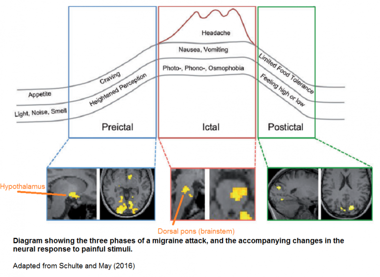
The hypothalamus couples with the brainstem to cause migraines

This 2016 German human study with one subject found:

“The hypothalamus to be the primary generator of migraine attacks which, due to specific interactions with specific areas in the higher and lower brainstem, could alter the activity levels of the key regions of migraine pathophysiology.”

The subject underwent daily fMRI scans, and procedures to evoke brain activity. She didn’t take any medications, and suffered three migraine attacks during the 31-day experimental period.

Neuroskeptic commented:

“The dorsal pons has previously been found to be hyperactive during migraine. It’s been dubbed the brain’s ‘migraine generator.’ Schulte and May’s data suggest that this is not entirely true – rather, it looks like the hypothalamus may be the true generator of migraine, while the brainstem could be a downstream mediator of the disorder.

A hypothalamic origin of migraines would help to explain some of the symptoms of the disorder, such as changes in appetite, that often accompany the headaches.”


The above graphic looks to me like the result of feedback mechanisms that either didn’t exist or inadequately handled the triggering event. Other examples of the hypothalamus lacking feedback or being involved in a deviated feedback loop include:

There are many unanswered questions with a one-person study, of course. Addressing the cause of this painful condition would find out when, where, and how a person’s hypothalamus became modified to express migraine tendencies.

I’d guess that migraine tendencies may appear as early as the first trimester of pregnancy, given that a highly functional hypothalamus is needed for survival and development in our earliest lives. Gaining as much familial and historical information as possible from the person would be necessary steps in therapies that address migraine causes.

http://blogs.discovermagazine.com/neuroskeptic/2016/05/22/pinpointing-origins-of-migraine/ “Pinpointing the Origins of Migraine in the Brain”

https://academic.oup.com/brain/article/139/7/1987/2464241 “The migraine generator revisited: continuous scanning of the migraine cycle over 30 days and three spontaneous attacks”


As mentioned in How to cure the ultimate causes of migraines? comments are turned off for this post due to it somehow becoming a magnet for spammers. Readers can comment on that post instead.
The persistence of epigenetic marks in Type 1 diabetes

This 2016 California human study found:

“A persistency of DNA methylation over time at key genomic loci associated with diabetic complications. Two sets of DNAs collected at least 16–17 years apart from the same participants are used to show the persistency of DNA-me over time.

Twelve annotated differentially methylated loci were common in both WB [whole blood] and Monos [blood monocytes], including thioredoxin-interacting protein (TXNIP), known to be associated with hyperglycemia and related complications.

The top 38 hyperacetylated promoters in cases included 15 genes associated with the nuclear factor kappa-light-chain-enhancer of activated B cells (NF-κB) inflammatory pathway, which is strongly associated with diabetic complications.”

The researchers built on a series of studies that showed how subjects with early intensive interventions didn’t develop further complications, whereas subjects with later intensive interventions:

“Continued to develop complications, such as nephropathy, retinopathy, and macrovascular diseases, at significantly higher rates.

This persistence of benefit from early application of intensive therapy, called ‘metabolic memory,’ is an enigma.”


These researchers needed to also consider a point of Enduring memories? Or continuous toxic stimulation? that:

“The lasting epigenomic effect would not be due to memory, but continuous stimulation by persistent pathogens or persistent components.”

Other studies that involved specific genes of this study include:

http://www.pnas.org/content/113/21/E3002.full “Epigenomic profiling reveals an association between persistence of DNA methylation and metabolic memory in the DCCT/EDIC type 1 diabetes cohort”

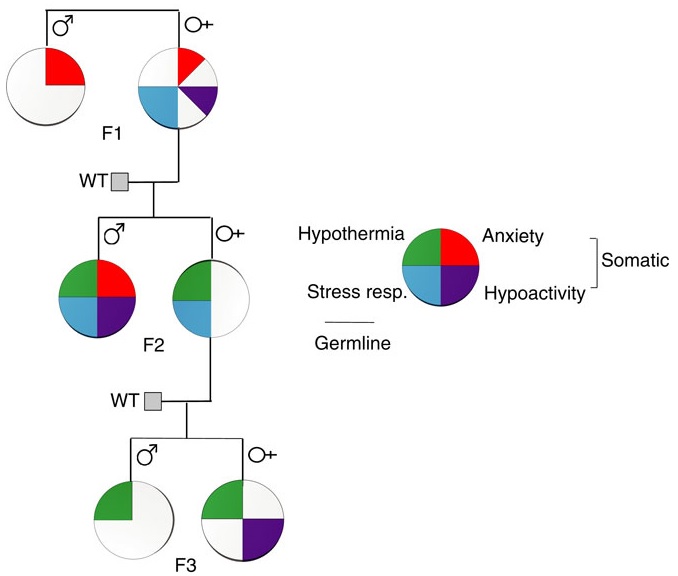
A limited study of parental transmission of anxiety/stress-reactive traits

BehavioralTraitsThis 2016 New York rodent study found:

“Parental behavioural traits can be transmitted by non-genetic mechanisms to the offspring.

We show that four anxiety/stress-reactive traits are transmitted via independent iterative-somatic and gametic epigenetic mechanisms across multiple generations.

As the individual traits/pathways each have their own generation-dependent penetrance and gender specificity, the resulting cumulative phenotype is pleiotropic. In the context of genetic diseases, it is typically assumed that this phenomenon arises from individual differences in vulnerability to the various effects of the causative gene. However, the work presented here reveals that pleiotropy can be produced by the variable distribution and segregated transmission of behavioural traits.”


A primary focus was how anxiety was transmitted from parents to offspring:

“The iterative propagation of the male-specific anxiety-like behaviour is most compatible with a model in which proinflammatory state is propagated from H [serotonin1A receptor heterozygote] F0 to F1 [children] females and in which the proinflammatory state is acquired by F1 males from their H mothers, and then by F2 [grandchildren] males from their F1 mothers.

We propose that increased levels of gestational MIP-1β [macrophage inflammatory protein 1β] in H and F1 mothers, together with additional proinflammatory cytokines and bioactive proteins, are required to produce immune system activation in their newborn offspring, which in turn promotes the development of the anxiety-like phenotype in males.

In particular, increase in the number of monocytes and their transmigration to the brain parenchyma in F1 and F2 males could be central to the development of anxiety.”


The researchers studied transmission of behavioral traits and epigenetic changes. Due to my quick take on the study title – “Behavioural traits propagate across generations..” – I had expectations of this study that weren’t born out. What could the researchers have done versus what they did?

The study design removed prenatal and postnatal parental behavioral transmission of behavioral traits and epigenetic changes as each generation’s embryos were implanted into foster wild-type (WT) mothers.

The study design substituted the foster mothers’ prenatal and postnatal parental environments for the biological parents’ environments. So we didn’t find out, for example:

  • To what extents the overly stress-reactive F1 female children’s prenatal environments and postnatal behaviors induced behaviors and/or epigenetic changes in their children; and
  • Whether the F2 grandchildren’s parental behaviors subsequently induced behaviors and/or epigenetic changes in the F3 great-grandchildren.

How did the study meet the overall goal of rodent studies: to help humans?

    1. Only a minority of humans experienced an early-life environment that included primary caregivers other than our biological parents.
    2. Very, very few of us experienced a prenatal environment other than our biological mothers.
    3. The study’s thorough removal of parental behavior was an outstanding methodology to confirm by falsifiability whether parental behavior was both an intergenerational and transgenerational epigenetic inheritance mechanism.
    4. Maybe the researchers filled in some gaps in previous rodent studies, such as determining what is or isn’t a “true transgenerational mechanism.”

As an example of a rodent study that more closely approximated human conditions, the behavior of a mother whose DNA was epigenetically changed by stress induced the same epigenetic changes to her child’s DNA when her child was stressed per One way that mothers cause fear and emotional trauma in their infants:

“Our results provide clues to understanding transmission of specific fears across generations and its dependence upon maternal induction of pups’ stress response paired with the cue to induce amygdala-dependent learning plasticity.”


How did parental behavioral transmission of behavioral traits and epigenetic changes become a subject not worth investigating? These traits and effects can be seen everyday in real-life human interactions, and in every human’s physiology.

But when investigating human correlates with behavioral epigenetic changes of rodents in the laboratory, parental behavioral transmission of behavioral traits is often treated the way this study treated it: as a confounder.

I doubt that people who have reached some degree of honesty about their early lives and concomitant empathy for others would agree with this prioritization. The papers of Transgenerational epigenetic inheritance week show the spectrum of opportunities to advance science that were intentionally missed.

http://www.nature.com/ncomms/2016/160513/ncomms11492/full/ncomms11492.html “Behavioural traits propagate across generations via segregated iterative-somatic and gametic epigenetic mechanisms”

Lack of oxygen’s epigenetic effects

This 2016 Finnish review subject was epigenetic effects of hypoxia:

“Ever since the Cambrian period, oxygen availability has been in the center of energy metabolism. Hypoxia stabilizes expression of hypoxia-inducible transcription factor-1α (HIF-1α), which controls expression of hundreds of survival genes related to enhanced energy metabolism and autophagy.

There are several other signals, mostly related to stresses, which can increase expression of HIF factors and thus improve cellular survival. However, a chronic activation of HIF factors can have detrimental effects, e.g. stimulate cellular senescence and tissue fibrosis commonly enhanced in age-related diseases.

Stabilization of HIF-1α increases expression of histone lysine demethylases (KDM). Hypoxia-inducible KDMs support locally the gene transcription induced by HIF-1α, although they can also control genome-wide chromatin landscape, especially KDMs which demethylate H3K9 and H3K27 sites (repressive epigenetic marks).”

Gene areas where HIF-1α is involved include:

  • “angiogenesis
  • autophagy
  • glucose uptake
  • glycolytic enzymes
  • immune responses
  • embryonic development
  • tumorigenesis
  • generation of miRNAs.”

HIF-1a signaling

Figure 1 above was instructive in that the reviewers pointed out the lack of a feedback mechanism in HIF-1α signaling. A natural lack of feedback to the HIF-1α signaling source contributed to diseases such as:

  • “age-related macular degeneration
  • cancer progression
  • chronic kidney disease
  • cardiomyopathies
  • adipose tissue fibrosis
  • inflammation
  • detrimental effects which are linked to epigenetic changes.”

The point was similar to a study referenced in The PRice “equation” for individually evolving: Which equation describes your life? that:

“Evolution may preferentially mitigate damage to a biological system than reduce the source of this damage.”


This review subject has many interdependencies and timings within a complex network. Contexts are important:

“Cross-talk between NF-κB [nuclear factor kappa B] and HIF-1α in inflammation might be organized in cell type and context-dependent manner.

It seems that ROS [reactive oxygen species] affect HIF-1α signaling in a context-dependent manner.

Hypoxia stimulated expression of KDM3A and KDM4B genes in different cellular contexts. Given that KDM3A and KDM4B are the major histone demethylases which remove repressive H3K9 sites, their role as transcriptional cofactors seems to be important in activation of HIF-1α signaling. Members of KDM4 subfamily have a crucial role in DNA repair systems, although responses seem to be enzyme-specific and appear in a context-dependent manner.

Acute hypoxia can stimulate cell-cycle arrest but does not provoke cellular senescence in all contexts.”

It wasn’t mentioned that hypoxia evokes cellular Adaptations to stress encourage mutations in a DNA area that causes diseases.

The review was tailored for the publishing journal Aging and Disease, and the subject was best summed up by:

“HIF-1α can control cellular fate in adult animals, either stimulating proliferation or triggering cellular senescence, by regulating the expression of different KDMs in a context-dependent manner.”


This review covered hypoxic conditions during human development that are clearly origins of many immediate and later-life diseases. However, cited remedies only addressed symptoms.

That these distant causes can no longer be addressed is a hidden assumption of research and treatment of effects of health problems. Aren’t such assumptions testable here in the current year?

http://www.aginganddisease.org/article/2016/2152-5250/ad-7-2-180.shtml “Hypoxia-Inducible Histone Lysine Demethylases: Impact on the Aging Process and Age-Related Diseases”

The inevitable effects of avoidable wars

“It was like Tom had tried to return home,” says Jacky Sweetnam. “But he didn’t quite make it.”

http://news.nationalpost.com/features/the-ghosts-of-vietnam-the-last-days-of-a-decorated-canadian-vet “The Ghosts of Vietnam”


In memoriam to my father who died twenty years ago last week. World War II ruined his life with undiagnosed PTSD, some of the effects of which affected his children.

His brother – my uncle – was a Navy hospital corpsman during the slaughter at Iwo Jima, and was even more afflicted.

Neither of them were ever treated.

Contending with epigenetic consequences of violence to women

This 2016 UK review subject was the interplay of genomic imprinting and intergenerational epigenetic information transfer:

“A range of evolutionary adaptations associated with placentation transfers disproportionate control of this process to the matriline, a period unique in mammalian development in that there are three matrilineal genomes interacting in the same organism at the same time (maternal, foetal, and postmeiotic oocytes).

Genomic imprinting is absent in egg laying mammals and only around 6 imprinted genes have been detected in a range of marsupial species; this is in contrast to eutherian mammals where around 150 imprinted genes have been described.

The interactions between the maternal and developing foetal hypothalamus and placenta can provide a template by which a mother can transmit potentially adaptive information concerning potential future environmental conditions to the developing brain.

In circumstances either where the early environment provides inaccurate cues to the environmental conditions prevailing when adult due to rapid environmental change or when disruptions to normal neural development occur, the mismatch between the environmental predictions made during early development and subsequent reality may mean that an organism may have a poorly adapted phenotype to its adult environment. An appreciation of these underlying evolutionary salient processes may provide a novel perspective on the [causal] mechanisms of a range of health problems.

The concept of a brain that is not pathological in the classical sense but it is simply mismatched to its environment has been most extensively studied in the context of ancestral and early developmental nutrition. However, this concept can be extended to provide insights into the development of a range of alternative neural phenotypes.”

The review’s final sentence was:

“Examination of the adaptive potential of a range of neural and cognitive deficits in the context of evolutionary derived foetocentric brain and placental development, epigenetics and environmental adaptation may provide novel insights into the development and potential treatment of a range of health, neurological, and cognitive disorders.”

One of the reviewers was cited in Epigenetic DNA methylation and demethylation with the developing fetus, which the review cited along with Epigenetic changes in the developing brain change behavior.


Researchers who avoid hypotheses that can’t be proven wrong could certainly test the subject matter of this review if they investigated their subjects’ histories.

For example, let’s say a patient/subject had symptoms where the “150 imprinted genes” were implicated. What are the chances a clinician or researcher would be informed by this review’s material and investigate the mother’s and grandmother’s histories?

For clinicians or researchers who view histories as irrelevant busywork: How many tens of millions of people alive today have mothers who were fetuses when their grandmothers were adversely affected by violence? Wouldn’t it be appropriate to assess possible historical contributions of:

“The mismatch between the environmental predictions made during early development and subsequent reality”

to the patient’s/subject’s current symptoms?

http://www.hindawi.com/journals/np/2016/6827135/ “Placental, Matrilineal, and Epigenetic Mechanisms Promoting Environmentally Adaptive Development of the Mammalian Brain”

A human study of pain avoidance

This 2016 UK human study found:

“People differ in how they learn to avoid pain, with some individuals refraining from actions that resulted in painful outcomes, whereas others favor actions that helped prevent pain.

Learning in our task was best explained as driven by an outcome prediction error that reflects the difference between expected and actual outcomes. Consistent with the expression of such a teaching signal, blood-oxygen level-dependent (BOLD) responses to outcomes in the striatum were modulated by expectation.

Positive learners showed significant functional connectivity between the insula and striatal regions, whereas negative learners showed significant functional connectivity between the insula and amygdala regions.

The degree to which a participant tended to learn from success in avoiding than experiencing shocks was predicted by the structure of a participants’ striatum, specifically by higher gray matter density where the response to shocks was consistent with a prediction error signal.

Higher gray matter density in the putamen (and lower gray matter density in the caudate) predicted better learning from shocks and poorer learning from success in avoiding shocks.”

The researchers termed the subjects’ pain responses “learning” instead of conditioning. The difference between the two terms in the experimental contexts was that the subjects weren’t presented with 100%-certain choices to avoid pain.

The experiments were also rigged to force choices at similar rates among subjects because:

“Participants who learned more from painful outcomes developed a propensity to avoid gambling, whereas participants who learned more from success in preventing pain developed a propensity to gamble.”


Human responses to pain don’t arise out of nowhere. The subjects’ pain histories were clearly relevant, but weren’t investigated.

The closest the study came to considering the subjects’ histories was:

“Before the experiment, participants completed an 80-item questionnaire composed of several measures of different mood and anxiety traits. Age, sex and mood and anxiety traits did not differ between participants later classified as positive and negative learners.”

Emotional content was neither included nor solicited. Emotions were inferred:

“Participants biased in favor of passive avoidance learning (i.e., learning what gambles should be avoided), striatal response to painful outcomes was consistent with an aversive prediction error, as seen in fear conditioning.”

As a result, there weren’t causal explanations for the subjects’ differing pain responses. How, when, and why did the behavioral, functional, and structural differences develop?


I didn’t see the level of detail needed to characterize striatal regions into the Empathy, value, pain, control: Psychological functions of the human striatum segments. I’d guess that the findings of “higher gray matter density in the putamen (and lower gray matter density in the caudate)” applied to the posterior putamen and the anterior caudate nucleus.

Two of the coauthors were also coauthors of If a study didn’t measure feelings, then its findings may not pertain to genuine empathy which I rated < 0 Detracted from science. The technique of Why do we cut short our decision-making process? was referenced.

http://www.pnas.org/content/early/2016/04/06/1519829113.full “Striatal structure and function predict individual biases in learning to avoid pain”

A one-sided review of stress

The subject of this 2016 Italian/New York review was the stress response:

“The stress response, involving the activation of the hypothalamic-pituitary-adrenocortical [HPA] axis and the consequent release of corticosteroid hormones, is indeed aimed at promoting metabolic, functional, and behavioral adaptations. However, behavioral stress is also associated with fast and long-lasting neurochemical, structural, and behavioral changes, leading to long-term remodeling of glutamate transmission, and increased susceptibility to neuropsychiatric disorders.

Of note, early-life events, both in utero and during the early postnatal life, trigger reprogramming of the stress response, which is often associated with loss of stress resilience and ensuing neurobehavioral (mal)adaptations.”


The reviewers’ intentional dismissal of the role of GABA in favor of the role of glutamate was a key point:

“The changes in neuronal excitability and synaptic plasticity induced by stress are the result of an imbalance of excitatory (glutamatergic) and inhibitory (GABAergic) transmission, leading to long-lasting (mal)adaptive functional modifications. Although both glutamate and GABA transmission are critically associated with stress-induced alteration of neuronal excitability, the present review will focus on the modulation of glutamate release and transmission induced by stress and glucocorticoids.”

No particular reason was given for this bias. I inferred from the review’s final sentence that the review’s sponsors and funding prompted this decision:

“In-depth studies of changes in glutamate transmission and dendrite remodeling induced by stress in early and late life will help to elucidate the biological underpinnings of the (mal)adaptive strategies the brain adopts to cope with environmental challenges in one’s life.”

The bias led to ignoring evidence for areas the reviewers posed as needing further research. An example of relevant research the reviewers failed to consider was the 2015 Northwestern University study I curated in A study that provided evidence for basic principles of Primal Therapy that found:

“In response to traumatic stress, some individuals, instead of activating the glutamate system to store memories, activate the extra-synaptic GABA system and form inaccessible traumatic memories.”

http://www.ncbi.nlm.nih.gov/pmc/articles/PMC4812483/ “Stress Response and Perinatal Reprogramming: Unraveling (Mal)adaptive Strategies”

Observing pain in others had long-lasting brain effects

This 2016 Israeli human study used whole-head magnetoencephalography (MEG) to study pain perception in military veterans:

Our findings demonstrate alterations in pain perception following extreme pain exposure, chart the sequence from automatic to evaluative pain processing, and emphasize the importance of considering past experiences in studying the neural response to others’ states.

Differences in brain activation to ‘pain’ and ‘no pain’ in the PCC [posterior cingulate cortex] emerged only among controls. This suggests that prior exposure to extreme pain alters the typical brain response to pain by blurring the distinction between painful and otherwise identical but nonpainful stimuli, and that this blurring of the ‘pain effect’ stems from increased responses to ‘no pain’ rather than from attenuated response to pain.”


Limitations included:

  • “The pain-exposed participants showed posttraumatic symptoms, which may also be related to the observed alterations in the brain response to pain.
  • We did not include pain threshold measurements. However, the participants’ sensitivity to experienced pain may have had an effect on the processing of observed pain.
  • The regions of interest for the examination of pain processing in the pain-exposed group were defined on the basis of the results identified in the control group.
  • We did not detect pain-related activations in additional regions typically associated with pain perception, such as the anterior insula and ACC. This may be related to differences between the MEG and fMRI neuroimaging approaches.”

The subjects self-administered oxytocin or placebo per the study’s design. However:

“We chose to focus on the placebo condition and to test group differences at baseline only, in light of the recent criticism on underpowered oxytocin administration studies, and thus all following analyses are reported for the placebo condition.”


A few questions:

  1. If observing others’ pain caused “increased responses to ‘no pain’,” wouldn’t the same effect or more be expected from experiencing one’s own pain?
  2. If there’s evidence for item 1, then why aren’t “increased responses to ‘no pain'” of affected people overtly evident in everyday life?
  3. If item 2 is often observed, then what are the neurobiological consequences for affected people’s suppression of “increased responses to ‘no pain’?”
  4. Along with the effects of item 3, what may be behavioral, emotional, and other evidence of this suppressed pain effect?
  5. What would it take for affected people to regain a normal processing of others’ “‘pain’ and ‘no pain’?”

https://www.researchgate.net/publication/299546838_Prior_exposure_to_extreme_pain_alters_neural_response_to_pain_in_others “Prior exposure to extreme pain alters neural response to pain in others” Thanks to one of the authors, Ruth Feldman, for providing the full study

Epigenetic contributions to hypertension

This 2016 Australian review subject was epigenetic contributions to hypertension:

“Hypertension (HT) affects more than 1 billion people globally and is a major risk factor for stroke, chronic kidney disease, and myocardial infarction.

Essential hypertension (EH) is a complex, polygenic condition with no single causative agent. There is increasing evidence that epigenetic modifications are as important as genetic predisposition in the development of EH.

Many epigenetic studies are, however, limited by the fact that only blood is studied rather than the effector tissues. The utility of blood methylation status in epigenetic research is yet to be determined. Furthermore, the polygenic complexity of HT and the limited knowledge on some of the non-coding RNAs makes it more challenging to decipher the exact mechanisms involved.”

The review had sections for hypertension studies on DNA methylation, histone modification, and microRNA/other non-coding RNA types. Here’s a sample of the findings:

“HSD11B2-mediated degradation of cortisol to cortisone is disrupted when the promoter region of the HSD11B2 gene is hypermethylated. The resulting imbalance in the active metabolites of cortisol and cortisone, tetrahydrocortisol, and tetrahydocortisone, respectively, promotes the onset of HT.

Histone modification affecting arterial pressure levels has been documented in a variety of human and animal tissues, including vascular smooth muscle. Vascular oxidative stress can contribute to endothelial dysfunction—a hallmark of HT—and the development of HT.

Two miRNAs (has-miR-181a and has-miR-663) with the ability to bind to the 3′ UTR of renin mRNA were found to be under-expressed in EH. These miRNAs were able to regulate the expression of a reporter gene and renin-mRNA itself, which explains over-expression of renin mRNA seen in EH kidney.”


The publisher, International Journal of Molecular Sciences, makes ALL of its articles open access. Another of its requirements is:

“The full experimental details must be provided so that the results can be reproduced.”

There also aren’t artificial limitations on either the length of the study or the number of supplementary files.

http://www.mdpi.com/1422-0067/17/4/451/htm “Epigenetic Modifications in Essential Hypertension”

What is epigenetic inheritance?

This 2016 review by Eric Nestler, a well-known and well-funded researcher, entitled Transgenerational Epigenetic Contributions to Stress Responses: Fact or Fiction? concluded:

“Further work is needed to understand whether and to what extent true epigenetic inheritance of stress vulnerability adds to the well-established and powerful influence of genetics and environmental exposures in determining an individual’s susceptibility versus resilience to stress throughout life.

There is growing evidence for at least some contribution of epigenetic regulation – perhaps achieved by miRNAs – in mediating part of the ability of parental behavioral experience to influence stress vulnerability in their offspring.”


The reviewer applied the terms involved to exclude behavioral inheritance mechanisms. The extent of what is “epigenetic inheritance” seemed to be lost in the process.

For example, his own 2011 research Paternal Transmission of Stressed-Induced Pathologies was cited for evidence that:

“Adult male mice subjected to chronic social defeat stress generate offspring that are more vulnerable to a range of stressful stimuli than the offspring of control mice.”

Yet that finding was dismissed in the review and in that study as behavioral:

“While epigenetic changes in sperm might be a small factor in transgenerational transmission of stress vulnerability, a large portion of the observed transmission may be behavioral.

The fact that most of the transgenerational transmission of stress vulnerability observed in our experiments was not seen with IVF argues against the preponderance of epigenetic mechanisms. Rather, our data would suggest that the bulk of the vulnerabilities are passed on to subsequent generations behaviorally.”


A few questions:

  1. If the experimental subjects had no more control over their behavioral stress-response effects than they had over their DNA methylation, histone modification, or microRNA stress-response effects, then why was such behavior not included in the “epigenetic mechanisms” term?
  2. How do behavioral inheritance mechanisms fall outside the “true epigenetic inheritance” term when behavioral stress-response effects are shown to be reliably transmitted generation after generation?
  3. Wouldn’t the cessation of behavioral inheritance mechanisms confirm their status by falsifiability? This was done with studies such as the 1995 Adoption reverses the long-term impairment in glucocorticoid feedback induced by prenatal stress.

Mechanisms of stress memories in plants

This 2016 Australian review’s subject was plant memory mechanisms:

“Plants are adept at rapidly acclimating to stressful conditions and are able to further fortify their defenses by retaining memories of stress to enable stronger or more rapid responses should an environmental perturbation recur.

The recovery process entails a balancing act between resetting and memory formation. During recovery, RNA metabolism, posttranscriptional gene silencing, and RNA-directed DNA methylation have the potential to play key roles in resetting the epigenome and transcriptome and in altering memory.”

Many of the principles applied to animals, and several animal studies were cited for illustration. Here’s one of the graphics:

F6.large


I disagreed with the Summary statement:

“Memory, in particular epigenetic memory, is likely a relatively rare event.”

The reviewers cited a 2015 Australian study Stress induced gene expression drives transient DNA methylation changes at adjacent repetitive elements which found the opposite conclusion with rice:

“Despite 21 days of starvation, resupplying phosphate for just 1 day reversed expression of 40% of induced genes, further increasing to 80% after 3 days and corresponding with a reestablished internal root phosphate concentration. Interestingly though, 80 genes remained differentially regulated even after 31 days of resupply.”

The cited study’s researchers attributed their epigenetic memory finding to several factors, including their study design:

“The majority of DNA methylation analyses performed in plants to date have focused on Arabidopsis, despite being relatively depleted of TEs [transposable elements] (15–20% of the genome) and being poorly methylated compared to other plant genomes.

To date, only a limited number of studies have comprehensively investigated the involvement of DNA methylation in response to adverse environmental conditions. Several studies have reported that changes in the environment can affect the methylation status of some regions of the genome, using low resolution and non-quantitative techniques. These studies have lacked the resolution to provide the specific context and genomic location of the changes in DNA methylation, thus offering limited insights into the potential role of stress-induced changes in DNA methylation.”

So, the current review judging “memory, in particular epigenetic memory” to be “a relatively rare event” probably had more to do with study designs rather than what actually occurs in nature. See one of the coauthor’s response below.

http://advances.sciencemag.org/content/2/2/e1501340.full “Reconsidering plant memory: Intersections between stress recovery, RNA turnover, and epigenetics”

Problematic research into epigenetic effects of paternal stress on male offspring

This 2016 Chinese rodent study and its accompanying commentary Don’t stress dad — it’s bad for your kids’ health were caught up in an agenda.

The first problem I noticed was that the hyperglycemic effects found only in the male offspring weren’t consistently labelled as sex-specific. Try to find that fact in the paywalled commentary with its intentionally misleading headline, or in the news coverage with headlines such as “Stressed mouse dads give their offspring high blood sugar.”

That the effects were male-only was briefly noted in the study, yet “male” was absent from the “stress-F1 mice” label used after the initial mention.

2015年12月22日-陆炎模式图单独导出-v2

The researchers provided no mechanisms that plausibly linked the effects to offspring sex. There was plenty of time between the May 3, 2015 submission and the February 18, 2016 publication to clarify this and other items. I wonder what the reviewer noted.

The second problem was that the highest number of male “stress-F1 mice” tested was only six. I didn’t see any disclosures of what led to the scarcity of subjects, or of the likely impact of using so few.

A related limitation was that the male “stress-F1 mice” were killed as young adults. Whether or not the hyperglycemic effects carried through to old age or to another generation wasn’t determined.


I’m leery of studies like this one that didn’t have a Limitations section, and especially so when the news coverage overlooked obvious limitations. It was difficult to place the findings in a context other than promoting that a male’s stress may also adversely affect their offspring.

One of the problems that research caught up in an agenda create is that non-headline findings are overlooked. Other than sex-specific effects, the study found that the putative preconception cause of hyperglycemia didn’t cause other symptoms:

  • “No significant growth defects were observed in male offspring from stress-F0 fathers (stress-F1 mice) during their early lives.
  • Insulin sensitivity was not changed in stress-F1 mice.
  • Serum glucagon, leptin, and pro-inflammatory cytokines (tumor necrosis factor α [TNFα], interleukin-6 [IL-6]) were unaffected.
  • Body weight, food intake, locomotor activity, CO2 production, O2 consumption, and respiratory exchange ratios also remained unchanged.
  • Liver weight, liver weight/body weight ratios, hepatic triglyceride content, and the histological phenotypes were also comparable.
  • The methylation pattern and expression of microRNAs were not affected in the fetal brains of stress-F1 mice.”

The handling of the study reminded me of Transgenerational epigenetic programming with stress and microRNA where most of the news coverage similarly focused on it being a male’s stress, not a female’s, that affected the developing embryo. The important part lost from news coverage of that study was it demonstrated how a damaging influence can begin immediately after conception, but the symptoms didn’t present until adulthood!

http://www.sciencedirect.com/science/article/pii/S1550413116300067 “Paternal Psychological Stress Reprograms Hepatic Gluconeogenesis in Offspring”

The current paradigm of child abuse limits pre-childhood causal research

As an adult, what would be your primary concern if you suspected that your early life had something to do with current problems? Would you be interested in effective treatments for causes of your symptoms?

Such information wasn’t available in this 2016 Miami review of the effects of child abuse. The review laid out the current paradigm mentioned in Grokking an Adverse Childhood Experiences (ACE) score, one that limits research into pre-childhood causes for later-life symptoms.

The review’s goal was to describe:

“How numerous clinical and basic studies have contributed to establish the now widely accepted idea that adverse early life experiences can elicit profound effects on the development and function of the nervous system.”

The hidden assumptions of almost all of the cited references were that these distant causes could no longer be addressed. Aren’t such assumptions testable today?

As an example, the Discussion section posed the top nine “most pressing unanswered questions related to the neurobiological effects of early life trauma.” In line with the current paradigm, the reviewer assigned “Are the biological consequences of ELS [early life stress] reversible?” into the sixth position.

If the current paradigm encouraged research into treatment of causes, there would probably already be plenty of evidence to demonstrate that directly reducing the source of damage would also reverse damaging effects. There would have been enough studies done so that the generalized question of reversibility wouldn’t be asked.

Aren’t people interested in treatments of originating causes so that their various symptoms don’t keep bubbling up? Why wouldn’t research paradigms be aligned accordingly?


The review also demonstrated how the current paradigm of child abuse misrepresented items like telomere length and oxytocin. Researchers on the bandwagon tend to forget about the principle Einstein expressed as:

“No amount of experimentation can ever prove me right; a single experiment can prove me wrong.”

That single experiment for telomere length arrived in 2016 with Using an epigenetic clock to distinguish cellular aging from senescence. The review’s seven citations for telomere length that all had findings “associated with” or “linked to” child abuse should now be viewed in a different light.

The same light shone on oxytocin with Testing the null hypothesis of oxytocin’s effects in humans and Oxytocin research null findings come out of the file drawer. See their references, and decide for yourself whether or not:

“Claimed research findings may often be simply accurate measures of the prevailing bias.”

http://www.cell.com/neuron/fulltext/S0896-6273%2816%2900020-9 “Paradise Lost: The Neurobiological and Clinical Consequences of Child Abuse and Neglect”


This post has somehow become a target for spammers, and I’ve disabled comments. Readers can comment on other posts and indicate that they want their comment to apply here, and I’ll re-enable comments.

Epigenetic regulation of natural killer cells

This 2016 German review focused on how epigenetic processes affected the natural killer cell part of the immune system:

“Natural killer (NK) cells recognize and eliminate tumor- and virus-infected cells, parasites as well as certain types of bacteria. NK cell activity is related to a complex interaction of activating and inhibiting receptors on the NK cell surface.

During the development of HPCs [hemopoietic progenitor cells] to mature NK cells, the DNA demethylation of KIR [killer cell immunoglobin-like receptors] genes leads to KIR expression. But DNA methylation does not just determine which KIR gene is expressed, it also determines which allele expresses the KIR gene. KIR genes are also regulated by microRNA.

KIR genes exhibit highly similar histone acetylation signatures, which are typically found in expressed genes. This fact puts the KIR genes into a state of readiness for transcription which is depending on the DNA methylation as critical epigenetic modification in the regulation of KIR gene expression.

Epigenetic modifications have been reported to be involved in the expression of NKG2D, which is one the most important activating NK cell receptor.”

The reviewers included a section on NK cell activity and external stimuli. They summarized:

“The significance of the described findings is limited by study designs. Although human NK cells were frequently used, in most cases treatment took place in ex vivo experiments.”

The reviewers also provided a good three-paragraph explanation of general epigenetic mechanisms.

http://www.mdpi.com/1422-0067/17/3/326/htm “Natural Killer Cells—An Epigenetic Perspective of Development and Regulation”