Infant DNA methylation and caregiving

This 2019 US human study attempted to replicate findings of animal studies that associated caregiver behavior with infant DNA methylation of the glucocorticoid receptor gene:

“Greater levels of maternal responsiveness and appropriate touch were related to less DNA methylation of specific regions in NR3c1 exon 1F, but only for females. There was no association with maternal responsiveness and appropriate touch or DNA methylation of NR3c1 exon 1F on prestress cortisol or cortisol reactivity. Our results are discussed in relation to programming models that implicate maternal care as an important factor in programing infant stress reactivity.”


The study had many undisclosed and a few disclosed limitations, one of which was:

“Our free-play session, while consistent with the length of free-play sessions in other studies, was short (5 min). It is unclear whether a longer length of time would have yielded significant different maternal responsiveness and appropriate touch data.”

The final sentence showed the study’s purpose was other than discovering factual evidence:

“Following replication of this work, it could ultimately be used in conjunction with early intervention, or home-visiting programs, to measure the strength of the intervention effect at the epigenetic level.”

https://onlinelibrary.wiley.com/doi/full/10.1002/imhj.21789 “DNA methylation of NR3c1 in infancy: Associations between maternal caregiving and infant sex” (not freely available)

What drives cellular aging?

This 2019 US/UK human cell study by the founder of the epigenetic clock method investigated epigenetic aging:

“It is widely assumed that extension of lifespan is a result of retardation of ageing. While there is no counter-evidence to challenge this highly intuitive association, supporting empirical evidence to confirm it is not easy to acquire.

The scarcity of empirical evidence is due in part to the lack of a good measure of age that is not based on time. In this regard, the relatively recent development of epigenetic clocks is of great interest.

At the cellular level more is known, but from the perspective of what epigenetic ageing is not, rather than what it is. While we still do not know what cellular feature is associated with epigenetic ageing, we can now remove:

  • somatic cell differentiation

from the list of possibilities and place it with

  • cellular senescence,
  • proliferation and
  • telomere length maintenance,

which represent cellular features that are all not linked to epigenetic ageing.”


The study used several agents, including rapamycin, to investigate the hypotheses. Rapamycin isn’t a panacea, however:

“The ability of rapamycin to suppress the progression of epigenetic ageing is very encouraging for many reasons not least because it provides a valuable point-of-entry into molecular pathways that are potentially associated with it. Evidently, the target of rapamycin, the mTOR complex is of particular interest.

The convergence of the GWAS observation with the experimental system described here is a testament of the strength of the skin & blood clock in uncovering biological features that are consistent between the human level and cellular level. It lends weight to the emerging view that the mTOR pathway may be the underlying mechanism that supports epigenetic ageing.”

The limitation section ended with:

“It is important to note that it is inadvisable (actively discouraged) to directly extrapolate the studies here, especially in terms of the magnitude of age suppression, to potential effects of rapamycin on humans.”

https://www.aging-us.com/article/101976/text “Rapamycin retards epigenetic ageing of keratinocytes independently of its effects on replicative senescence, proliferation and differentiation”

Another important transgenerational epigenetic inheritance study

This 2019 Washington State University rodent study from Dr. Michael Skinner’s lab found:

“A cascade of epigenetic alterations initiated in PGCs [primordial germ cells of F3 males] appears to be required to alter epigenetic programming during spermatogenesis to modify the sperm epigenome involved in transgenerational epigenetic inheritance phenomenon.

Following fertilization there is a DNA methylation erasure to generate stem cells in the early embryo, which then remethylate in a cell type-specific manner. DNA methylation erasure is thought to, in part, reset deleterious epigenetics in the germline. However, imprinted gene DNA methylation sites and induced transgenerational epimutations appear to be protected from this DNA methylation erasure.

A germline with an altered epigenome has the capacity to alter the early embryo’s stem cell’s epigenome and transcriptome that can subsequently impact epigenomes and transcriptomes of all derived somatic cells. Therefore, an altered sperm epigenome has the capacity to transmit phenotypes transgenerationally. Experiments have demonstrated that epigenetic inheritance can also be transmitted through the female germline.

Previously, agricultural fungicide vinclozolin was found to promote transgenerational inheritance of sperm differential DNA methylation regions (DMRs) termed epimutations that help mediate this epigenetic inheritance. The current study was designed to investigate developmental origins of transgenerational DMRs during gametogenesis.

The current study with vinclozolin-induced transgenerational inheritance demonstrates that sperm DMRs also originate during both spermatogenesis and earlier stages of germline development, but at distinct developmental stages. Fetal exposure initiates a developmental cascade (i.e., distinct developmental origins) of aberrant epigenetic programming, and does not simply induce a specific number of DMRs that are maintained throughout development.”

https://www.tandfonline.com/doi/pdf/10.1080/15592294.2019.1614417?needAccess=true “Transgenerational sperm DNA methylation epimutation developmental origins following ancestral vinclozolin exposure”


The study’s main hypotheses were:

“Following fertilization, the hypothesis is that transgenerational epimutations modify early embryonic transcriptomes and epigenomes to re-establish the cascade for the next generation.

As the individual develops, all somatic cells have altered epigenomes and transcriptomes to promote disease susceptibility later in life.”

Researchers: adopt these hypotheses, and apply them to human studies.

1. Don’t get off track by requiring that the same phenotype must be observed in each generation for there to be transgenerational epigenetic inheritance, because:

“Fetal exposure..does not simply induce a specific number of DMRs that are maintained throughout development.”

Animal transgenerational studies have shown that epigenetic inheritance mechanisms may both express different phenotypes for each generation, and entirely skip a phenotype in one or more generations!

2. Don’t limit your study designs to F1 children as did:

3. Don’t stop at F2 grandchildren as did:

4. Continue studies on to F3 great-grandchildren who had no direct exposure to altering stimulus. Keep in the forefront of your research proposals that there are probably more than 10,000,000 F3 descendants of DES-exposed women just in the US!

The transgenerational impact of Roundup exposure

This 2019 Washington rodent study from Dr. Michael Skinner’s lab found adverse effects in the grand-offspring and great-grand-offspring following their ancestor’s exposure during pregnancy to the world’s most commonly used herbicide:

“Using a transient exposure of gestating F0 generation female rats found negligible impacts of glyphosate on the directly exposed F0 generation, or F1 generation offspring pathology. In contrast, dramatic increases in pathologies in the F2 generation grand-offspring, and F3 transgenerational great-grand-offspring were observed.

The transgenerational pathologies observed include prostate disease, obesity, kidney disease, ovarian disease, and parturition (birth) abnormalities:

  1. Prostate disease in approximately 30% of F3 generation glyphosate lineage males, a three-fold increase in disease rate over controls.
  2. A transgenerational (F3 generation) obese phenotype was observed in approximately 40% of the glyphosate lineage females and 42% of the glyphosate lineage males.
  3. An increased incidence of kidney disease observed in the F3 generation glyphosate lineage females affecting nearly 40% of females.
  4. A significant increase in ovarian disease observed in the F2 [48% vs. 21% for controls] and F3 [36% vs. 15% for controls] generation glyphosate lineage females.
  5. During the gestation of F2 generation mothers with the F3 generation fetuses, dramatic parturition abnormalities were observed in the glyphosate lineage. The frequency of unsuccessful parturition was 35%. To further investigate the parturition abnormalities an outcross of F3 generation glyphosate lineage males with a wildtype female was performed. There were parturition abnormalities observed with a frequency of 30%.

Classic and current toxicology studies only involve direct exposure of the individual, while impacts on future generations are not assessed. The ability of glyphosate and other environmental toxicants to impact our future generations needs to be considered, and is potentially as important as the direct exposure toxicology done today for risk assessment.”


Why isn’t coverage of this study the top story of world news organizations? Is what’s reported more important than reliable evidence of generational consequences to environmental experiences?

Current toxicology practices are a scientific disgrace:

  • What are hypotheses of practices that test only effects on somatic cells, and don’t look for generational effects on germ cells?
  • Are tests selected for their relative convenience instead of chosen for their efficacy?

Why don’t sponsors fund and researchers perform human studies of transgenerational epigenetic inheritance? For example, from Burying human transgenerational epigenetic evidence:

“From the late 1930s through the early 1970s, DES was given to nearly two million pregnant women in the US alone.

Fourth [F3] generation effects of prenatal exposures in humans have not been reported.

Zero studies of probably more than 10,000,000 F3 great-grandchildren of DES-exposed women just here in the US!

There will be abundant human evidence to discover if sponsors and researchers will take their fields seriously.

https://www.nature.com/articles/s41598-019-42860-0.pdf “Assessment of Glyphosate Induced Epigenetic Transgenerational Inheritance of Pathologies and Sperm Epimutations: Generational Toxicology”

Non-emotional memories

This 2019 US review covered memory mechanisms:

“With memory encoding reliant on persistent changes in the properties of synapses, a key question is how can memories be maintained from days to months or a lifetime given molecular turnover? It is likely that positive feedback loops are necessary to persistently maintain the strength of synapses that participate in encoding.

These levels are not isolated, but linked by shared components of feedback loops.”


Despite the review’s exhaustive discussion, the reviewers never came to the point. The word cloud I made of the review’s most frequent thirty words had little to do with why memory occurs:

  • Why do some stimuli evoke a memory in response?
  • Why are almost all of the stimuli an organism receives not remembered?

Much of the discussion was baseless because it excluded emotion. Many of the citations’ memory findings relied on emotion, though.

For example, in the subsection Roles of persistent epigenetic modifications for maintaining LTF [long-term facilitation], LTP [long-term potentiation], and LTM [long-term memory]:

  • Histone acetylation is increased after fear conditioning in the hippocampus and amygdala.
  • Correspondingly, inhibition of histone deacetylase enhances fear conditioning and LTP.
  • Following fear conditioning, histone phosphorylation is also increased.
  • DNA methylation is also up-regulated in the hippocampus and amygdala after fear conditioning, and inhibition of DNA methylation blocks fear LTM.”

http://learnmem.cshlp.org/content/26/5/133.full “How can memories last for days, years, or a lifetime? Proposed mechanisms for maintaining synaptic potentiation and memory”

Statistical inferences vs. biological realities

A 2019 UCLA study introduced a derivative of the epigenetic clock named GrimAge:

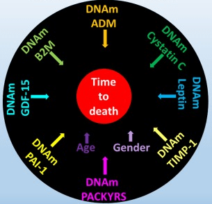
“DNAm GrimAge, a linear combination of chronological age, sex, and DNAm-based surrogate biomarkers for seven plasma proteins and smoking pack-years, outperforms all other DNAm-based biomarkers, on a variety of health-related metrics.

An age-adjusted version of DNAm GrimAge, which can be regarded as a new measure of epigenetic age acceleration (AgeAccelGrim), is associated with a host of age-related conditions, lifestyle factors, and clinical biomarkers. Using large scale validation data from three ethnic groups, we demonstrate that AgeAccelGrim stands out among pre-existing epigenetic clocks in terms of its predictive ability for time-to-death, time-to-coronary heart disease, time-to-cancer, its association with computed tomography data for fatty liver/excess fat, and early age at menopause.”

https://www.ncbi.nlm.nih.gov/pmc/articles/PMC6366976/ “DNA methylation GrimAge strongly predicts lifespan and healthspan”


A miserable attempt at reporting the study’s findings included angles of superstition, fear-of-the-future, and suspicion-by-spurious-association:

“The research has already captured the attention of the life insurance industry. After all, a solid death date could mean real savings when it comes to pricing policies.

The hope is that if and when legitimate anti-aging drugs are developed, GrimAge could be used to test their effectiveness. In a world with functional anti-aging drugs, “doctors could test [your GrimAge number] and say, ‘You know what, you’re aging too quickly. Take this,'” Horvath said.”

https://onezero.medium.com/a-new-test-predicts-when-youll-die-give-or-take-a-few-years-2d08147c8ea6 “A New Test Predicts When You’ll Die (Give or Take a Few Years)”


A detailed blog post from Josh Mitteldorf provided scientific coverage of the study:

“Methylation sites associated with smoking history predicted how long the person would live more accurately than the smoking history itself. Even stranger, the methylation marks most closely associated with smoking were found to be a powerful indication of future health even when the sample was confined to non-smokers.

The DNAm GrimAge clock was developed in two stages, a correlation of a correlation. Curiously, the indirect computation yields the better result.

Horvath’s finding that secondary methylation indicators are more accurate than the underlying primary indicator from which they were derived is provocative, and calls out for a new understanding.”

https://joshmitteldorf.scienceblog.com/2019/03/05/dnam-grimage-the-newest-methylation-clock “DNAm GrimAge—the Newest Methylation Clock”


When there are logical disconnects in findings like the above, it’s time to examine underlying premises. As noted in Group statistics don’t necessarily describe an individual, an assumption required by statistical analyses is that each measured item in the sample is interchangeable with the next.

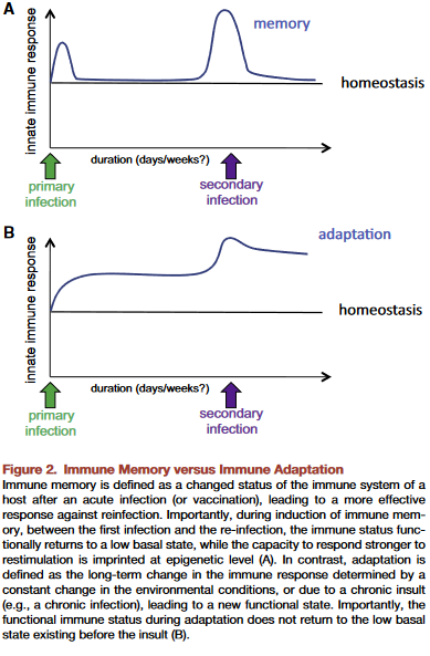
This presumption is often false, producing individually inapplicable results. For example, Immune memory vs. immune adaptation included this description of the adaptive immune system:

“To be effective, highly specific immune response requires huge diversity of receptors and antibodies, which is achieved by somatic rearrangement of gene segments. Recombination results in millions of TCR [T cell receptor] and antibody variants able to recognize and neutralize millions of various antigens.”

Standard statistics of millions of T cell receptor and antibody variants won’t represent their individually unique properties. But individual differences are both their purpose and benefit to us.

The GrimAge study’s overreach was most apparent in stratifying educational attainment to develop correlations. As mentioned in Does a societal mandate cause DNA methylation? such statistics are poor evidence of each individual’s biological realities.

Neither derivatives of group statistics, nor correlations of correlations, seem to be the techniques needed to understand biological causes of effects. Another commentary on the GrimAge study mentioned but glossed over this point:

“It remains a mystery why exactly the epigenetic clocks work, and whether age-related changes in DNA methylation contribute to the cause of aging or are a result of it.”

Immune memory vs. immune adaptation

This 2019 Dutch/German/Romanian perspective aimed for a better understanding of immune systems:

“Based on molecular, immunological, and evolutionary arguments, we propose that innate immune memory is a primitive form of immune memory present in all living organisms, while adaptive immune memory is an advanced form of immune memory representing an evolutionary innovation in vertebrates.

Innate immune responses have the capacity to be trained, and thereby exert a new type of immunological memory upon reinfection. The central feature of trained innate immune cells is their ability to mount a qualitatively and quantitatively different transcriptional response when challenged with microbes or danger signals. Evidence supports convergence of multiple regulatory layers for mediating innate immune memory, including changes in chromatin organization, DNA methylation, and probably non-coding RNAs such as microRNAs and/or long non-coding RNAs.

Two properties of adaptive immune response are mediated by two fundamentally different types of mechanisms:

  1. Higher magnitude and speed of the response is mediated by epigenetic programming.
  2. Specificity of the response is insured by gene recombination of TCR [T cell receptor] and BCR [B cell receptor] and clonal expansion of specific cell subpopulations upon antigen recognition.

To be effective, highly specific immune response requires huge diversity of receptors and antibodies, which is achieved by somatic rearrangement of gene segments. Recombination results in millions of TCR and antibody variants able to recognize and neutralize millions of various antigens.


This paper included speculations such as “Evidence supports..probably non-coding RNAs” quoted above, and the penultimate sentence:

“One can envision that vaccines that are capable of inducing both forms of immune memory at the same time would be more effective.”

100% factual evidence is preferred. Overall information can only be as accurate as the least accurate information.

This review highlighted a goal for humans to have both a functional innate immune system and a functional adaptive immune system. The lead author coauthored A dietary supplement that trains the innate immune system and a study referenced in Eat your oats.

https://www.sciencedirect.com/science/article/pii/S1931312818306334 “Innate and Adaptive Immune Memory: an Evolutionary Continuum in the Host’s Response to Pathogens” (not freely available)

Our brains are shaped by our early environments

This 2019 McGill paper reviewed human and animal studies on brain-shaping influences from the fetal period through childhood:

“In neonates, regions of the methylome that are highly variable across individuals are explained by the genotype alone in 25 percent of cases. The best explanation for 75 percent of variably methylated regions is the interaction of genotype with different in utero environments.

A meta-analysis including 45,821 individuals with attention-deficit/hyperactivity disorder and 9,207,363 controls suggests that conditions such as preeclampsia, Apgar score lower than 7 at 5 minutes, breech/transverse presentations, and prolapsed/nuchal cord – all of which involve some sort of poor oxygenation during delivery – are significantly associated with attention-deficit/hyperactivity disorder. The dopaminergic system seems to be one of the brain systems most affected by perinatal hypoxia-ischemia.

Exposure to childhood trauma activates the stress response systems and dysregulates serotonin transmission that can adversely impact brain development. Smaller cerebral, cerebellar, prefrontal cortex, and corpus callosum volumes were reported in maltreated young people as well as reduced hippocampal activity.

Environmental enrichment has a series of beneficial effects associated with neuroplasticity mechanisms, increasing hippocampal volume, and enhancing dorsal dentate gyrus-specific differences in gene expression. Environmental enrichment after prenatal stress decreases depressive-like behaviors and fear, and improves cognitive deficits.”


The reviewers presented strong evidence until the Possible Factors for Reversibility section, which ended with the assertion:

“All these positive environmental experiences mentioned in this section could counterbalance the detrimental effects of early life adversities, making individuals resilient to brain alterations and development of later psychopathology.”

The review’s penultimate sentence recognized that research is seldom done on direct treatments of causes:

“The cross-sectional nature of most epigenetic studies and the tissue specificity of the epigenetic changes are still challenges.”

Cross-sectional studies won’t provide definitive data on cause-and-effect relationships.

The question yet to be examined is: How can humans best address these early-life causes to ameliorate their lifelong effects?

https://onlinelibrary.wiley.com/doi/full/10.1111/dmcn.14182 “Early environmental influences on the development of children’s brain structure and function” (not freely available)

A therapy to reverse cognitive decline

This 2018 human study presented the results of 100 patients’ personalized therapies for cognitive decline:

“The first examples of reversal of cognitive decline in Alzheimer’s disease and the pre-Alzheimer’s disease conditions MCI (Mild Cognitive Impairment) and SCI (Subjective Cognitive Impairment) have recently been published..showing sustained subjective and objective improvement in cognition, using a comprehensive, precision medicine approach that involves determining the potential contributors to the cognitive decline (e.g., activation of the innate immune system by pathogens or intestinal permeability, reduction in trophic or hormonal support, specific toxin exposure, or other contributors), using a computer-based algorithm to determine subtype and then addressing each contributor using a personalized, targeted, multi-factorial approach dubbed ReCODE for reversal of cognitive decline.

An obvious criticism of the initial studies is the small number of patients reported. Therefore, we report here 100 patients, treated by several different physicians, with documented improvement in cognition, in some cases with documentation of improvement in electrophysiology or imaging, as well.”

https://www.omicsonline.org/open-access/reversal-of-cognitive-decline-100-patients-2161-0460-1000450-105387.html “Reversal of Cognitive Decline: 100 Patients”


The lead author commented on Josh Mitteldorf’s informative post A cure for Alzheimer’s? Yes, a cure for Alzheimer’s!:

  1. “We have a paper in press, due to appear 10.22.18 (open access, JADP, I’ll send a copy as soon as available), showing 100 patients with documented improvement – some with MRI volumetrics improved, others with quantitative EEG improvements, others with evoked response improvements, and all with quantitative cognitive assessment improvement. Some are very striking – 12 point improvements in MoCA [Montreal Cognitive Assessment], for example – others less so, but all also have subjective improvement. Hopefully this will address some of the criticisms that we haven’t documented improvement in enough people.
  2. We were just turned down again for a randomized, controlled clinical trial, so on the one hand, we are told repeatedly that no one will believe that this approach works until we publish a randomized, controlled study, and on the other hand, we’ve been turned down (first in 2011/12, and now in 2018), with the complaint that we are trying to address more than one variable in the trial (as if AD is a single-variable disease!). Something of a catch-22. We are now resubmitting (unfortunately, the IRBs are not populated by functional medicine physicians, so they are used to seeing old-fashioned drug studies), and we’ll see what happens.
  3. I’ve been extending the studies to other neurodegenerative diseases, and it has been impressive how much of a programmatic response there seems to be in these ‘diseases.’
  4. I agree with you that there are many features in common with aging itself.
  5. You made a good point that APP [amyloid precursor protein] is a dependence receptor, and in fact it functions as an integrating dependence receptor, responding to numerous inputs (Kurakin and Bredesen, 2015).
  6. In the book and the publications, we don’t claim it is a “cure” since we don’t have pathological evidence that the disease process is gone. What we claim is ‘reversal of cognitive decline’ since that is what we document.
  7. As I mentioned in the book, AD is turning out to be a protective response to multiple insults, and this fits well with the finding that Abeta has an antimicrobial effect (Moir and Tanzi’s work). It is a network-downsizing, protective response, which is quite effective – some people live with the ongoing degenerative process for decades.
  8. We have seen several cases now in which a clinical trial of an anti-amyloid antibody made the person much worse in a time-dependent manner (each time there was an injection, the person would get much worse for 5-10 days, then begin to improve back toward where he/she was, but over time, marked decline occurred), and this makes sense for the idea that the amyloid is actually protecting against pathogens or toxins or some other insult.
  9. It is important to note that we’ve never claimed that all people get better – this is not what we’ve seen. People very late in the process, or who don’t follow the protocol, or who don’t address the various insults, do not improve. It is also turning out to be practitioner dependent – some are getting the vast majority of people to improve, others very few, so this is more like surgery than old-fashioned prescriptive medicine – you have to do a somewhat complicated therapeutic algorithm and get it right for best results.
  10. I’m very interested in what is needed to take the next step in people who have shown improvement but who started late in the course. For example, we have people now who have increased MoCA from 0 to 9 (or 0 to 3, etc.), with marked subjective improvement but plateauing at less than normal. These people had extensive synaptic and cellular loss prior to the program. So what do we need to raise the plateau? Stem cells? Intranasal trophic support? Something else?
  11. I haven’t yet seen a mono-etiologic theory of AD or a mono-therapeutic approach that has repeatedly positive results, so although I understand that there are many theories and treatments, there doesn’t seem to be one etiology to the disease, nor does there seem to be one simple treatment that works for most. It is much more like a network failure.”

At a specific level:

  • “There doesn’t seem to be one etiology to the disease,
  • Nor does there seem to be one simple treatment that works for most.
  • We don’t have pathological evidence that the disease process is gone.”

For general concepts, however:

  • “AD is turning out to be a protective response to multiple insults.
  • It is a network-downsizing, protective response, which is quite effective.
  • The amyloid is actually protecting against pathogens or toxins or some other insult.”

For a framework of an AD cure to be valid, each source of each insult that evoked each “protective response” should be traced.

Longitudinal studies would be preferred inside this framework. These study designs would investigate evidence of each insult’s potential modifying effect on each “protective response” that could affect the cumulative disease trajectory of each individual.

In many cases, existing study designs would be adequate if they extended their periods to the end of the subjects’ natural lifetimes. One AD-relevant example would be extending the prenatally-restraint-stressed model used in:

The framework would also encourage extending studies to at least three generations to investigate evidence for transgenerational effects, as were found in:

Epigenetic transgenerational inheritance mechanisms that lead to prostate disease

This 2019 Washington State University rodent study found:

“Ancestral exposure to toxicant vinclozolin induces an epigenetic transgenerational increase in susceptibility to prostate pathology in F3 [male great-grandchildren] generation rats. These results are in agreement with previous studies which found a transgenerational increase in rates of prostatic:

  • Epithelial atrophy;
  • Cystic hyperplasia; and
  • Prostatitis

in transgenerational F3 and F4 [male great-great-grandchildren] generations after exposure of F0 [great-great-grandmother] generation pregnant rats to vinclozolin. These effects were accompanied by transgenerational changes in mRNA expression in F3 generation ventral prostate epithelial cells.

A number of previous transgenerational studies have shown no ventral prostate histopathology or disease detected. Therefore, observations suggest ancestral exposure specificity in the ability to induce transgenerational inheritance of prostate disease.

There was also no increase in prostate histopathology in directly exposed F1 [male children] or F2 [male grandchildren] generation vinclozolin lineage rats compared to controls.

prostate pathology

The mechanism by which epigenetic transgenerational inheritance affects prostate epithelium involves control of gene expression by DNA methylation and lncRNAs. It will be necessary to determine exact gene targets of these epigenetic modifications to determine further mechanisms.

Future studies need to investigate if similar mechanisms are at work in human males who have adult-onset BPH or prostate cancer. Ancestral exposures to toxicants and epigenetic transgenerational inheritance may contribute to development of prostate disease in men today.”


This study’s above bolded sentence added to evidence that epigenetic effects may skip generations. A study by the same group, Epigenetic transgenerational inheritance of ovarian disease, found in females:

There was no increase in ovarian disease in direct fetal exposed F1 or germline exposed F2 generation vinclozolin or DDT lineage rats compared to controls.

A disturbance in the paradigm of child abuse referenced other studies that found generation-skipping effects.

Researchers are closer to discovering evidence for precise mechanisms of epigenetic transgenerational inheritance. It’s well past time that other researchers performing studies like Burying human transgenerational epigenetic evidence:

  • Turn things around;
  • Take their work seriously; and
  • Truly investigate human evidence for epigenetic transgenerational inheritance.

What are more important research and funding priorities than such human studies?

https://www.nature.com/articles/s41598-019-38741-1 “Environmental Toxicant Induced Epigenetic Transgenerational Inheritance of Prostate Pathology and Stromal-Epithelial Cell Epigenome and Transcriptome Alterations: Ancestral Origins of Prostate Disease”

An hour of the epigenetic clock

Starting the fifth year of this blog with a 2018 presentation by the founder of the epigenetic clock method describing the state of the art up through July 2018. The webinar was given on the release day of The epigenetic clock now includes skin study.


Segments before the half-hour mark provide an introduction to the method and several details about the concurrently-released study. The Q&A section starts a little before the hour mark.

Epigenetic causes of sexual orientation and handedness?

This 2018 Austrian human study subject was various associations of prenatal testosterone levels to fetal development:

“The available evidence suggests, albeit not conclusively, that prenatal testosterone levels may be one cause for the association of sexual orientation with handedness. Associations among women were consistent with predictions of the Geschwind–Galaburda theory (GGT), whereas those among men were consistent with predictions of the callosal hypothesis. However, research on the associations between sexual orientation and handedness appears to be compromised by various methodological and interpretational problems which need to be overcome to arrive at a clearer picture.

The GGT posits that high prenatal testosterone levels cause a delay in the fetal development of the left cerebral hemisphere which results in a right-hemisphere dominance and hence in a tendency for left-handedness. According to the GGT, high prenatal testosterone levels entail not only a masculinization of the female fetus, but also a feminization of the male fetus (contrary to neurohormonal theory). Overall, the male fetus is subjected to higher levels of intrauterine testosterone than the female fetus. The GGT is thus consistent with the higher prevalence of left-handedness among men than among women.

The callosal hypothesis applies to men only and assumes, in line with neurohormonal theory, that low prenatal testosterone levels are associated with later homosexuality. According to the CH, high prenatal testosterone enhances processes of cerebral lateralization through mechanisms of axonal pruning, thereby resulting in stronger left-hemisphere dominance and a smaller corpus callosum. Consistent with this, women have a larger corpus callosum than men.”


The study’s Limitations section included the following:

  1. “Limitations of the current study pertain to the self-report nature of our data. Behavioral data may provide differing results from those obtained here.
  2. Assessment of sexual orientation relied on a single-item measure. Utilization of rating scales (e.g., the Kinsey Sexual Orientation Scale) or of multi-item scales, and assessing different components of sexual orientation, would have allowed for a more fine-grained analysis and for a cross-validation of sexual orientation ratings with sexual attraction.
  3. Albeit both our samples were large, the proportions of bisexual and homosexual individuals were, expectedly, only small, as were effects of lateral preferences. Thus, in analysis we could not differentiate bisexual from homosexual individuals. Bisexual and homosexual individuals may differ with regard to the distribution of lateral preferences.
  4. Some effect tests in this study have been underpowered. Independent replications with even larger samples are still needed.”

The largest unstated limitation was no fetal measurements. When a fetus’ epigenetic responses and adaptations aren’t considered, not only can the two competing hypotheses not be adequately compared, but causes for the studied phenotypic programming and other later-life effects will also be missed.

https://link.springer.com/article/10.1007/s10508-018-1346-9 “Associations of Bisexuality and Homosexuality with Handedness and Footedness: A Latent Variable Analysis Approach”

Eat your oats!

Here’s some motivation to replenish your oats supply.

From a 2013 Canadian human review:

“Review of human studies investigating the post-prandial blood-glucose lowering ability of oat and barley food products” https://www.nature.com/articles/ejcn201325

“Change in glycaemic response (expressed as incremental area under the post-prandial blood-glucose curve) was greater for intact grains than for processed foods. For processed foods, glycaemic response was more strongly related to the β-glucan dose alone than to the ratio of β-glucan to the available carbohydrate.”

The review found that people don’t have to eat a lot of carbohydrates to get the glycemic-response benefits of β-glucan. Also, eating ~3 grams of β-glucan in whole oats and barley will deliver the same glycemic-response benefits as eating ~4 grams of β-glucan in processed oats and barley.

However, the glycemic index used in the review is a very flawed measure. What’s the point of indexing healthy choices like whole grains to unhealthy choices that healthy people aren’t going to make anyway?


The reviewer somewhat redeemed herself by participating in a 2018 review:

“Processing of oat: the impact on oat’s cholesterol lowering effect” https://www.ncbi.nlm.nih.gov/pmc/articles/PMC5885279/

“For a similar dose of β-glucan:

  1. Liquid oat-based foods seem to give more consistent, but moderate reductions in cholesterol than semi-solid or solid foods where the results are more variable;
  2. The quantity of β-glucan and the molecular weight at expected consumption levels (∼3 g day) play a role in cholesterol reduction; and
  3. Unrefined β-glucan-rich oat-based foods (where some of the plant tissue remains intact) often appear more efficient at lowering cholesterol than purified β-glucan added as an ingredient.”

The review’s sections 3. Degree of processing and functionality and 4. Synergistic action of oat constituents were informative:

“Both in vitro and in vivo studies clearly demonstrated the beneficial effect of oat on cholesterolemia, which is unlikely to be due exclusively to β-glucan, but rather to a combined and synergetic action of several oat compounds acting together to reduce blood cholesterol levels.”


Another use of β-glucan is to improve immune response. Here’s a 2016 Netherlands study where the researchers used β-glucan to get a dozen people well after making them sick with lipopolysaccharide as is often done in animal studies:

β-Glucan Reverses the Epigenetic State of LPS-Induced Immunological Tolerance” https://www.ncbi.nlm.nih.gov/pmc/articles/PMC5927328/

“The innate immune ‘training stimulus’ β-glucan can reverse macrophage tolerance ex vivo.”

I’ve curated other research on β-glucan’s immune-response benefits in:

Adverse epigenetic effects of prenatal and perinatal anesthesia

This 2018 Chinese animal review subject was prenatal and perinatal anesthesia’s adverse epigenetic effects on a fetus/neonate:

“Accumulating evidence from rodent and primate studies has demonstrated that in utero or neonatal exposure to commonly used inhaled and intravenous general anesthetics is associated with neural degeneration and subsequent neurocognitive impairments, manifested in learning and memory disabilities.

So far, conflicting data exist about the effect of anesthetic agents on neurodevelopment in humans and no definite conclusion has been given yet.”

The inhibitors in the above graphic counter anesthesia’s effects on the fetus/neonate, summarized as:

“Epigenetic targeting of DNA methyltransferases and/or histone deacetylases may have some therapeutic value.”


Do physicians consider possible epigenetic alterations of a newborn’s chromatin structure and gene expression when they administer anesthesia to mothers during childbirth?

https://www.ncbi.nlm.nih.gov/pmc/articles/PMC6079265/ “Epigenetic Alterations in Anesthesia-Induced Neurotoxicity in the Developing Brain”

Epigenetic clock statistics and methods

This 2018 Chinese study was a series of statistical and methodological counter-arguments to a previous epigenetic clock study finding that:

“Only [CpG] sites mapping to the ELOVL2 promoter constitute cell and tissue-type independent aDMPs [age-associated differentially methylated positions].”

The study used external data sets and the newer epigenetic clock’s fibroblast data in its analyses to find:

“While we agree that specific sites mapping to ELOVL2 are special aDMPs in the sense that their effect sizes are particularly large across a number of different tissue-types, our analysis suggests that most aDMPs are valid across multiple different tissue types, suggesting that shared aDMPs are common.”

The details of each of the study’s counter-arguments were compelling. For example:

“We analyzed Illumina 850k data from an EWAS profiling blood, buccal and cervical samples from a common set of 263 women. Because blood is a complex mixture of many immune-cell subtypes, and buccal and cervical samples are highly contaminated by immune cells, we identified aDMPs in each tissue after adjustment for batch effects and cell-type heterogeneity.

Using either an FDR [false discovery rate] < 0.05 or Bonferroni adjusted P-value < 0.05 thresholds, the overlap of aDMPs between the 3 tissues was highly significant, mimicking the result obtained on blood cell subtypes. We observed a total of 2200 aDMPs in common between blood, buccal and cervix, an overlap which cannot be explained by random chance.”

The study’s Discussion section provided qualifications and limitations such as:

“It is important to point out that even if age-associated DNAm changes are widespread across the genome, downstream functional effects may be rare. While specific aDMPs may be shared between tissue-types, it is only in specific tissues or cell-types that any associated functional deregulation may be of biological and clinical significance.

https://www.aging-us.com/article/101666/text “Cell and tissue type independent age-associated DNA methylation changes are not rare but common”


The November 2018 issue of Aging also contained other articles of interest:

https://www.aging-us.com/article/101626/text “Accelerated DNA methylation age and the use of antihypertensive medication among older adults”

“DNAmAge and AA [age acceleration] may not be able to capture the preventive effects of AHMs [antihypertensive medications] that reduce cardiovascular risks and mortality.”

https://www.aging-us.com/article/101633/text “Azithromycin and Roxithromycin define a new family of senolytic drugs that target senescent human fibroblasts”

“Azithromycin preferentially targets senescent cells, removing approximately 97% of them with great efficiency. This represents a near 25-fold reduction in senescent cells.”

https://www.aging-us.com/article/101647/text “Disease or not, aging is easily treatable”

“Aging consists of progression from (pre)-pre-diseases (early aging) to diseases (late aging associated with functional decline). Aging is NOT a risk factor for these diseases, as aging consists of these diseases: aging and diseases are inseparable.”