Work your voluntary muscles today

This 2020 review by the Aging as a disease research group highlighted their specialty:

“A theory that fits both the aging and the rejuvenation data suggests that aging is caused primarily by the functional (and notably, experimentally reversible) inactivation of resident stem cells, which precipitates deteriorated tissue maintenance and repair and leads to the loss of organ homeostasis.

The damaged and unrepaired tissues suffer changes in their biochemistry, including the molecular crosstalk with resident stem cells, which further inhibits productive, regenerative responses. The inflammatory and fibrotic secretome can then propagate systemically, affecting the entire organism.

Skeletal muscle accounts for almost 40% of the total adult human body mass. This tissue is indispensable for vital functions such as respiration, locomotion, and voluntary movements and is among the most age-sensitive in mammals.

Muscle is capable of active repair in response to daily wear and tear, intense exercises, or injuries. Muscle regeneration relies on the adult muscle stem cells, also called satellite cells.

Rather than a significant decline in the total number with age, most of the data support a dramatic lack of activation of muscle stem cells after injury and a concomitant lack in the formation of progenitors that are needed for repair.

Multiple experimental approaches have been used for tissue rejuvenation and/or systemic rejuvenation; these include ablation of senescent cells and re-calibration of key signaling pathways that are needed for productive stem cell responses. To test the success in experimental rejuvenation, 1-4 approaches are typically applied, and skeletal muscle is well-suited for assaying each one.”

https://www.ncbi.nlm.nih.gov/pmc/articles/PMC7007696/ “Skeletal muscle as an experimental model of choice to study tissue aging and rejuvenation”


The review had a short section on inflammation details. Not enough, and there’s no tissue repair. Continuing unchecked is a systemic issue that led the reviewers to their paradigm of aging as a disease.

The review concluded with a subject that’s taught in high school, and should be understood at least before college graduation. It’s curious that an item like sample size required emphasis. Maybe research that doesn’t adhere to basics is a current issue?

Aging as an unintended consequence

The coauthors of 2018’s The epigenetic clock theory of aging reviewed progress that’s been made todate in understanding epigenetic clock mechanisms.

1. Proven DNA methylation features of epigenetic clocks:

  1. “Methylation of cytosines is undoubtedly a binary event.
  2. The increase in epigenetic age is contributed by changes of methylation profiles in a very small percent of cells in a population.
  3. The clock ticks extremely fast in early post-natal years and much slower after puberty.
  4. Clock CpGs have specific locations in the genome.
  5. It applies to prenatal biological samples and embryonic stem cells.

While consistency with all the five attributes does not guarantee veracity of a model, inconsistency with any one will signal the unlikely validity of a hypothesis.”

2. Regarding what epigenetic clocks don’t measure:

“The effects of

  • Telomere maintenance,
  • Cellular senescence,
  • DNA damage signaling,
  • Terminal differentiation and
  • Cellular proliferation

have all been tested and found to be unrelated to epigenetic ageing.”

3. Regarding cyclical features:

Both the epigenetic and circadian clocks are present in all cells of the body, but their ticking rates are regulated. Both these clocks lose synchronicity when cells are isolated from tissues and grown in vitro.

These similarities compel one to ponder potential links between them.”

This was among the points that Linear thinking about biological age clocks missed.

4. The reviewers discussed 3 of the 5 treatment elements in Reversal of aging and immunosenescent trends:

“It is not known at this stage whether the rejuvenating effect is mediated through the regeneration of the thymus or a direct effect of the treatment modality on the body. Also, it is not known if the effect is mediated by all three compounds or one or two of them.

What we know at this stage does not allow the formation of general principles regarding the impact of hormones on epigenetic age, but their involvement in development and maintenance of the body argue that they do indeed have a very significant impact on the epigenetic clock.”

Not sure why they omitted 3000 IU vitamin D and 50 mg zinc, especially since:

“It is not known if the effect is mediated by all three [five] compounds or one or two of them.”

5. They touched on the specialty of Aging as a disease researchers with:

“Muscle stem cells isolated from mice were epigenetically much younger independently of the ages of the tissue / animal from which they were derived.

The proliferation and differentiation of muscle stem cells cease upon physical maturation. These activities are initiated in adult muscles only in response to injury.

6. The reviewers agreed with those researchers in the Conclusion:

“Epigenetic ageing begins from very early moments after the embryonic stem cell stage and continues uninterrupted through the entire lifespan. The significance of this is profound as the question of why we age has been attributed to many different things, most commonly to ‘wear-and-tear.’

The ticking of the epigenetic clock from the embryonic state challenges this perspective and supports the notion that ageing is an unintended consequence of processes that are necessary for

  • The development of the organism and
  • Tissue homeostasis thereafter.”


https://journals.sagepub.com/doi/10.1177/1535370220918329 “Current perspectives on the cellular and molecular features of epigenetic ageing” (not freely available)

Linear thinking about biological age clocks

This 2020 review by a Hong Kong company’s researchers compared and contrasted measures of biological age:

“More than a dozen aging clocks use molecular features to predict an organism’s age, each of them utilizing different data types and training procedures. We offer a detailed comparison of existing mouse and human aging clocks, discuss their technological limitations and the underlying machine learning algorithms. We also discuss promising future directions of research.

Biomarkers placed on an intuitive plane of Accuracy vs Utility. Bubble size depends on the number of clocks based on a corresponding aging biomarker.

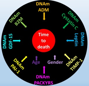
Currently, DNAm [DNA methylation] is the most accurate and the most frequently used biomarker in biohorology. However, it is harder to apply a DNAm clock compared to clocks based on clinical blood tests. Moreover, DNAm marks often take a long time to emerge in response to aging interventions.

Chromatin structure and telomeres, while intriguing, are too labor intensive and error-prone to be practical.”

https://www.sciencedirect.com/science/article/pii/S1568163719302582 “Biohorology and biomarkers of aging: current state-of-the-art, challenges and opportunities”


We think about chronological age linearly. The reviewers hinted at but didn’t directly assess the extent to which techniques such as linear regression may also influence people to think linearly about biological age.

We experience cyclical changes every day (like sleep), month, season, and longer periods. The reviewers didn’t mention techniques that incorporate our cyclical experiences or assess cyclical biological age.

1. The reviewers pointed out some biological age clock linearity flaws:

“Most aging clocks base their BA [biological age] definitions either on CA [chronological age] or mortality risk. Mortality risk in its turn is derived from demographic tables and can be assumed to be a function of CA in most animals, including human.

Thus, aging clocks are ultimately treating CA as a substitute BA with the caveat that deviations from the actual CA signify better or worse physical fitness when compared to age matched controls. Such a design has several flaws.”

2. They pointed out non-linear characteristics of chromosomal telomere length:

“DNA lesions caused by oxidative stress are repaired less efficiently in telomeric regions, which causes frailty and subsequent telomere shortening. Oxidative stress levels may fluctuate due to habitat, life style, inflammatory diseases – factors that do not necessarily represent replicative clock ticking.

Telomere length typically fluctuates within ±2-4% per month. This led scientists to hypothesize that telomere attrition is an oscillatory process.”

Since cell components show cyclical phases, why wouldn’t cells and each higher living structural level likewise demonstrate cyclical phases? That avenue wasn’t explored.

3. They mentioned the non-linearity of epigenetic clocks:

“If an organism’s DNAm profile is not directly linked to the thermodynamic root of aging [entropy] but instead is a downstream product of competing processes, the applicability of DNAm aging clock methodology is at risk. In this case different aging clocks may not be equally good for different experiment settings.

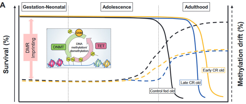
While genetic, pharmacological and dietary interventions with proven effect on life expectancy change the methylation state of the age-associated CpG sites, they do so in different ways. Caloric restriction is more efficient in preventing methylation loss at hypomethylated sites and methylation gain at hypermethylated sites than rapamycin.

These findings imply that DNAm profiles do not simply gravitate towards the average with age and that there is no single pathway through which all aging processes are imbued into an organism’s epigenetic landscape.”

4. Genetic and epigenetic regulatory pathways were presented with linear thinking:

“Protein structures encapsulating DNA and regulating its accessibility (chromatin and histones) have also been shown to change with age. Moreover, DNAm machinery and histone modifications are interlinked and change throughout aging concordantly.

For example, DNA methyltransferases are attracted by the H3K36me mark. With aging it is less tightly regulated, and thus, more sporadic DNAm occurs, which ultimately translates to epigenetic clock ticking.”


An individual’s capability to regulate their own aging phenotype wasn’t addressed, only externally applied “aging interventions.” Diseases were considered chronological-“age-associated.”

Biological aging was neither viewed as a disease nor as an unintended consequence. If these researchers don’t grasp the foundations of their field of study, why do they work in the biological aging field? It isn’t just math.

  • Could this paper reflect one company’s desire to frame arguments in favor of the company’s offered solution?
  • Could this paper reflect a “chronological age is the cause” meme that satisfied organizational imperatives for sponsors like the Buck Institute for Research on Aging?
  • Or could it be that the reviewers had other paradigms?

What do you think?

Aging as a disease

This 2020 interview was with UC Berkeley researchers:

“Lack of cure goes hand in hand with inability to accept that this [aging] is disease. For example, there was some resistance to accept tuberculosis as the actual disease. When there was no antibiotics or cure against it, people tended to discard it and said, oh, it’s just nerves, you need to go to a sanatorium and relax.

It used to be that, please do not diagnose that there’s bacterial meningitis, because there is no cure. Whatever else you can come up with, do it first. Now, diagnose it as fast as possible, so we can put patients on antibiotics immediately. My prediction is that the same will happen to aging.

We and others have demonstrated that you can, from the outside, either by some signal or blood therapy, parabiosis, something like that, some intervention, jump-start aged resident stem cells in tissue and get them to behave as, by whatever means you’re measuring it, young or a lot closer to young than they would normally be. Intrinsic capacity of them to act that way is there.

As we grow old, the environment of differentiated niche stem cells does not provide productive instruction. It provides counterproductive instruction, which, overall, tells them just to remain quiescent and do nothing.

It’s not a program to kill you. It’s the lack of a program to keep you young and healthy for longer than 90 years.

If your program was that whenever you’re a damaged, differentiated cell, you simply trigger apoptosis and activate stem cells to make new cells, we would live much longer and healthy. The program right now is to resist being dead and replaced as much as you can for as long as you can.

So cells produce too much TGF beta [transforming growth factor-β] because it helps them to keep functioning even when they’re damaged. That too much TGF beta, ironically, inhibits resident stem cells, so they are not replacing old cells with new ones. It’s almost like you have old bureaucrats that are running an organization and do not want to be replaced.

Our thoughts are probably different from most people, because we go to the data and the data show that they’re not really fully what authors wrote in the abstract or conclusion. When you look at that, my thought is that much more work needs to be done before it [partial cellular reprogramming] could be even thought to be commercialized.”

https://www.lifespan.io/news/apheresis-with-profs-irina-michael-conboy/ “Irina & Michael Conboy – Resetting Aged Blood to Restore Youth”


Keep in mind that although the interviewers’ organization had changed, their advocacy position as displayed in A blood plasma aging clock persisted. One of the interviewees is on the interviewers’ organization scientific advisory board, and they also have an interest in downgrading competing approaches.

Despite caveats, this interview was these researchers’ perspective in their decades-long investigations of aging. I included a graphic and below quote from Organismal aging and cellular senescence to note how their paradigm compared with other aging researchers:

“In our view, recent evidence that

  • Senescence is based on an unterminated developmental growth program and finding that
  • The concept of post-mitotic senescence requires activation of expansion, or ‘growth’ factors as a second hit,

favor the assumption that aging underlies a grating of genetic determination similarly to what is summarized above under the pseudo-programmed causative approach.”

An out-of-date review of epigenetic transgenerational inheritance

This December 3, 2019, French review title was “Transgenerational Inheritance of Environmentally Induced Epigenetic Alterations during Mammalian Development”:

“We attempt to summarize our current knowledge about transgenerational inheritance of environmentally induced epigenetic changes. While the idea that information can be inherited between generations independently of DNA’s nucleotide sequence is not new, the outcome of recent studies provides a mechanistic foundation for the concept.

Systematic resetting of epigenetic marks between generations represents the largest hurdle to conceptualizing epigenetic inheritance. Our understanding of rates and causes of epimutations remains rudimentary.

Environmental exposure to toxicants could promote changes in germline cells at any developmental stage, with more dramatic effects being observed during embryonic germ cell reprogramming. Epigenetic factors and their heritability should be considered during disease risk assessment.”


This review showed an inexplicable lack of thorough research. 2017 was its latest citation of epigenetic transgenerational inheritance studies from Washington State University labs of Dr. Michael Skinner. I’ve curated six of the labs’ 2019 studies!

  1. Transgenerational diseases caused by great-grandmother DDT exposure;
  2. Another important transgenerational epigenetic inheritance study;
  3. The transgenerational impact of Roundup exposure;
  4. Epigenetic transgenerational inheritance mechanisms that lead to prostate disease;
  5. A transgenerational view of the rise in obesity; and
  6. Epigenetic transgenerational inheritance extends to the great-great-grand offspring.

This lack led to – among other items – equivocal statements where current definitive evidence could have been cited. This review was submitted on October 31, 2019, and all above studies were available.


The publisher provided insight into the peer review process via https://www.mdpi.com/2073-4409/8/12/1559/review_report:

  • Peer reviewer 1: “Taking into account that this is not my main area of expertise..Do the authors really believe in that?”
  • Peer reviewer 2 provided a one-paragraph non-review.
  • Peer reviewer 3: “The authors are missing a large sector of what types of environmental factors can influence methylation and do not acknowledge that other sources exist.”

Authors responded with changes or otherwise addressed peer reviewer comments.

https://www.mdpi.com/2073-4409/8/12/1559/htm “Transgenerational Inheritance of Environmentally Induced Epigenetic Alterations during Mammalian Development”

An epigenetic clock review by committee

This 2019 worldwide review of epigenetic clocks was a semi-anonymous mishmash of opinions, facts, hypotheses, unwarranted extrapolations, and beliefs. Diversity of viewpoints among the 21 coauthors wasn’t evident.

1. Citations of coauthors’ works seemed excessive, and they apologized for omissions. However:

  • Challenge 5 was titled “Single-cell analysis of aging changes and disease” and
  • Table 1 “Major biological and analytic issues with epigenetic DNA methylation clocks” had single-cell analysis as the Proposed solution to five Significant issues.

Yet studies such as High-Resolution Single-Cell DNA Methylation Measurements Reveal Epigenetically Distinct Hematopoietic Stem Cell Subpopulations were unmentioned.

2. Some coauthors semi-anonymously expressed faith that using current flawed methodologies in the future – only more thoroughly, with newer equipment, etc. – would yield better results. If all 21 coauthors were asked their viewpoints of Proposed solutions to the top three Significant issues of epigenetic clocks, what would they emphasize when quoted?

3. Techniques were praised:

“Given the precision with which DNA methylation clock age can be estimated and evolving measures of biological, phenotype-, and disease-related age (e.g., PhenoAge, GrimAge)..”

Exactly why these techniques have at times produced inexplicable results wasn’t examined, though. Two examples:

  • In Reversal of aging and immunosenescent trends, Levine PhenoAge methodology estimated that the 51-65 year old subjects’ biological ages at the beginning of the study averaged 17.5 years less than their chronological age. Comparing that to Horvath average biological age of 3.95 years less raised the question: exactly why did PhenoAge show such a large difference?
  • The paper mentioned GrimAge methodology findings about “smoking-related changes.” But it didn’t explain why GrimAge methylation findings most closely associated with smoking history also accurately predicted future disease risk with non-smokers.

Eluding explanations for these types of findings didn’t help build confidence in methodologies.

4. A more readable approach to review by committee could have coauthors – in at least one section – answer discussion questions, as Reversing epigenetic T cell exhaustion did with 18 experts.

https://genomebiology.biomedcentral.com/articles/10.1186/s13059-019-1824-y “DNA methylation aging clocks: challenges and recommendations”

A GWAS meta-analysis of two epigenetic clocks

This 2019 UK human study conducted a meta-analysis of genome-wide association studies of two epigenetic clocks using 13,493 European-ancestry individuals aged between ten and 98 years:

“Horvath-EAA, described in previous publications as ‘intrinsic’ epigenetic age acceleration (IEAA), can be interpreted as a measure of cell-intrinsic ageing that exhibits preservation across multiple tissues, appears unrelated to lifestyle factors, and probably indicates a fundamental cell ageing process that is largely conserved across cell types.

In contrast, Hannum-EAA, referred to in previous studies as ‘extrinsic’ epigenetic age acceleration (EEAA), can be considered a biomarker of immune system ageing, explicitly incorporating aspects of immune system decline such as age-related changes in blood cell counts, correlating with lifestyle and health-span related characteristics, and thus yielding a stronger predictor of all-cause mortality.

The meta-analysis of Horvath-EAA identified ten independent associated SNPs [single nucleotide polymorphisms], doubling the number reported to date, and highlighted 21 genes involved in Horvath-based epigenetic ageing. Four of the ten Horvath-EAA-associated SNPs are mQTL [methylation quantitative trait loci] for CpGs used in the Horvath/Hannum epigenetic clocks. A possible interpretation of this is that the functional mechanism by which these SNPs influence the rate of biological ageing is via altering methylation levels.

Father’s age at death, a rough proxy for lifespan, was nominally significantly correlated with both EAA measures, and parents’ age at death was additionally correlated with Hannum-EAA. Aside from these, genetic correlations with age-related traits were surprisingly few: it is possible that this could reflect an overly conservative correction for the multiple tests carried out, or low statistical power, rather than a genuine lack of correlations.

Genetic correlation analysis should be restricted to GWAS with a heritability Z-score of 4 or more, on the grounds of interpretability and power, so the Horvath-based results particularly should be interpreted with caution.”


A non-apologetic way to explain the above graphic is that NONE of these 218 “health and behavioral traits” were any more associated with the studied genetic measurements than would be expected by chance!

Fervent believers in the GWAS methodology’s capability to exactly predict individual phenotypes eventually become victims of the scientific method. These GWAS researchers griped about “overly conservative correction, or low statistical power” and other predictable shortfalls, and ended a long limitations statement with:

“While we have identified a number of SNPs and genes significantly associated with EAA, including genes already known to be related to ageing, the analyses presented here fall short of providing a mechanistic explanation for how these variants and genes act to influence biological age.”

Outside of beliefs, it’s hard to understand why research money keeps pouring into the GWAS dead end. If these researchers and their employing institution and sponsors want to make a difference in human lives, they need to get busy in other areas.

These researchers were employed by the same institution that couldn’t be bothered to scrape together six more weeks of funds to study the transgenerational damaging effects of acetaminophen – an analgesic available to billions of people – in Epigenetics research that was designed to fall one step short of wonderful.

https://journals.plos.org/plosgenetics/article?id=10.1371/journal.pgen.1008104 “A meta-analysis of genome-wide association studies of epigenetic age acceleration”

Organismal aging and cellular senescence

I’ll curate this 2019 German review through its figures:

“With the discovery of beneficial aspects of cellular senescence and evidence of senescence being not limited to replicative cellular states, a redefinition of our comprehension of aging and senescence appears scientifically overdue.

Figure 1. Current determinants and relevant open questions, marking the processes of aging and senescence as discussed in the text. Aspects represented in green are considered as broadly accepted or scientifically consolidated. Novel aspects that are yet unproven, or are under debate, are highlighted in red.

SASP = senescence-associated secretory phenotype. AASP = putative aging-associated secretory phenotype as suggested in the text.

Figure 2. Theories on the causality and purpose of aging. Graphically summarized are four contrasting concepts crystallized from current evidence addressing the inductive driving force of aging. Apart from a stochastic deleteriome, there are arguments for a pseudo-programmed, programmed or at least partially programmed nature of aging.

Figure 3. Comparative representation of the aging and senescence processes highlighting different levels of interaction and putative sites of interventions.

(1) As discussed in the text, causative mechanisms of aging are still not well understood, however, multiple factors including genetic, epigenetic and stress-related effects seem to have an orchestrated role in the progression of aging. Senescence on the other hand, is seen as a programmed response to different kinds of stressors, which proceed in defined stages. Whether, in analogy, aging also follows a defined program or sequential stages is not known.

(2) Senescence involves autocrine and paracrine factors, which are responsible for a ‘seno-infection’ or bystander effect in neighboring cells. There is currently no direct evidence for a similar factor composition propagating the aging process via a kind of ‘gero-infection’.

(3) Accumulation of senescent cells has been described as a hallmark of aging; however, whether they are a causative factor or a consequence of tissue and organismal aging is still unknown. As discussed in the text, it appears possible that aging and senescence mutually influence each other through positive feedback at this level, leading to accelerated tissue damage and aging.

(4,5) Clearance of senescent or aging cells might constitute putative targets for interventional approaches aimed to reduce or reverse the impact of aging and improve cell and tissue homeostasis by inducing a ‘rejuvenation’ process.

Figure 4. Pathological and beneficial functions of aging and senescence, according to current knowledge. In red are represented pathological consequences and in green beneficial functions of aging and senescence.

The impact of aging has mainly been described at the organismal level, since a complete cellular functional profile has not yet been established. Accordingly, whether beneficial consequences of the aging process exist at the cellular level is unclear.”


The assertion of Figure 3 (2) that:

“There is currently no direct evidence for a similar factor composition propagating the aging process via a kind of ‘gero-infection.”

was shown to be false in Reevaluate findings in another paradigm:

“It was demonstrated that increased aging occurred as a result of lack of gonadotropin-releasing hormone and that increased lifespan resulted from its provision during aging.

In this manner:

  1. Aging of hypothalamic microglia leads to
  2. Aging of the hypothalamus, which leads to
  3. Aging elsewhere in the body.

So here we have a multi-level interaction:

  1. Activation of NF-κB leads to
  2. Cellular aging, leading to
  3. A diminished production of GnRH, which then
  4. Acts (through cells with a receptor for it, or indirectly as a result of changes to GnRH-receptor-possessing cells) to decrease lifespan.

So the age state of hypothalamic cells, at least with respect to NF-κB activation, is communicated to other cells via reduced output of GnRH.”


The reviewers’ position on Figure 2 was:

“In our view, recent evidence that

  • Senescence is based on an unterminated developmental growth program and the finding that
  • The concept of post-mitotic senescence requires the activation of expansion, or ‘growth’ factors as a second hit,

favor the assumption that aging underlies a grating of genetic determination similarly to what is summarized above under the pseudo-programmed causative approach.”

Their position on Figure 4’s beneficial effects of aging began with the sentence:

“If we assume that aging already starts before birth, it can be considered simply a developmental stage, required to complete the evolutionary program associated with species-intrinsic biological functions such as reproduction, survival, and selection.”

Cited studies included:

https://www.mdpi.com/2073-4409/8/11/1446 “Dissecting Aging and Senescence-Current Concepts and Open Lessons”

A strawman argument against epigenetic clocks

This 2019 review of epigenetic clocks by Washington cancer researchers ignored the elephant in the room: Their epigenetic drift paradigm is generally inapplicable to humans because the vast majority of our cells don’t divide/proliferate. They repeatedly returned to an argument for randomness as a cause for aging and disease:

“A time-dependent stochastic event process, like epigenetic drift, could lead to cancer formation through the accumulation of random epigenetic alterations that, through chance, eventually alter epigenetic driver gene expression leading to a clone of cells destined to become cancer.

It is plausible that the stochastic process inherent in epigenetic drift can induce aberrant methylation events that accumulate in normal cells and eventually induce cancer formation.

Epigenetic drift relates to a biological process that changes the DNA methylome with age via stochastic gains or losses of DNA methylation. Epigenetic drift can be understood in terms of errors in DNA methylation maintenance during DNA-replication.

The phenomenon of (epi)genetic drift is generally associated with phenotypic neutrality.

For patients who develop cancer around age 80, the most likely initiation time for the founder adenoma cell is predicted to be very early in life, roughly between the ages 15 to 20 years. This unexpected and provocative finding suggests that the optimal age-range for prevention of colorectal cancer may be in adolescence and early adulthood (and ideally through lifelong) dietary and lifestyle interventions.”


The reviewers’ strawman arguments intentionally mischaracterized aspects of the epigenetic clock:

1. The epigenetic clock founder’s actual view on aging was in The epigenetic clock theory of aging:

“The proposed epigenetic clock theory of ageing views biological ageing as an unintended consequence of both developmental programmes and maintenance programmes, the molecular footprints of which give rise to DNAm age estimators.”

The reviewers omitted this intrinsic view of aging, which didn’t fit into the above graphic.

2. Another misrepresentation was:

“In contrast to epigenetic clocks, epigenetic drift refers to a stochastic process that involves both gains and losses of the methylation state of CpG dinucleotides over time.”

A reader of the original 2013 epigenetic clock study would understand that epigenetic clocks measure “both gains and losses of methylation” as in:

“The 193 positively and 160 negatively correlated CpGs get hypermethylated and hypomethylated with age, respectively.”

3. These reviewers omitted recent epigenetic clock significant developments. For example, there was no mention of the GrimAge study, although it was published before this review was submitted.

4. Epigenetic drift as the cause of aging and disease has abundant contrary evidence. These reviewers tossed in a little toward the end of their directed narrative:

“We found only a small number of drift-related CpG island-gene pairs for which drift correlated positively and significantly with gene expression.

The functional consequences of epigenetic drift need to be further elucidated.”

However, they never acknowledged the elephant in the room!

https://cancerres.aacrjournals.org/content/early/2019/11/06/0008-5472.CAN-19-0924 “Epigenetic aging: more than just a clock when it comes to cancer” (not freely available)

Caloric restriction’s epigenetic effects

This 2019 US review subject was caloric restriction (CR) without malnutrition:

“Cellular adaptation that occurs in response to dietary patterns can be explained by alterations in epigenetic mechanisms such as DNA methylation, histone modifications, and microRNA. Epigenetic reprogramming of the underlying chronic low-grade inflammation by CR can lead to immuno-metabolic adaptations that enhance quality of life, extend lifespan, and delay chronic disease onset.

Short- and long-term CRs produce significant changes in different tissues and across species, in some animal models even with sex-specific effects. Early CR onset may cause a different and even an opposite effect on physiological outcomes in animal models such as body weight.”

https://academic.oup.com/advances/article-abstract/10/3/520/5420411 “Epigenetic Regulation of Metabolism and Inflammation by Calorie Restriction” (not freely available)


1. The review didn’t present evidence to equate survival (left axis) with methylation drift (right axis) per the above graphic. Methylation drift should point in the opposite direction of survival, if anything.

2. No mention was made of the epigenetic clock method of measuring age acceleration, although it’s been available since 2013 and recent diet studies have used it. The sole citation of an age acceleration study was from 2001, which was unacceptable for a review published in 2019.

3. The review provided many cellular-level details about the subject. However, organism-level areas weren’t sufficiently evidenced:

A. Arguments for an effect usually include explanations for no effect as well as for opposite effects. The reviewers didn’t provide direct evidence for why, if caloric restriction extended lifespan, caloric overabundance produced shorter lifespans.

B. Caloric restriction evidence was presented as if only it was responsible for organism-level effects. Other mechanisms may have been involved.

An example of such a mechanism was demonstrated in a 2007 rodent study Reduced Oxidant Stress and Extended Lifespan in Mice Exposed to a Low Glycotoxin Diet which compared two 40%-calorie-restricted diets.

The calories and composition of both diets were identical. However, advanced glycation end product (AGE) levels were doubled in standard chow because heating temperatures were “sufficiently high to inadvertently cause standard mouse chow to be rich in oxidant AGEs.”

The study found that a diet with lower chow heating temperatures increased lifespan and health span irrespective of caloric restriction!

  • The low-AGE calorie-restricted diet group lived an average of 15% longer (>20 human equivalent years) than the CR group.
  • 40% of the low-AGE calorie-restricted diet group were still alive when the last CR group member died.
  • The CR group also had significantly more: 1) oxidative stress damage; 2) glucose and insulin metabolism problems; and 3) kidney, spleen, and liver injuries.

A drug that countered effects of a traumatizing mother

This 2019 US rodent study concerned transmitting poor maternal care to the next generation:

“The quality of parental care received during development profoundly influences an individual’s phenotype, including that of maternal behavior. Infant experiences with a caregiver have lifelong behavioral consequences.

Maternal behavior is a complex behavior requiring the recruitment of multiple brain regions including the nucleus accumbens, bed nucleus of the stria terminalis, ventral tegmental area, prefrontal cortex, amygdala, and medial preoptic area. Dysregulation within this circuitry can lead to altered or impaired maternal responsiveness.

We administered zebularine, a drug known to alter DNA methylation, to dams exposed during infancy to the scarcity-adversity model of low nesting resources, and then characterized the quality of their care towards their offspring.

  1. We replicate that dams with a history of maltreatment mistreat their own offspring.
  2. We show that maltreated-dams treated with zebularine exhibit lower levels of adverse care toward their offspring.
  3. We show that administration of zebularine in control dams (history of nurturing care) enhances levels of adverse care.
  4. We show altered methylation and gene expression in maltreated dams normalized by zebularine.

These findings lend support to the hypothesis that epigenetic alterations resulting from maltreatment causally relate to behavioral outcomes.

Maternal behavior is an intergenerational behavior. It is important to establish the neurobiological underpinnings of aberrant maternal behavior and explore treatments that can improve maternal behavior to prevent the perpetuation of poor maternal care across generations.”


The study authors demonstrated intergenerational epigenetic effects, and missed an opportunity to also investigate transgenerational epigenetically inherited effects. They cited reference 60 for the first part of the above quotation, but the cited reviewer misused the transgenerational term by applying it to grand-offspring instead of the great-grand-offspring.

There were resources available to replicate the study authors’ previous findings, which didn’t show anything new. Why not use such resources to uncover evidence even more applicable to humans by extending experiments to great-grand-offspring that would have no potential germline exposure to the initial damaging cause?

Could a study design similar to A limited study of parental transmission of anxiety/stress-reactive traits have been integrated? That study’s thorough removal of parental behavior would be an outstanding methodology to confirm by falsifiability whether parental behavior is both an intergenerational and a transgenerational epigenetic inheritance mechanism.

Rodent great-grand-offspring can be studied in < 9 months. It takes > 50 years for human studies to reach the great-grand-offspring transgenerational generation.

  • Why not attempt to “prevent the perpetuation of poor maternal care across generations?”
  • Isn’t it a plausible hypothesis that humans “with a history of maltreatment mistreat their own offspring?”
  • Isn’t it worth the extra effort to extend animal research to investigate this unfortunate chain?

https://www.nature.com/articles/s41598-019-46539-4 “Pharmacological manipulation of DNA methylation normalizes maternal behavior, DNA methylation, and gene expression in dams with a history of maltreatment”

What drives cellular aging?

This 2019 US/UK human cell study by the founder of the epigenetic clock method investigated epigenetic aging:

“It is widely assumed that extension of lifespan is a result of retardation of ageing. While there is no counter-evidence to challenge this highly intuitive association, supporting empirical evidence to confirm it is not easy to acquire.

The scarcity of empirical evidence is due in part to the lack of a good measure of age that is not based on time. In this regard, the relatively recent development of epigenetic clocks is of great interest.

At the cellular level more is known, but from the perspective of what epigenetic ageing is not, rather than what it is. While we still do not know what cellular feature is associated with epigenetic ageing, we can now remove:

  • somatic cell differentiation

from the list of possibilities and place it with

  • cellular senescence,
  • proliferation and
  • telomere length maintenance,

which represent cellular features that are all not linked to epigenetic ageing.”


The study used several agents, including rapamycin, to investigate the hypotheses. Rapamycin isn’t a panacea, however:

“The ability of rapamycin to suppress the progression of epigenetic ageing is very encouraging for many reasons not least because it provides a valuable point-of-entry into molecular pathways that are potentially associated with it. Evidently, the target of rapamycin, the mTOR complex is of particular interest.

The convergence of the GWAS observation with the experimental system described here is a testament of the strength of the skin & blood clock in uncovering biological features that are consistent between the human level and cellular level. It lends weight to the emerging view that the mTOR pathway may be the underlying mechanism that supports epigenetic ageing.”

The limitation section ended with:

“It is important to note that it is inadvisable (actively discouraged) to directly extrapolate the studies here, especially in terms of the magnitude of age suppression, to potential effects of rapamycin on humans.”

https://www.aging-us.com/article/101976/text “Rapamycin retards epigenetic ageing of keratinocytes independently of its effects on replicative senescence, proliferation and differentiation”

Statistical inferences vs. biological realities

A 2019 UCLA study introduced a derivative of the epigenetic clock named GrimAge:

“DNAm GrimAge, a linear combination of chronological age, sex, and DNAm-based surrogate biomarkers for seven plasma proteins and smoking pack-years, outperforms all other DNAm-based biomarkers, on a variety of health-related metrics.

An age-adjusted version of DNAm GrimAge, which can be regarded as a new measure of epigenetic age acceleration (AgeAccelGrim), is associated with a host of age-related conditions, lifestyle factors, and clinical biomarkers. Using large scale validation data from three ethnic groups, we demonstrate that AgeAccelGrim stands out among pre-existing epigenetic clocks in terms of its predictive ability for time-to-death, time-to-coronary heart disease, time-to-cancer, its association with computed tomography data for fatty liver/excess fat, and early age at menopause.”

https://www.ncbi.nlm.nih.gov/pmc/articles/PMC6366976/ “DNA methylation GrimAge strongly predicts lifespan and healthspan”


A miserable attempt at reporting the study’s findings included angles of superstition, fear-of-the-future, and suspicion-by-spurious-association:

“The research has already captured the attention of the life insurance industry. After all, a solid death date could mean real savings when it comes to pricing policies.

The hope is that if and when legitimate anti-aging drugs are developed, GrimAge could be used to test their effectiveness. In a world with functional anti-aging drugs, “doctors could test [your GrimAge number] and say, ‘You know what, you’re aging too quickly. Take this,'” Horvath said.”

https://onezero.medium.com/a-new-test-predicts-when-youll-die-give-or-take-a-few-years-2d08147c8ea6 “A New Test Predicts When You’ll Die (Give or Take a Few Years)”


A detailed blog post from Josh Mitteldorf provided scientific coverage of the study:

“Methylation sites associated with smoking history predicted how long the person would live more accurately than the smoking history itself. Even stranger, the methylation marks most closely associated with smoking were found to be a powerful indication of future health even when the sample was confined to non-smokers.

The DNAm GrimAge clock was developed in two stages, a correlation of a correlation. Curiously, the indirect computation yields the better result.

Horvath’s finding that secondary methylation indicators are more accurate than the underlying primary indicator from which they were derived is provocative, and calls out for a new understanding.”

https://joshmitteldorf.scienceblog.com/2019/03/05/dnam-grimage-the-newest-methylation-clock “DNAm GrimAge—the Newest Methylation Clock”


When there are logical disconnects in findings like the above, it’s time to examine underlying premises. As noted in Group statistics don’t necessarily describe an individual, an assumption required by statistical analyses is that each measured item in the sample is interchangeable with the next.

This presumption is often false, producing individually inapplicable results. For example, Immune memory vs. immune adaptation included this description of the adaptive immune system:

“To be effective, highly specific immune response requires huge diversity of receptors and antibodies, which is achieved by somatic rearrangement of gene segments. Recombination results in millions of TCR [T cell receptor] and antibody variants able to recognize and neutralize millions of various antigens.”

Standard statistics of millions of T cell receptor and antibody variants won’t represent their individually unique properties. But individual differences are both their purpose and benefit to us.

The GrimAge study’s overreach was most apparent in stratifying educational attainment to develop correlations. As mentioned in Does a societal mandate cause DNA methylation? such statistics are poor evidence of each individual’s biological realities.

Neither derivatives of group statistics, nor correlations of correlations, seem to be the techniques needed to understand biological causes of effects. Another commentary on the GrimAge study mentioned but glossed over this point:

“It remains a mystery why exactly the epigenetic clocks work, and whether age-related changes in DNA methylation contribute to the cause of aging or are a result of it.”

A therapy to reverse cognitive decline

This 2018 human study presented the results of 100 patients’ personalized therapies for cognitive decline:

“The first examples of reversal of cognitive decline in Alzheimer’s disease and the pre-Alzheimer’s disease conditions MCI (Mild Cognitive Impairment) and SCI (Subjective Cognitive Impairment) have recently been published..showing sustained subjective and objective improvement in cognition, using a comprehensive, precision medicine approach that involves determining the potential contributors to the cognitive decline (e.g., activation of the innate immune system by pathogens or intestinal permeability, reduction in trophic or hormonal support, specific toxin exposure, or other contributors), using a computer-based algorithm to determine subtype and then addressing each contributor using a personalized, targeted, multi-factorial approach dubbed ReCODE for reversal of cognitive decline.

An obvious criticism of the initial studies is the small number of patients reported. Therefore, we report here 100 patients, treated by several different physicians, with documented improvement in cognition, in some cases with documentation of improvement in electrophysiology or imaging, as well.”

https://www.omicsonline.org/open-access/reversal-of-cognitive-decline-100-patients-2161-0460-1000450-105387.html “Reversal of Cognitive Decline: 100 Patients”


The lead author commented on Josh Mitteldorf’s informative post A cure for Alzheimer’s? Yes, a cure for Alzheimer’s!:

  1. “We have a paper in press, due to appear 10.22.18 (open access, JADP, I’ll send a copy as soon as available), showing 100 patients with documented improvement – some with MRI volumetrics improved, others with quantitative EEG improvements, others with evoked response improvements, and all with quantitative cognitive assessment improvement. Some are very striking – 12 point improvements in MoCA [Montreal Cognitive Assessment], for example – others less so, but all also have subjective improvement. Hopefully this will address some of the criticisms that we haven’t documented improvement in enough people.
  2. We were just turned down again for a randomized, controlled clinical trial, so on the one hand, we are told repeatedly that no one will believe that this approach works until we publish a randomized, controlled study, and on the other hand, we’ve been turned down (first in 2011/12, and now in 2018), with the complaint that we are trying to address more than one variable in the trial (as if AD is a single-variable disease!). Something of a catch-22. We are now resubmitting (unfortunately, the IRBs are not populated by functional medicine physicians, so they are used to seeing old-fashioned drug studies), and we’ll see what happens.
  3. I’ve been extending the studies to other neurodegenerative diseases, and it has been impressive how much of a programmatic response there seems to be in these ‘diseases.’
  4. I agree with you that there are many features in common with aging itself.
  5. You made a good point that APP [amyloid precursor protein] is a dependence receptor, and in fact it functions as an integrating dependence receptor, responding to numerous inputs (Kurakin and Bredesen, 2015).
  6. In the book and the publications, we don’t claim it is a “cure” since we don’t have pathological evidence that the disease process is gone. What we claim is ‘reversal of cognitive decline’ since that is what we document.
  7. As I mentioned in the book, AD is turning out to be a protective response to multiple insults, and this fits well with the finding that Abeta has an antimicrobial effect (Moir and Tanzi’s work). It is a network-downsizing, protective response, which is quite effective – some people live with the ongoing degenerative process for decades.
  8. We have seen several cases now in which a clinical trial of an anti-amyloid antibody made the person much worse in a time-dependent manner (each time there was an injection, the person would get much worse for 5-10 days, then begin to improve back toward where he/she was, but over time, marked decline occurred), and this makes sense for the idea that the amyloid is actually protecting against pathogens or toxins or some other insult.
  9. It is important to note that we’ve never claimed that all people get better – this is not what we’ve seen. People very late in the process, or who don’t follow the protocol, or who don’t address the various insults, do not improve. It is also turning out to be practitioner dependent – some are getting the vast majority of people to improve, others very few, so this is more like surgery than old-fashioned prescriptive medicine – you have to do a somewhat complicated therapeutic algorithm and get it right for best results.
  10. I’m very interested in what is needed to take the next step in people who have shown improvement but who started late in the course. For example, we have people now who have increased MoCA from 0 to 9 (or 0 to 3, etc.), with marked subjective improvement but plateauing at less than normal. These people had extensive synaptic and cellular loss prior to the program. So what do we need to raise the plateau? Stem cells? Intranasal trophic support? Something else?
  11. I haven’t yet seen a mono-etiologic theory of AD or a mono-therapeutic approach that has repeatedly positive results, so although I understand that there are many theories and treatments, there doesn’t seem to be one etiology to the disease, nor does there seem to be one simple treatment that works for most. It is much more like a network failure.”

At a specific level:

  • “There doesn’t seem to be one etiology to the disease,
  • Nor does there seem to be one simple treatment that works for most.
  • We don’t have pathological evidence that the disease process is gone.”

For general concepts, however:

  • “AD is turning out to be a protective response to multiple insults.
  • It is a network-downsizing, protective response, which is quite effective.
  • The amyloid is actually protecting against pathogens or toxins or some other insult.”

For a framework of an AD cure to be valid, each source of each insult that evoked each “protective response” should be traced.

Longitudinal studies would be preferred inside this framework. These study designs would investigate evidence of each insult’s potential modifying effect on each “protective response” that could affect the cumulative disease trajectory of each individual.

In many cases, existing study designs would be adequate if they extended their periods to the end of the subjects’ natural lifetimes. One AD-relevant example would be extending the prenatally-restraint-stressed model used in:

The framework would also encourage extending studies to at least three generations to investigate evidence for transgenerational effects, as were found in:

An hour of the epigenetic clock

Starting the fifth year of this blog with a 2018 presentation by the founder of the epigenetic clock method describing the state of the art up through July 2018. The webinar was given on the release day of The epigenetic clock now includes skin study.


Segments before the half-hour mark provide an introduction to the method and several details about the concurrently-released study. The Q&A section starts a little before the hour mark.