Little evidence for mitochondrial DNA methylation

This 2018 Japanese rodent study used three different techniques to detect mitochondrial DNA methylation:

“Whilst 5-methylcytosine (5mC) is a major epigenetic mark in the nuclear DNA in mammals, whether or not mitochondrial DNA (mtDNA) receives 5mC modification remains controversial.

We used bisulfite sequencing, McrBC digestion analyses and liquid chromatography mass spectrometry, which are distinctly differing methods for detecting 5mC. We analysed mtDNAs from mouse ESCs [embryonic stem cells] and from mouse liver and brain tissues.

Taken together, we propose that 5mC is not present at any specific region(s) of mtDNA and that levels of the methylated cytosine are fairly low, provided the modification occurs. It is thus unlikely that 5mC plays a universal role in mtDNA gene expression or mitochondrial metabolism.”


Bisulfite sequencing infers the presence of CpG (CG above) and non-CpG (CH above) methylation through unconverted residues:

“Synthetic and native mtDNA gave similar patterns, suggesting that the resistance of cytosines to bisulfite conversion is not due to methylation.”


It seems that epigenetic changes to mitochondrial DNA occur primarily through histone modifications. Lysine acetylation is gnarly and dynamic is one paper that detailed aspects of this functionality in mitochondria.

https://www.nature.com/articles/s41598-018-24251-z “Accurate estimation of 5-methylcytosine in mammalian mitochondrial DNA”

The purpose of epigenetic mechanisms

The concluding remarks of this 2018 Chinese review were:

“Using heterochromatin as a model, we have reviewed here the mechanisms behind the establishment and maintenance of silent chromatin domains. We conclude that almost every component of the chromatin environment, including DNA elements, RNAs, histones and other chromatin proteins, plays a role in the process of shaping and maintaining epigenetic states.

Epigenetic mechanisms have evolved..to solve the problem of orchestrating the differentiation of cells with the same genome. Just as any stable system must preserve some degree of flexibility, crosstalk and feedback among all elements in the system are mechanistically required.

We emphasize that:

  1. Epigenetic information is inherited [from parent cell to child cell] in a relatively stable but imprecise fashion;
  2. Multiple cis and trans factors are involved in the maintenance of epigenetic information during mitosis; and
  3. The maintenance of a repressive epigenetic state requires both recruitment and self-reinforcement mechanisms.”


Studies I’ve curated in 2018 whose methodologies may have benefited from investigating multiple epigenetic mechanisms included:

Only DNA methylation:

Only microRNAs:

A review of studies that investigated DNA methylation and microRNAs but not histone modifications:

https://link.springer.com/article/10.1007/s11427-018-9276-7 “Recruitment and reinforcement: maintaining epigenetic silencing” (not freely available)

Genomic imprinting and growth

This 2018 UK paper reviewed genomic imprinting:

“Since their discovery nearly 30 years ago, imprinted genes have been a paradigm for exploring the epigenetic control of gene expression. Moreover, their roles in early life growth and placentation are undisputed.

However, it is becoming increasingly clear that imprinted gene function has a wider role in maternal physiology during reproduction – both by modulating fetal and placental endocrine products that signal to alter maternal energy homeostasis, and by altering maternal energetic set points, thus producing downstream actions on nutrient provisioning.”

“Imprinted genes in the conceptus produce products that alter maternal resource allocation by:

  1. altering the transport capacity of the placenta;
  2. increasing fetal demand for resources by their action on the intrinsic growth rate; and
  3. signalling to the mother by the production of fetal/placental hormones that modify maternal metabolism.”

Other studies/reviews I’ve curated that covered genomic imprinting are:

http://jeb.biologists.org/content/jexbio/221/Suppl_1/jeb164517.full.pdf “Genomic imprinting, growth and maternal-fetal interactions”


This post has somehow become a target for spammers, and I’ve disabled comments. Readers can comment on other posts and indicate that they want their comment to apply here, and I’ll re-enable comments.

How well do single-mother rodent studies inform us about human fathers?

Two items before getting to the review:

This 2018 Australian review subject was paternal intergenerational and transgenerational transmission of biological and behavioral phenotypes per this partial outline:

“Evidence for non-genetic inheritance of behavioral traits in human populations

  • Intergenerational inheritance modulating offspring phenotypes following paternal exposure to trauma
  • Epigenetic inheritance via the germline following paternal environmental exposures
  • Limitations of research on epigenetic inheritance in human populations

The transgenerational impact of stressful paternal environments

  • Impact of paternal stress on affective behaviors and HPA-axis regulation of progeny
  • Influence of paternal stress exposure on offspring cognition
  • Role of sperm-borne microRNAs in the epigenetic inheritance of stress

Sexually dimorphic aspects of paternal transgenerational epigenetic inheritance”

The review was comprehensive, and filled in the above outline with many details towards the goal of:

“This exciting new field of transgenerational epigenomics will facilitate the development of novel strategies to predict, prevent and treat negative epigenetic consequences on offspring health, and psychiatric disorders in particular.”

The reviewers also demonstrated that current intergenerational and transgenerational research paradigms exclude a father’s child care behavior.


The fact that studies use rat and mouse species where fathers don’t naturally provide care for their offspring has warped the translation of findings to humans. The underlying question every animal study must answer is: how can its information be used to help humans? I asked in A limited study of parental transmission of anxiety/stress-reactive traits:

“How did parental behavioral transmission of behavioral traits and epigenetic changes become a subject not worth investigating? These traits and effects can be seen everyday in real-life human interactions, and in every human’s physiology.

Who among us doesn’t still have biological and behavioral consequences from our experiences of our father’s child care actions and inactions? Why can’t researchers and sponsors investigate these back to their sources that may include grandparents and great-grandparents?

Such efforts weren’t apparent in the review’s 116 cited references that included:

The reviewer in the latter has been instrumental in excluding behavioral inheritance mechanisms from these research paradigms, leading to my questions:

  1. “If the experimental subjects had no more control over their behavioral stress-response effects than they had over their DNA methylation, histone modification, or microRNA stress-response effects, then why was such behavior not included in the “epigenetic mechanisms” term?
  2. How do behavioral inheritance mechanisms fall outside the “true epigenetic inheritance” term when behavioral stress-response effects are shown to be reliably transmitted generation after generation?
  3. Wouldn’t the cessation of behavioral inheritance mechanisms confirm their status by falsifiability as was similarly done with studies such as the 1995 Adoption reverses the long-term impairment in glucocorticoid feedback induced by prenatal stress?”

Translating rodent studies into human mothers’ behavioral transmission of biological and behavioral phenotypes isn’t hampered by the studied species’ traits as it is for human fathers. But sponsors have to have the guts to support human research that may not produce politically-correct findings.


http://www.translatingtime.org provides an inter-species comparative timeline. For example, an input of:

  • Species 1: Human
  • Process: Lifespan
  • Location: Whole Organism
  • Days (post-conception): 270
  • Species 2: Mouse

produces a list of event predictions. Note how many significant events occur before humans are born at day 270, assuming everything goes right with our developmental processes! Also, the model predictions for humans end at post-conception day 979, three weeks short of when we celebrate our second birthday.

https://www.nature.com/articles/s41380-018-0039-z “Transgenerational epigenetic influences of paternal environmental exposures on brain function and predisposition to psychiatric disorders” (not freely available) Thanks to Dr. Shlomo Yeshurun for providing a full copy.

Dealing with big data in epigenetic studies

There’s been a long-standing need for tools and mathematical techniques which effectively deal with the large amount of data present in epigenetic studies. Complete experimental conditions and results aren’t accurately described when researchers fail to transform large sets of data into information.

This 2018 Baltimore review/promotional paper described an approach that promised to resolve the following data issues.

1. Epigenetic changes occur in many ways and areas, and they often aren’t isolated from each other:

“Fully characterizing the polymorphic and stochastic nature of DNA methylation requires specification of joint probability distributions of methylation patterns formed by sets of spatially coupled CpG sites.”

2. The absence of DNA methylation or gene expression provides signals that should be processed into information. A study of DNA methylation and age reported this situation as:

“Due to the methods applied in the present study, not all the effects of DNA methylation on gene expression could be detected; this limitation is also true for previously reported results.

The textbook case of DNA methylation regulating gene expression (the methylation of a promoter and silencing of a gene) remains undetected in many cases because in an array analysis, an unexpressed gene shows no signal that can be distinguished from background and is therefore typically omitted from the analysis.”

The current review described the problem as:

“These techniques assign zero probabilities to unobserved methylation patterns despite their biological plausibility, which results in underestimating the true biological heterogeneity of methylation patterns.”

3. A subset of the above is that unknown or random past causes and effects of epigenetic changes aren’t adequately modeled:

“We demonstrated..that the empirical approach to joint methylation analysis..does not perform well when dealing with highly stochastic methylation data.”


The paper’s approach is tailored for whole genome bisulfite sequencing (WGBS), the “gold-standard experimental technique for studying DNA methylation.” It’s named informME and is publicly available at https://github.com/GarrettJenkinson/informME.

https://bmcbioinformatics.biomedcentral.com/track/pdf/10.1186/s12859-018-2086-5 “An information-theoretic approach to the modeling and analysis of whole-genome bisulfite sequencing data”

Maternal obesity causes fetal liver damage

This 2018 US baboon study was on fetal effects from maternal obesity before and during pregnancy:

“Approximately 64% of women of childbearing age in the USA [are] overweight or obese. The baboon is a well-characterized animal model sharing many physiological, metabolic, and genetic characteristics with humans allowing direct translation of findings to human pregnancy.

Our study shows that fetal exposure to the MO [maternal obesity] intrauterine environment results in dysregulation of fetal hepatic genes central to metabolism.

These findings were further supported by identification of miRNAs that were inversely expressed with key genes in these pathways..suggest important early molecular mechanisms by which MO programs fetal hepatic lipid metabolism.

Future studies are required in MO post-natal offspring to determine the extent to which the fetal phenotype persists, and the degree to which this increases offspring risk of cardiometabolic disorders in later life.”


The study provided many measurements that may be relevant to humans. Other consequential measurements were missing that may have made the study’s findings even more applicable to humans:

  • No placental measurements other than weight. The organ through which the fetus received its nutrients, signaled its needs, modulated its growth rate, and developed its organs, was only measured by weight?
  • No other epigenetic analyses such as DNA methylation and histone modifications.

Were these omitted due to limited resources?

http://onlinelibrary.wiley.com/doi/10.1113/JP275422/pdf “Primate fetal hepatic responses to maternal obesity: epigenetic signalling pathways and lipid accumulation”

RNA and neurodegenerative diseases

This 2018 Chinese paper reviewed the associations among long non-coding RNA and four neurodegenerative diseases:

“lncRNAs are widely implicated in various physiological and pathological processes, such as epigenetic regulation, cell cycle regulation, cell differentiation regulation, cancer, and neurodegenerative diseases, through their interactions with chromatin, protein, and other RNAs. Numerous studies have suggested that lncRNAs are closely linked with the occurrence and development of a variety of diseases, especially neurodegenerative diseases, of which the etiologies are complicated and the underlying mechanisms remain elusive.

We focus on how lncRNA dysfunctions are involved in the pathogenesis of Alzheimer’s disease, Parkinson’s disease, Huntington’s disease, and amyotrophic lateral sclerosis.”


Table 1 showed specific lncRNAs that acted as “bodyguards” in inherited Huntington’s disease, “culprits” in Alzheimer’s disease, and as both in Parkinson’s disease. The table didn’t include lncRNAs associated with amyotrophic lateral sclerosis although the review text mentioned several.

https://www.sciencedirect.com/science/article/pii/S2162253117303104 “Long Non-coding RNAs, Novel Culprits, or Bodyguards in Neurodegenerative Diseases”

Cell senescence and DNA methylation

This 2018 Baltimore cell study found:

“Based on similarities in overall methylation patterns in replicative senescence and cancers, it is hypothesized that tumor-promoting DNA methylation in cancers derives from cells escaping senescence.

We show that the tumor-associated methylation changes evolve independently of senescence and are pro-survival events with functional implications contrasting that in senescence.

In our analyses, although overall global gains and losses in DNA methylation are similar, at individual genomic regions the methylation patterns are very different for senescence versus transformation.”

https://www.sciencedirect.com/science/article/pii/S1535610818300084 “DNA Methylation Patterns Separate Senescence from Transformation Potential and Indicate Cancer Risk” (not freely available)


I hesitated to use the study’s main graphic:
because the “Stochastic” labeling of the upper branch didn’t represent the vector’s meaning. The In Brief and the Summary sections contributed to the misrepresentation by stating:

“transformation-associated methylation changes arise stochastically.”

which wasn’t the study’s main finding:

“Our data outlined in the above sections strongly suggest against this senescence bypass hypothesis.”

Although the experimental design and methods evoked randomness:

“Immortalization on the path to malignant transformation involves stochastic epigenetic patterns from which cells contributing to transformation may evolve.”

the graphic’s upper branch vector represented the cells’ evolutionary responses. The Significance section best characterized what the study found:

“Tumor-associated methylation changes evolve independently of senescence and are pro-survival events.”

Would anyone at John Hopkins argue, as the graphic’s upper branch labeling suggested, that cellular aging is a predominantly random process? NO!


1. Epigenetics research and evolution promoted understanding the graphic’s upper branch vector:

“Evolution is an ongoing set of iterative interactions between organisms and the environment. Directionality is introduced by the agency of organisms themselves.”

2. The current study provided another data point about the uselessness of convenient but non-etiologic, inconsequential measurements of global methylation:

“Although overall global gains and losses in DNA methylation are similar, at individual genomic regions the methylation patterns are very different.”

3. The current study was congruent with the below finding of Using an epigenetic clock to distinguish cellular aging from senescence regarding the differentiation of cellular aging from senescence:

“Cellular ageing is distinct from cellular senescence and independent of DNA damage response and telomere length.”

Viruses target epigenetic processes

This 2018 Colorado review subject was general and specific ways viruses target epigenetic processes:

“We describe viral mechanisms and virus-host interactions by which DNA tumor viruses regulate host DNA methylation to evade antiviral immunity.

It is well known that most endogenous retroviruses and retrotransposons in the human genome are inactivated by DNA hypermethylation. In addition to endogenous retroviruses, the genomes of DNA viruses, such as human papillomavirus (HPV), herpes simplex virus 1 (HSV-1), adenovirus, and hepatitis B virus (HBV), are also frequently methylated and silenced in infected cells.

A recently described mechanism for viruses to epigenetically subvert host immunity is repression of immune-related gene expression by induction of DNA hypermethylation. Some host genes are not silenced simply through promoter hypermethylation or histone deacetylation alone, and therefore, viruses may have evolved mechanisms to ensure host gene downregulation through multiple epigenetic modifications.”

http://www.mdpi.com/1999-4915/10/2/82/htm “DNA Tumor Virus Regulation of Host DNA Methylation and Its Implications for Immune Evasion and Oncogenesis”


A second 2018 New York study focused on the Zika virus and DNA methylation:

“We studied the impact of ZIKV infection on the DNA methylation pattern across the entire genome in selected neural cell types. The virus unexpectedly alters the DNA methylome of neural progenitors, astrocytes, and differentiated neurons at genes that have been implicated in the pathogenesis of a number of brain disorders.

It remains open, however, whether the methylation changes come first or whether the viral infection dysregulates epigenetic regulatory genes prior to any epigenetic shift.”

http://msystems.asm.org/content/3/1/e00219-17 “Zika Virus Alters DNA Methylation of Neural Genes in an Organoid Model of the Developing Human Brain”

What are the chances?

This 2018 UC Davis anthropology study was on dice changes over two centuries:

“In Roman times, many dice were visibly lopsided..It did not matter what the objects were made of (metal, clay, bone, antler and ivory), or whether they were precisely symmetrical or consistent in size or shape, because, like the weather, rolls were predetermined by gods or other supernatural elements.

Dice, like many material objects, reflect a lot about people’s changing worldviews, Eerkens said. In this case, we believe it follows changing ideas about chance and fate.”


Think of a significant event in your life. Was it brought about by:

  1. Fate?
  2. Karma, divine intervention?
  3. A prayer, belief, placebo-effect process?
  4. Randomness?
  5. A coin-flip, card-draw, dice-roll decision process?
  6. A weighted-probability decision process?
  7. Chosen behavior, thoughts, and feelings?
  8. Unconscious behavior, thoughts, and feelings?
  9. Culturally-guided motivations?
  10. Non-arbitrary influences of other parties?

Which one or more of these factors would you now prefer to have been involved?

https://www.ucdavis.edu/news/it-not-how-you-play-game-how-dice-were-made “It’s Not How You Play the Game, but How the Dice Were Made”

Epigenetic mechanisms of muscle memory

This 2018 UK human study detailed epigenetic muscle memory:

“We aimed to investigate an epigenetic memory of earlier hypertrophy in adult human skeletal muscle using a within measures design, by undertaking:

  1. Resistance exercise induced muscle growth (loading) [3 days a week for 7 weeks], followed by;
  2. Cessation of resistance exercise, to return muscle back towards baseline levels (unloading) [7 weeks], and;
  3. A subsequent later period of resistance exercise induced muscle hypertrophy (reloading) [3 days a week for 7 weeks].”

The findings were:

“Frequency of genome-wide hypomethylation is the largest after reloading induced hypertrophy where lean muscle mass is enhanced.

Hypomethylation is maintained from earlier load induced hypertrophy even during unloading where muscle mass returns back towards baseline, and is inversely associated with gene expression.

A single bout of acute resistance exercise evokes hypomethylation of genes that have enhanced gene expression in later reload induced hypertrophy.”

https://www.nature.com/articles/s41598-018-20287-3 “Human Skeletal Muscle Possesses an Epigenetic Memory of Hypertrophy”


The study provided another example of how our bodies remember. It began with only eight male 27.6 ± 2.4 year-old subjects, though, and one of them dropped out.

See the discussion of a 2017 Netherlands human study in Are Underpowered Studies Ever Justified? with comments on studies with few subjects, such as:

“The problem occurs when people do small quantitative studies, but draw conclusions nonetheless, simply adding a disclaimer to the discussion (which they don’t put in the abstract, or the press release).”

“Underpowered studies may only be useful to check if the experiment works out wrt understanding instructions, do the programs run, etc, but not as much for testing and estimating effects.”

“The problem with underpowered studies is that all estimates can vary erratically between samples. Combined with the desire of many researchers (and universities’ press offices) to find sensational patterns, this means that evidence from underpowered studies is ‘asymmetrically’ likely to be considered more conclusive. As in, something that seems really cool will probably be considered more conclusive than something that’s disappointing. Highly powered studies don’t afford this flexibility.”

Non-CpG DNA methylation

This 2017 Korean review compared and contrasted CpG and non-CpG DNA methylation:

“Non-CpG methylation is restricted to specific cell types, such as pluripotent stem cells, oocytes, neurons, and glial cells. Accumulation of methylation at non-CpG sites and CpG sites in neurons seems to be involved in development and disease etiology.

Non-CpG methylation is established during postnatal development of the hippocampus and its levels increase over time. Similarly, non-CpG methylation is scarcely detected in human fetal frontal cortex, but is dramatically increased in later life. This increase in non-CpG methylation occurs simultaneously with synaptic development and increases in synaptic density.

In contrast, CpG methylation occurs during early development and does not increase over time.

Neurons have considerably higher levels of non-CpG methylation than glial cells. The human male ES [embryonic stem] cell line (H1) is more highly methylated than the female ES cell line (H9).

Among the different types of non-CpG methylation (CpA [adenosine], CpT [thymine], and CpC [another cytosine]), methylation is most common at CpA sites. For instance, in human iPS [induced pluripotent stem] cells, 5mCs are found in approximately 68.31%, 7.81%, 1.99%, and 1.05% of CpG, CpA, CpT, and CpC sites, respectively.”


The reviewers’ referenced statement:

“CpG methylation occurs during early development and does not increase over time.”

was presented outside of its context. The 2013 cited source’s statement was restricted to “selected loci” in the rodent hippocampus:

“Consistent with a recent study of the cortex, time-course analyses revealed that CpH [non-CpG] methylation at the selected loci was established during postnatal development of the hippocampus and was then present throughout life, whereas CpG methylation was established during early development.”

Epigenetic study methodologies improved in 2017 had more information on CpA methylation.

http://www.mdpi.com/2073-4425/8/6/148/htm “CpG and Non-CpG Methylation in Epigenetic Gene Regulation and Brain Function”

Epigenetics research and evolution

This 2017 UK essay was a longish review of how epigenetics and other research has informed evolutionary theory:

“There are several processes by which directed evolutionary change occurs – targeted mutation, gene transposition, epigenetics, cultural change, niche construction and adaptation.

Evolution is an ongoing set of iterative interactions between organisms and the environment. Directionality is introduced by the agency of organisms themselves.”

A few takeaway items concerned:

“It is of course the functional phenotype that is ‘seen’ by natural selection. DNA sequences are not directly available for selection other than through their functional consequences.

The comparative failure of genome-wide association studies to reveal very much about the genetic origins of health and disease is one of the most important empirical findings arising from genome sequencing.

Environmental epigenetic impacts on biology and disease include:

  • Worldwide differences in regional disease frequencies
  • Low frequency of genetic component of disease as determined with genome wide association studies (GWAS)
  • Dramatic increases in disease frequencies over past decades
  • Identical twins with variable and discordant disease frequency
  • Environmental exposures associated with disease
  • Regional differences and rapid induction events in evolution

The above list was from the cited 2016 review “Developmental origins of epigenetic transgenerational inheritance” https://www.ncbi.nlm.nih.gov/pmc/articles/PMC4933018


Further points about behavior’s role in evolution:

“Differential mutation rates are not essential to enable organisms to guide their own evolution.

If organisms have agency and, within obvious limits, can choose their lifestyles, and if these lifestyles result in inheritable epigenetic changes, then it follows that organisms can at least partially make choices that can have long-term evolutionary impact.”

These discussions provided support for the central question of The PRice “equation” for individually evolving: Which equation describes your life?:

“Applying the “How does a phenotype influence its own change?” question to a person:

How can a person remedy their undesirable traits – many of which are from their ancestral phenotype – and acquire desirable traits?”

http://www.mdpi.com/2079-7737/6/4/47/htm “Was the Watchmaker Blind? Or Was She One-Eyed?”

The pain societies instill into children

The human subjects of this 2017 Swiss study had previously been intentionally traumatized by Swiss society:

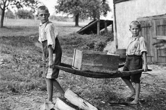
“Swiss former indentured child laborers (Verdingkinder) were removed as children from their families by the authorities due to different reasons (poverty, being born out of wedlock) and were placed to live and work on farms. This was a practice applied until the 1950s and many of the Verdingkinder were subjected to childhood trauma and neglect during the indentured labor.

DNA methylation modifications indicated experiment-wide significant associations with the following complex posttraumatic symptom domains: dissociation, tension reduction behavior and dysfunctional sexual behavior.”


https://bmcresnotes.biomedcentral.com/articles/10.1186/s13104-017-3082-y “A pilot investigation on DNA methylation modifications associated with complex posttraumatic symptoms in elderly traumatized in childhood”


Imagine being taken away from your family during early childhood for no other reason than your parents weren’t married.

Consider just a few of the painful feelings such a child had to deal with then and ever since:

  • I’m unloved.
  • Alone.
  • No one can help me.

Imagine some of the ways a child had to adapt during their formative years because of this undeserved punishment:

  • How fulfilling it would be to believe that they were loved, even by someone they couldn’t see, touch, or hear.
  • How fulfilling it would be to get attention from someone, anyone.
  • How a child became conditioned to do things by themself without asking for help.

The study described a minute set of measurements of the subjects’ traumatic experiences and their consequential symptoms. The researchers tried to group this tiny sample of the subjects’ symptoms into a new invented category.


Another example was provided in Is IQ an adequate measure of the quality of a young man’s life?:

“During this time period [between 1955 and 1990], because private adoptions were prohibited by Swedish law, children were taken into institutional care by the municipalities shortly after birth and adopted at a median age of 6 mo, with very few children adopted after 12 mo of age.”

Swedish society deemed local institutional care the initial destination for disenfranchised infants, regardless of whether suitable families were willing and able to adopt the infants. What happened to infants who weren’t adopted by age 1?

Did Swedish society really need any further research to know that an adoptive family’s care would be better for a child than living in an institution?


It’s hard to recognize when our own thoughts, feelings, and behavior provide evidence of childhood pain that’s still with us.

Let’s not hope and believe that the societies we live in will resolve adverse effects of childhood trauma its members caused. Other people may guide us, but each of us has to individually get our life back:

“What is the point of life if we cannot feel and love others? Without feeling, life becomes empty and sterile.

It, above all, loses its meaning.

Every society has its horror stories. People who have reached some degree of honesty about their early lives and concomitant empathy for others can document these terrible circumstances and events.

Have traumatic effects on children from societal policies ceased?

A review of biological variability

This 2017 UK/Spanish review subject was biological variability:

“No two cells in a cellular population are the same, and no two individuals of a multi-cellular species are identical-not even if they share the same genetic makeup like monozygotic twins or cloned animals.

Epigenetic and gene expression variability are key contributors to phenotypic differences. There are many possible sources of epigenetic and transcriptional variability, which can be divided into three main categories:

  1. individual-intrinsic factors;
  2. environmental factors; and
  3. random fluctuations, also referred to as stochasticity.”

Most of the review cited cell studies. The reviewers cited their own studies in the Introduction section, for example:

“These studies were among the first to classify disease status or aggressiveness based on variability, where the classical comparison of mean DNA methylation or gene expression levels was not informative.”

to help support a later observation:

“It is critical to obtain a measurement of variability that is independent of the mean to ensure to not confound changes in variability with shifts in mean.”


The review didn’t cover a pertinent aspect of the subject: how standard research approaches miss detecting biological variability.

For example, from Changing an individual’s future behavior even before they’re born that referred to the methodology of genome-wide association studies (GWAS):

“When phenotypic variation results from alleles that modify phenotypic variance rather than the mean, this link between genotype and phenotype will not be detected.”

Another omission was the point made in A study of DNA methylation and age:

“Due to the methods applied in the present study, not all the effects of DNA methylation on gene expression could be detected; this limitation is also true for previously reported results.

The textbook case of DNA methylation regulating gene expression (the methylation of a promoter and silencing of a gene) remains undetected in many cases because in an array analysis, an unexpressed gene shows no signal that can be distinguished from background and is therefore typically omitted from the analysis.”


The reviewers also didn’t cover variability in phenotypic behaviors. I’ll repeat my thoughts from A limited study of parental transmission of anxiety/stress-reactive traits:

“How did parental behavioral transmission of behavioral traits and epigenetic changes become a subject not worth investigating? These traits and effects can be seen everyday in real-life human interactions, and in every human’s physiology.

Perhaps these omissions reflected the reviewers’ focus on their specialties?

Perhaps it isn’t politically correct to discuss or fund research on aspects of biological variability that would advance science by falsifying preferred previous findings? Or advance science by measuring the extent of parental involvement in shaping their offspring’s behavioral and biological variability?

What do you think?

http://onlinelibrary.wiley.com/doi/10.1002/bies.201700148/full “Epigenetic and Transcriptional Variability Shape Phenotypic Plasticity”


This post has somehow become a target for spammers, and I’ve disabled comments. Readers can comment on other posts and indicate that they want their comment to apply here, and I’ll re-enable comments.