Eat your oats!

Here’s some motivation to replenish your oats supply.

From a 2013 Canadian human review:

“Review of human studies investigating the post-prandial blood-glucose lowering ability of oat and barley food products” https://www.nature.com/articles/ejcn201325

“Change in glycaemic response (expressed as incremental area under the post-prandial blood-glucose curve) was greater for intact grains than for processed foods. For processed foods, glycaemic response was more strongly related to the β-glucan dose alone than to the ratio of β-glucan to the available carbohydrate.”

The review found that people don’t have to eat a lot of carbohydrates to get the glycemic-response benefits of β-glucan. Also, eating ~3 grams of β-glucan in whole oats and barley will deliver the same glycemic-response benefits as eating ~4 grams of β-glucan in processed oats and barley.

However, the glycemic index used in the review is a very flawed measure. What’s the point of indexing healthy choices like whole grains to unhealthy choices that healthy people aren’t going to make anyway?


The reviewer somewhat redeemed herself by participating in a 2018 review:

“Processing of oat: the impact on oat’s cholesterol lowering effect” https://www.ncbi.nlm.nih.gov/pmc/articles/PMC5885279/

“For a similar dose of β-glucan:

  1. Liquid oat-based foods seem to give more consistent, but moderate reductions in cholesterol than semi-solid or solid foods where the results are more variable;
  2. The quantity of β-glucan and the molecular weight at expected consumption levels (∼3 g day) play a role in cholesterol reduction; and
  3. Unrefined β-glucan-rich oat-based foods (where some of the plant tissue remains intact) often appear more efficient at lowering cholesterol than purified β-glucan added as an ingredient.”

The review’s sections 3. Degree of processing and functionality and 4. Synergistic action of oat constituents were informative:

“Both in vitro and in vivo studies clearly demonstrated the beneficial effect of oat on cholesterolemia, which is unlikely to be due exclusively to β-glucan, but rather to a combined and synergetic action of several oat compounds acting together to reduce blood cholesterol levels.”


Another use of β-glucan is to improve immune response. Here’s a 2016 Netherlands study where the researchers used β-glucan to get a dozen people well after making them sick with lipopolysaccharide as is often done in animal studies:

“β-Glucan Reverses the Epigenetic State of LPS-Induced Immunological Tolerance” https://www.ncbi.nlm.nih.gov/pmc/articles/PMC5927328/

“The innate immune ‘training stimulus’ β-glucan can reverse macrophage tolerance ex vivo.”

I’ve curated other research on β-glucan’s immune-response benefits in:

Adverse epigenetic effects of prenatal and perinatal anesthesia

This 2018 Chinese animal review subject was prenatal and perinatal anesthesia’s adverse epigenetic effects on a fetus/neonate:

“Accumulating evidence from rodent and primate studies has demonstrated that in utero or neonatal exposure to commonly used inhaled and intravenous general anesthetics is associated with neural degeneration and subsequent neurocognitive impairments, manifested in learning and memory disabilities.

So far, conflicting data exist about the effect of anesthetic agents on neurodevelopment in humans and no definite conclusion has been given yet.”

The inhibitors in the above graphic counter anesthesia’s effects on the fetus/neonate, summarized as:

“Epigenetic targeting of DNA methyltransferases and/or histone deacetylases may have some therapeutic value.”


Do physicians consider possible epigenetic alterations of a newborn’s chromatin structure and gene expression when they administer anesthesia to mothers during childbirth?

https://www.ncbi.nlm.nih.gov/pmc/articles/PMC6079265/ “Epigenetic Alterations in Anesthesia-Induced Neurotoxicity in the Developing Brain”

A top-down view of biological goal-directed mechanisms

This 2016 US/Italy article was written from a perspective of regenerative bioengineering:

“Higher levels beyond molecular can have their own unique dynamics that offer better (e.g. more parsimonious and potent) explanatory power than models made at lower levels. Biological systems may be best amenable to models that include information structures (organ shape, size, topological arrangements and complex anatomical metrics) not defined at molecular or cellular level but nevertheless serving as the most causally potent ‘knobs’ regulating large-scale outcomes.

Top-down models can be as quantitative as familiar bottom-up systems biology examples, but they are formulated in terms of building blocks that cannot be defined at the level of gene expression and treat those elements as bona fide causal agents (which can be manipulated by interventions and optimization techniques). The near-impossibility of determining which low-level components must be tweaked in order to achieve a specific system-level outcome is a problem that plagues most complex systems.

The current paradigm in biology of exclusively tracking physical measurable and ignoring internal representation and information structures in patterning contexts quite resemble the ultimately unsuccessful behaviourist programme in psychology and neuroscience. For example, even if stem cell biologists knew how to make any desired cell type from an undifferentiated progenitor, the task of assembling them into a limb would be quite intractable.

Current state of the art in the field of developmental bioelectricity is that it is known, at the cellular level, how resting potentials are transduced into downstream gene cascades, as well as which transcriptional and epigenetic targets are sensitive to change in developmental bioelectrical signals. What is largely missing however is a quantitative understanding of how global dynamics of bioelectric circuits make decisions that orchestrate large numbers of individual cells, spread out over considerable anatomical distances, towards specific pattern outcomes.”


Regenerative research is gathering evidence for goal-directed memory and learning that doesn’t meet current definitions. For example:

salamander

“A tail grafted to the flank of a salamander slowly remodels to a limb, a structure more appropriate for its new location, illustrating shape homeostasis towards a normal amphibian body plan. Even tail tip cells (in red) slowly become fingers, showing that remodelling is not driven by only local information.”

These reviewers compared their findings to several existing research and real-world-operations domains. Other models may also benefit from concepts of:

“Quantitative, predictive, mechanistic understanding of goal-directed morphogenesis.”

https://royalsocietypublishing.org/doi/full/10.1098/rsif.2016.0555 “Top-down models in biology: explanation and control of complex living systems above the molecular level”


I came across this article as a result of its citation in The Body Electric blog post.

“Levin drops a hint that there are photo-sensitive drugs that can control ion gates that can be used to translate a projected geometric image into a pattern of membrane potentials. He argues that the patterns encode ‘blueprints’ rather than a ‘construction manual’ based on the fact that the program is adaptive in the face of physical barriers and disruptions.”

Epigenetic clock statistics and methods

This 2018 Chinese study was a series of statistical and methodological counter-arguments to a previous epigenetic clock study finding that:

“Only [CpG] sites mapping to the ELOVL2 promoter constitute cell and tissue-type independent aDMPs [age-associated differentially methylated positions].”

The study used external data sets and the newer epigenetic clock’s fibroblast data in its analyses to find:

“While we agree that specific sites mapping to ELOVL2 are special aDMPs in the sense that their effect sizes are particularly large across a number of different tissue-types, our analysis suggests that most aDMPs are valid across multiple different tissue types, suggesting that shared aDMPs are common.”

The details of each of the study’s counter-arguments were compelling. For example:

“We analyzed Illumina 850k data from an EWAS profiling blood, buccal and cervical samples from a common set of 263 women. Because blood is a complex mixture of many immune-cell subtypes, and buccal and cervical samples are highly contaminated by immune cells, we identified aDMPs in each tissue after adjustment for batch effects and cell-type heterogeneity.

Using either an FDR [false discovery rate] < 0.05 or Bonferroni adjusted P-value < 0.05 thresholds, the overlap of aDMPs between the 3 tissues was highly significant, mimicking the result obtained on blood cell subtypes. We observed a total of 2200 aDMPs in common between blood, buccal and cervix, an overlap which cannot be explained by random chance.”

The study’s Discussion section provided qualifications and limitations such as:

“It is important to point out that even if age-associated DNAm changes are widespread across the genome, downstream functional effects may be rare. While specific aDMPs may be shared between tissue-types, it is only in specific tissues or cell-types that any associated functional deregulation may be of biological and clinical significance.“

https://www.aging-us.com/article/101666/text “Cell and tissue type independent age-associated DNA methylation changes are not rare but common”


The November 2018 issue of Aging also contained other articles of interest:

https://www.aging-us.com/article/101626/text “Accelerated DNA methylation age and the use of antihypertensive medication among older adults”

“DNAmAge and AA [age acceleration] may not be able to capture the preventive effects of AHMs [antihypertensive medications] that reduce cardiovascular risks and mortality.”

https://www.aging-us.com/article/101633/text “Azithromycin and Roxithromycin define a new family of senolytic drugs that target senescent human fibroblasts”

“Azithromycin preferentially targets senescent cells, removing approximately 97% of them with great efficiency. This represents a near 25-fold reduction in senescent cells.”

https://www.aging-us.com/article/101647/text “Disease or not, aging is easily treatable”

“Aging consists of progression from (pre)-pre-diseases (early aging) to diseases (late aging associated with functional decline). Aging is NOT a risk factor for these diseases, as aging consists of these diseases: aging and diseases are inseparable.”

Chronological age by itself is an outdated clinical measurement

This 2018 editorial in the New England Journal of Medicine concerned a clinical trial of an osteoporosis treatment:

“When measurement of bone density was first introduced 25 years ago, absolute bone mineral density (g per square centimeter) was considered as too onerous for clinicians to understand. Ultimately, these events led to a treatment gap in patients who had strong clinical risk factors for an osteoporotic fracture (particularly age) but had T scores in the osteopenic range.

The average age of the participants in the current trial was approximately 3.5 years older than that in the Fracture Intervention Trial. Owing to the interaction between age and bone mineral density, the results of the current trial should not be extrapolated to younger postmenopausal women (50 to 64 years of age) with osteopenia.

This trial reminds us that risk assessment and treatment decisions go well beyond bone mineral density and should focus particularly on age and a history of previous fractures.”

https://www.nejm.org/doi/pdf/10.1056/NEJMe1812434 “A Not-So-New Treatment for Old Bones”


This editorial provided some history of how a still-generally-accepted set of diagnostic measurements were selected for their relative convenience instead of chosen for their efficacy. Add chronological age to such ineffective measurements.

Let’s recognize better aging and diagnostic measurements, then incorporate them. How else will we advance past the above uninformative averaging and unhelpful recommendation based on chronological age?

The time has passed for physicians and clinicians to consider only chronological age when evaluating a patient’s clinical age. More effective human age measurements covering the entire person as well as their body’s components include:

F2.large

A slanted view of the epigenetic clock

The founder of the epigenetic clock technique was interviewed for MIT Technology Review:

“We need to find ways to keep people healthier longer,” he says. He hopes that refinements to his clock will soon make it precise enough to reflect changes in lifestyle and behavior.”


The journalist attempted to dumb the subject down “for the rest of us” with distortions such as the headline. The varying correlation of epigenetic age to chronological age was somewhat better reported in the story:

“The epigenetic clock is more accurate the younger a person is. It’s especially inaccurate for the very old.”

The journalist inappropriately used luck as a synonym for randomness/stochasticity:

“He estimates that about 40% of the ticking rate is determined by genetic inheritance, and the rest by lifestyle and luck.”

A third example of less-than-straightforward journalism started with:

“Such personalization raises questions about fairness. If your epigenetic clock is ticking faster through no fault of your own..”

Were MIT Technology Review readers unable to comprehend a straightforward story on the epigenetic clock? What was the purpose of slants and distortions in an introductory article?

https://www.technologyreview.com/s/612256/want-to-know-when-youre-going-to-die/ “Want to know when you’re going to die?”

Epigenetic transgenerational inheritance of ovarian disease

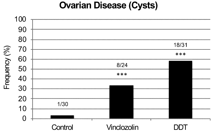
This 2018 Washington rodent study investigated ovarian disease in F3 great-granddaughters caused by their F0 great-grandmothers’ exposures to DDT or vinclozolin while pregnant:

“Two of the most prevalent ovarian diseases affecting women’s fertility and health are Primary Ovarian Insufficiency (POI) and Polycystic Ovarian Syndrome (PCOS). POI is characterized by a marked reduction in the primordial follicle pool of oocytes and the induction of menopause prior to age 40. POI currently affects approximately 1% of female population. While genetic causes can be ascribed to a minority of patients, around 90% of POI cases are considered idiopathic, with no apparent genetic link nor known cause.

PCOS is a multi-faceted disease that affects 6-18% of women. It is characterized by infrequent ovulation or anovulation, high androgen levels in the blood, and the presence of multiple persistent ovarian cysts.

For both PCOS and POI other underlying causes such as epigenetic transgenerational inheritance of disease susceptibility have seldom been considered. Epigenetic transgenerational inheritance is defined as “the germline transmission of epigenetic information and phenotypic change across generations in the absence of any continued direct environmental exposure or genetic manipulation.” Epigenetic factors include:

    • DNA methylation,
    • Histone modifications,
    • Expression of noncoding RNA,
    • RNA methylation, and
    • Alterations in chromatin structure.

The majority of transgenerational studies have examined sperm transmission of epigenetic changes due to limitations in oocyte numbers for efficient analysis.

There was no increase in ovarian disease in direct fetal exposed F1 [grandmothers] or germline exposed F2 [mothers] generation vinclozolin or DDT lineage rats compared to controls.

F3 generation ovarian disease

The transgenerational molecular mechanism is distinct and involves the germline (sperm or egg) having an altered epigenome that following fertilization may modify the embryonic stem cells epigenome and transcriptome. This subsequently impacts the epigenetics and transcriptome of all somatic cell types derived from these stem cells.

Therefore, all somatic cells in the transgenerational [F3] animal have altered epigenomes and transcriptomes and those sensitive to this alteration will be susceptible to develop disease. The F3 generation can have disease while the F1 and F2 generations do not, due to this difference in the molecular mechanisms involved.

The epimutations and gene expression differences observed are present in granulosa cells in the late pubertal female rats at 22-24 days of age, which is long before any visible signs of ovarian disease are detectable. This indicates that the underlying factors that can contribute to adult-onset diseases like PCOS and POI appear to be present early in life.

Ancestral exposure to toxicants is a risk factor that must be considered in the molecular etiology of ovarian disease.”


1. The study highlighted a great opportunity for researchers of any disease that frequently has an “idiopathic” diagnosis. It said a lot about research priorities that “around 90% of POI cases are considered idiopathic, with no apparent genetic link nor known cause.”

It isn’t sufficiently explanatory for physicians to continue using categorization terminology from thousands of years ago. Science has progressed enough with measured evidence to discard the “idiopathic” category and express probabilistic understanding of causes.

2. One of this study’s coauthors made a point worth repeating in The imperative of human transgenerational studies: What’s keeping researchers from making a significant difference in their fields with human epigenetic transgenerational inheritance studies?

3. Parts of the study’s Discussion section weren’t supported by its evidence. The study didn’t demonstrate:

  • That “all somatic cells in the transgenerational animal have altered epigenomes and transcriptomes”; and
  • The precise “molecular mechanisms involved” that exactly explain why “the F3 generation can have disease while the F1 and F2 generations do not.”

https://www.tandfonline.com/doi/abs/10.1080/15592294.2018.1521223 “Environmental Toxicant Induced Epigenetic Transgenerational Inheritance of Ovarian Pathology and Granulosa Cell Epigenome and Transcriptome Alterations: Ancestral Origins of Polycystic Ovarian Syndrome and Primary Ovarian Insuf[f]iency” (not freely available)

The imperative of human transgenerational studies

The coauthor of:

pointed out the opportunity for the researchers of A seasonal epigenetic effect of conception on BMI to have their work make a difference in their field:

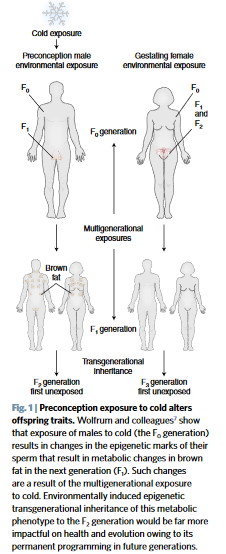
“The ability of environmental epigenetics to promote an adaptive phenotype to cold has impacts on evolution. However, the impacts would be far greater if the phenomenon was transgenerational.

Future studies are now needed to determine whether the cold-induced thrifty metabolic phenotype is transmitted to subsequent generations. If exposure not only impacts the health of offspring, but also of all subsequent generations, the impact is significant.”


Every human alive today has observable lasting epigenetic effects caused by environmental factors:

  • During the earliest parts of our lives;
  • From our parents’ exposures and experiences before we’re conceived – many of which are inadequately researched; and
  • Potentially from some of our earlier ancestors’ exposures and experiences.

Aren’t animal studies’ evidence for epigenetic transgenerational inheritance sufficient to compel serious human follow-on research efforts by research sponsors and study designers?

The same comments about epigenetic effects caused by temperature potentially inherited by multiple human generations can also be made about other environmental factors, such as:

  • Nutrition,
  • Toxins – the commentator’s usual area of study, and
  • Stress.

I hope that these researchers value their professions enough to make a difference with this or other areas of their expertise. And that sponsors won’t thwart researchers’ desires for difference-making science by putting them into endless funding queues.

https://www.nature.com/articles/s41591-018-0187-3 “Preconception cold-induced epigenetic inheritance” (not freely available)

Reversing epigenetic changes with CRISPR/Cas9

This 2018 Chinese review highlighted areas in which CRISPR/Cas9 technology has, is, and could be applied to rewrite epigenetic changes:

“CRISPR/Cas9-mediated epigenome editing holds a great promise for epigenetic studies and therapeutics.

It could be used to selectively modify epigenetic marks at a given locus to explore mechanisms of how targeted epigenetic alterations would affect transcription regulation and cause subsequent phenotype changes. For example, inducing histone methylation or acetylation at the Fosb locus in the mice brain reward region, nucleus accumbens, could affect relevant transcription network and thus control behavioral responses evoked by drug and stress.

Epigenome editing has the potential for epigenetic treatment, especially for the disorders with abnormal gene imprinting or epigenetic marks. Targeted epigenetic silencing or reactivation of the mutant allele could be a potential therapeutic approach for diseases such as Rett syndrome and Huntington’s disease.

Noncoding RNA plays important roles in gene imprinting and chromatin remodeling. CRISPR/Cas9 has been shown to be potential for manipulating noncoding RNA expression, including microRNA, long noncoding RNA, and miRNA families and clusters.

In vivo overexpression of the Yamanaka factors have proven to be able to fully or partially help somatic cells to regain pluripotency in situ. These rejuvenated cells would subsequently differentiate again to replace the lost cell types.”


The last paragraph was described in The epigenetic clock theory of aging as a promising technique:

“To date, the most effective in vitro intervention against epigenetic ageing is achieved through expression of Yamanaka factors, which convert somatic cells into pluripotent stem cells, thereby completely resetting the epigenetic clock.”

The reviewers cited three references for in vivo studies of this technique. Overall, I didn’t see that any of the review’s references were in vivo human studies.

https://www.ncbi.nlm.nih.gov/pmc/articles/PMC6079388/ “Novel Epigenetic Techniques Provided by the CRISPR/Cas9 System”

The epigenetic clock now includes skin

The originator of the 2013 epigenetic clock improved its coverage with this 2018 UCLA human study:

“We present a new DNA methylation-based biomarker (based on 391 CpGs) that was developed to accurately measure the age of human fibroblasts, keratinocytes, buccal cells, endothelial cells, skin and blood samples. We also observe strong age correlations in sorted neurons, glia, brain, liver, and bone samples.

The skin & blood clock outperforms widely used existing biomarkers when it comes to accurately measuring the age of an individual based on DNA extracted from skin, dermis, epidermis, blood, saliva, buccal swabs, and endothelial cells. Thus, the biomarker can also be used for forensic and biomedical applications involving human specimens.

The biomarker applies to the entire age span starting from newborns, e.g. DNAm of cord blood samples correlates with gestational week.

Furthermore, the skin & blood clock confirms the effect of lifestyle and demographic variables on epigenetic aging. Essentially it highlights a significant trend of accelerated epigenetic aging with sub-clinical indicators of poor health.

Conversely, reduced aging rate is correlated with known health-improving features such as physical exercise, fish consumption, high carotenoid levels. As with the other age predictors, the skin & blood clock is also able to predict time to death.

Collectively, these features show that while the skin & blood clock is clearly superior in its performance on skin cells, it crucially retained all the other features that are common to other existing age estimators.”

http://www.aging-us.com/article/101508/text “Epigenetic clock for skin and blood cells applied to Hutchinson Gilford Progeria Syndrome and ex vivo studies”


An introduction to the study highlighted several items:

“Although the skin-blood clock was derived from significantly less samples (~900) than Horvath’s clock (~8000 samples), it was found to more accurately predict chronological age, not only across fibroblasts and skin, but also across blood, buccal and saliva tissue. A potential factor driving this improved accuracy in blood could be related to the approximate 18-fold increase in genomic coverage afforded by using Illumina 450k/850k beadarrays.

It serves as a roadmap for future clock studies, pointing towards the importance of constructing tissue or cell-type specific epigenetic clocks, to more accurately measure biological aging in the given tissue/cell-type, and therefore with the potential to be more informative of disease-risk or the success of disease interventions in the tissue or cell-type of interest.”

http://www.aging-us.com/article/101533/text “Epigenetic clocks galore: a new improved clock predicts age-acceleration in Hutchinson Gilford Progeria Syndrome patients”

Epigenetic factors affecting female rat sexual behavior

This 2018 Baltimore/Montreal rodent study found:

“If sexually naïve females have their formative sexually rewarding experiences paired with the same male, they will recognize that male and display mate-guarding behavior towards him in the presence of a female competitor. Female rats that display mate-guarding behavior also show enhanced activation of oxytocin and vasopressin neurons in the supraoptic and paraventricular hypothalamic nucleus.

We examined the effect of a lysine-specific demethylase-1 inhibitor to block the action of demethylase enzymes and maintain the methylation state of corresponding genes. Female rats treated with the demethylase inhibitor failed to show any measure of mate guarding, whereas females treated with vehicle displayed mate guarding behavior. Demethylase inhibitor treatment also blocked the ability of familiar male cues to activate oxytocin and vasopressin neurons, whereas vehicle-treated females showed this enhanced activation.”

General principles and their study-specific illustrations were:

“Histone modifications are a key element in gene regulation through chromatin remodeling. Histone methylation / demethylation does not have straightforward transcriptional outcomes as do other histone modifications, like acetylation, which is almost invariably associated with transcriptional activation.

What is of vital importance in regards to histone methylation / demethylation is the pattern of methylation that is established. Patterns of methylation incorporate both methylated and demethylated residues, and are what ultimately play a role in transcriptional outcomes.

In the present study, inhibiting LSD1 demethylase enzymes disrupted the ability of cells to properly establish histone methylation / demethylation patterns, thus creating a deficit in the cells’ ability to transcribe the gene products necessary for the enhanced induction of OT, AVP, and the subsequent mate-guarding behaviors we observed. This study is the first to demonstrate a definitive role of epigenetic histone modifications in a conditioned sexual response.”

https://www.sciencedirect.com/science/article/pii/S0031938418303421 “Inhibition of lysine-specific demethylase enzyme disrupts sexually conditioned mate guarding in the female rat” (not freely available)

Hijacking the epigenetic clock paradigm

This 2018 German human study’s last sentence was:

“Additionally we found an association between DNAm [DNA methylation] age acceleration and rLTL [relative leukocyte telomere length], suggesting that this epigenetic clock, at least partially and possibly better than other epigenetic clocks, reflects biological age.”

Statements in the study that contradicted, qualified, and limited the concluding sentence included:

“The epigenetic clock seems to be mostly independent from the mitotic clock as measured by the rLTL.

It could be possible that associations are confounded due to short age ranges or non-continuous age distribution, as displayed in the BASE-II cohort (no participants between the age of 38 and 59 years). [see the below graphic]

The BASE-II is a convenience sample and participants have been shown to be positively selected with respect to education, health and cognition.

Samples in which DNAm age and chronological age differed more than three standard deviations from the mean were excluded (N=19).

While the original publication employed eight CpG sites for DNAm age estimation, we found that one of these sites did not significantly improve chronological age prediction in BASE-II. Thus, we reduced the number of sites considered to seven in the present study and adapted the algorithm to calculate DNAm age.

  • Horvath described a subset of 353 methylation sites predicting an individual’s chronological age with high accuracy..
  • Even though the available methods using more CpG sites to estimate DNAm age predict chronological age with higher accuracy..
  • It is not clear how much of the deviation between chronological age and DNAm age reflects measurement error/low number of methylation sites and which proportion can be attributed to biological age.

Due to the statistical method employed, we encountered a systematic deviation of DNAm age in our dataset.”


Findings that aren’t warranted by the data is an all-too-common problem with published research. This study illustrated how researcher hypothesis-seeking behavior – that disregarded what they knew or should have known – can combine with a statistics package to produce almost any finding.

It reminded me of A skin study that could have benefited from preregistration that made a similar methodological blunder:

The barbell shape of the subjects’ age distribution wouldn’t make sense if the researchers knew they were going to later use the epigenetic clock method.

The researchers did so, although the method’s instructive study noted:

“The standard deviation of age has a strong relationship with age correlation”

and provided further details in “The age correlation in a data set is determined by the standard deviation of age” section.

Didn’t the researchers, their organizations, and their sponsors realize that this study’s problematic design and performance could misdirect readers away from the valid epigenetic clock evidence they referenced? What purposes did it serve for them to publish this study?

https://academic.oup.com/biomedgerontology/advance-article-abstract/doi/10.1093/gerona/gly184/5076188 “Epigenetic clock and relative telomere length represent largely different aspects of aging in the Berlin Aging Study II (BASE-II)” (not freely available)

The role of recall neurons in traumatic memories

This 2018 Swiss rodent study found:

“Our data show that:

  • A subset of memory recall–induced neurons in the DG [dentate gyrus] becomes reactivated after memory attenuation,
  • The degree of fear reduction positively correlates with this reactivation, and
  • The continued activity of memory recall–induced neurons is critical for remote fear memory attenuation.

Although other brain areas such as the prefrontal cortex and the amygdala are likely to be implicated in remote fear memories and remain to be investigated, these results suggest that fear attenuation at least partially occurs in memory recall–induced ensembles through updating or unlearning of the original memory trace of fear.

These data thereby provide the first evidence at an engram-specific level that fear attenuation may not be driven only by extinction learning, that is, by an inhibitory memory trace different from the original fear trace.

Rather, our findings indicate that during remote fear memory attenuation both mechanisms likely coexist, albeit with the importance of the continued activity of memory recall–induced neurons experimentally documented herein. Such activity may not only represent the capacity for a valence change in DG engram cells but also be a prerequisite for memory reconsolidation, namely, an opportunity for learning inside the original memory trace.

As such, this activity likely constitutes a physiological correlate sine qua non for effective exposure therapies against traumatic memories in humans: the engagement, rather than the suppression, of the original trauma.”

The researchers also provided examples of human trauma:

“We dedicate this work to O.K.’s father, Mohamed Salah El-Dien, and J.G.’s mother, Wilma, who both sadly passed away during its completion.”


So, how can this study help humans? The study had disclosed and undisclosed limitations:

1. Humans aren’t lab rats. We can ourselves individually change our responses to experiential causes of ongoing adverse effects. Standard methodologies can only apply external treatments.

2. It’s a bridge too far to go from neural activity in transgenic mice to expressing unfounded opinions on:

“A physiological correlate sine qua non for effective exposure therapies against traumatic memories in humans.”

Human exposure therapies have many drawbacks, in addition to being applied externally to the patient on someone else’s schedule. A few others were discussed in The role of DNMT3a in fear memories:

  • “Inability to generalize its efficacy over time,
  • Potential return of adverse memory in the new/novel contexts,
  • Context-dependent nature of extinction which is widely viewed as the biological basis of exposure therapy.”

3. Rodent neural activity also doesn’t elevate recall to become an important goal of effective human therapies. Clearly, what the rodents experienced should have been translated into human reliving/re-experiencing, not recall! Terminology used in animal studies preferentially has the same meaning with humans, since the purpose of animal studies is to help humans.

4. The researchers acknowledged that:

“Other brain areas such as the prefrontal cortex and the amygdala are likely to be implicated in remote fear memories and remain to be investigated.”

A study that provided evidence for basic principles of Primal Therapy determined another brain area:

“The findings imply that in response to traumatic stress, some individuals, instead of activating the glutamate system to store memories, activate the extra-synaptic GABA system and form inaccessible traumatic memories.”

The study I curated yesterday, Organ epigenetic memory, demonstrated organ memory storage. It’s hard to completely rule out that other body areas may also store traumatic memories.

The wide range of epigenetic memory storage vehicles is one reason why effective human therapies need to address the whole person, the whole body, and each individual’s entire history.

http://science.sciencemag.org/content/360/6394/1239 “Reactivation of recall-induced neurons contributes to remote fear memory attenuation” (not freely available)

Here’s one of the researchers’ outline:


This post has somehow become a target for spammers, and I’ve disabled comments. Readers can comment on other posts and indicate that they want their comment to apply here, and I’ll re-enable comments.

Organ epigenetic memory

This 2018 Japanese review subject was the relationships of organ memory and non-communicable diseases:

“Organ memory is the engraved phenotype of altered organ responsiveness acquired by a time-dependent accumulation of organ stress responses. This phenomenon is known as “metabolic memory” or “legacy effect,” which is similar to neuronal and immune memory.

Not only is the epigenetic change of key genes involved in the formation of organ memory but the alteration of multiple factors, including low molecular weight energy metabolites, immune mediators, and tissue structures, is involved as well. These factors intercommunicate during every stress response and carry out incessant remodeling in a certain direction in a spiral fashion through positive feedback mechanisms.

The systematic review revealed that each intervention type, that is:

  • Glucose lowering,
  • Blood pressure lowering, or
  • LDL-cholesterol lowering,

possessed unique characteristics of the memory phenomenon. Most of the observational periods of these studies lasted for > 10 years. Memory phenomenon was suggested to last for a long time and is thought to have a considerable effect on the clinical course of NCDs [non-communicable diseases].

Organs cannot possess consciousness, so it might not be appropriate to consider whether a recalling process exists in organs. However, the properties of organs are incessantly altered by external stimuli loaded on organs as if it is updating.

It is clinically important to investigate whether organ memory can be updated by our behaviors. Once organ memory is established in an organ, organ memory in each organ can influence one another and affect organ memory in a different organ.

Epigenome-modification enzymes, such as histone deacetylases and DNA methyltransferases, and transcription factors seem to be essential for the epigenetic regulation of gene expression, which is involved in the generation of organ memory. Cellular metabolism can epigenetically modulate the expression of genes that are related to the progression of diseases.”

https://www.nature.com/articles/s41440-018-0081-x “Organ memory: a key principle for understanding the pathophysiology of hypertension and other non-communicable diseases” (not freely available)


1. The reviewers asserted:

“Organs cannot possess consciousness, so it might not be appropriate to consider whether a recalling process exists in organs.”

Memory studies don’t require this consciousness to investigate even brain areas and functions. Researchers observe memory by measuring stimulus/response items like neuron activation and various levels of behavior. Consciousness is an emergent property.

2. Regarding recall: An organ’s “engraved phenotype of altered organ responsiveness” may not have recall itself, but it doesn’t have a separate existence apart from its body. An organ can’t be removed from its body for very long and still be part of its body.

When an organ is in its normal state as part of a body, it has access to recall-like functions via “inter-organ communication of organ memory.” The review also mentioned:

“Organ memory in each organ can influence one another and affect organ memory in a different organ.

Evolution didn’t support unnecessary duplication for a kidney’s memory to include recall because it’s part of a body that includes a brain that has recall. Evolution didn’t duplicate functions of a kidney’s memory in a brain, either.

Flawed epigenetic measurements of behavioral experiences

This 2018 New York rodent study not only wasted resources but also speciously attempted to extrapolate animal study findings to humans:

“While it is clear that behavioral experience modulates epigenetic profiles, it is less evident how the nature of that experience influences outcomes and whether epigenetic/genetic “biomarkers” could be extracted to classify different types of behavioral experience.

Male and female mice were subjected to either:

  • a Fixed Interval (FI) schedule of food reward, or
  • a single episode of forced swim followed by restraint stress, or
  • no explicit behavioral experience

after which global expression levels of two activating (H3K9ac and H3K4me3) and two repressive (H3K9me2 and H3k27me3) post-translational histone modifications (PTHMs), were measured in hippocampus (HIPP) and frontal cortex (FC).

A random subset of 5 of the 12 animals from each sex/behavioral experience group were used for these analyses. FC and HIPP were dissected from each of those 5 brains and homogenized for subsequent analyses. Thus, sample size for PTHM expression levels was n = 5 for each region/sex/behavioral treatment group and all PTHM expression level analyses utilized the homogenized tissue.

The specific nature of the behavioral experience differentiated profiles of PTHMs in a sex- and brain region-dependent manner, with all 4 PTHMs changing in parallel in response to different behavioral experiences. Global PTHMs may provide a higher-order pattern recognition function.”


The researchers knew or should have known that measuring “global expression levels” in “homogenized tissue” of “n = 5” subjects was flawed, and they did it anyway. They acknowledged some of the numerous study design defects with qualifiers such as:

“Even though these were global levels of histone modifications (and thus not indicative of changes at specific genes or sites on genes)..

As FS-RS behavioral experience was completed before FI behavioral experience, a longer overall post-behavior experience time (approximately 1 week) elapsed for this group, resulting in some differences in overall timing between these experiences and global PTHM assessment. However, extending the duration of the FS-RS experience (i.e., repeated exposures) would also have led to habituation..”

Did they purposely make these mistakes because of the “biomarkers” paradigm?

What would they have found if they had followed their judgments and training to design a better study? Experience-dependent histone modifications that differed by gender and brain region was certainly a promising research opportunity.

As for extrapolating the cited animal study findings to humans? Ummm..NO!

https://www.ncbi.nlm.nih.gov/pmc/articles/PMC6060276/ “Different Behavioral Experiences Produce Distinctive Parallel Changes in, and Correlate With, Frontal Cortex and Hippocampal Global Post-translational Histone Levels”